Related Research Articles
Panniculitis is a group of diseases whose hallmark is inflammation of subcutaneous adipose tissue. Symptoms include tender skin nodules, and systemic signs such as weight loss and fatigue.

Lupus vulgaris are painful cutaneous tuberculosis skin lesions with nodular appearance, most often on the face around the nose, eyelids, lips, cheeks, ears and neck. It is the most common Mycobacterium tuberculosis skin infection. The lesions may ultimately develop into disfiguring skin ulcers if left untreated.
Discoid lupus erythematosus (DLE) is an uncommon autoimmune disease of the basal cell layer of the skin. It occurs in humans and cats, more frequently occurring in dogs. It was first described in dogs by Griffin and colleagues in 1979. DLE is one form of cutaneous lupus erythematosus (CLE). DLE occurs in dogs in two forms: a classical facial predominant form or generalized with other areas of the body affected. Other non-discoid variants of CLE include vesicular CLE, exfoliative CLE and mucocutaenous CLE. It does not progress to systemic lupus erythematosus (SLE) in dogs. SLE can also have skin symptoms, but it appears that the two are either separate diseases. DLE in dogs differs from SLE in humans in that plasma cells predominate histologically instead of T lymphocytes. Because worsening of symptoms occurs with increased ultraviolet light exposure, sun exposure most likely plays a role in DLE, although certain breeds are predisposed. After pemphigus foliaceus, DLE is the second most common autoimmune skin disease in dogs.
Desquamative gingivitis is an erythematous (red), desquamatous (shedding) and ulcerated appearance of the gums. It is a descriptive term and can be caused by several different disorders.

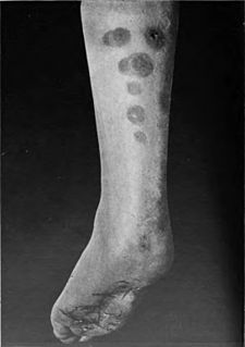
Discoid lupus erythematosus is the most common type of chronic cutaneous lupus (CCLE), an autoimmune skin condition on the lupus erythematosus spectrum of illnesses. It presents with red, inflamed, coin-shaped patches of skin with a scaling and crusty appearance, most often on the scalp, cheeks, and ears. Hair loss may occur if the lesions are on the scalp. The lesions can then develop severe scarring, and the centre areas may appear lighter in color with a rim darker than the normal skin. These lesions can last for years without treatment.

Lupus erythematosus is a collection of autoimmune diseases in which the human immune system becomes hyperactive and attacks healthy tissues. Symptoms of these diseases can affect many different body systems, including joints, skin, kidneys, blood cells, heart, and lungs. The most common and most severe form is systemic lupus erythematosus.

Splinter hemorrhages are tiny blood clots that tend to run vertically under the nails. Splinter hemorrhages are not specific to any particular condition, and can be associated with subacute infective endocarditis, scleroderma, trichinosis, systemic lupus erythematosus (SLE), rheumatoid arthritis, psoriatic nails, antiphospholipid syndrome, haematological malignancy, and trauma. At first they are usually plum-colored, but then darken to brown or black in a couple of days. In certain conditions, clots can migrate from the affected heart valve and find their way into various parts of the body. If this happens in the finger, it can cause damage to the capillaries resulting in a splinter hemorrhage.
Pemphigus erythematosus is simply a localized form of pemphigus foliaceus with features of lupus erythematosus.
Chilblain lupus erythematosus is a chronic, unremitting form of lupus erythematosus with the fingertips, rims of ears, calves, and heels affected, especially in women.
Tumid lupus erythematosus is a rare, but distinctive entity in which patients present with edematous erythematous plaques, usually on the trunk.
Subacute cutaneous lupus erythematosus is a clinically distinct subset of cases of lupus erythematosus that is most often present in white women aged 15 to 40, consisting of skin lesions that are scaly and evolve as poly-cyclic annular lesions or plaques similar to those of plaque psoriasis.
Neonatal lupus erythematosus is the occurrence of systemic lupus erythematosus (SLE) symptoms in an infant born from a mother with SLE, most commonly presenting with a rash resembling subacute cutaneous lupus erythematosus, and sometimes with systemic abnormalities such as complete heart block or hepatosplenomegaly.
Palisaded neutrophilic and granulomatous dermaititis is usually associated with a well-defined connective tissue disease, lupus erythematosus or rheumatoid arthritis most commonly, and often presents with eroded or ulcerated symmetrically distributed umbilicated papules or nodules on the elbows.
Tufted folliculitis presents with doll's hair-like bundling of follicular units, and is seen in a wide range of scarring conditions including chronic staphylococcal infection, chronic lupus erythematosus, lichen planopilaris, Graham-Little syndrome, folliculitis decalvans, acne keloidalis nuchae, immunobullous disorders, and dissecting cellulitis.

Lupus, technically known as systemic lupus erythematosus (SLE), is an autoimmune disease in which the body's immune system mistakenly attacks healthy tissue in many parts of the body. Symptoms vary between people and may be mild to severe. Common symptoms include painful and swollen joints, fever, chest pain, hair loss, mouth ulcers, swollen lymph nodes, feeling tired, and a red rash which is most commonly on the face. Often there are periods of illness, called flares, and periods of remission during which there are few symptoms.

Cutaneous lymphoid hyperplasia refers to a groups of benign cutaneous disorders characterized by collections of lymphocytes, macrophages, and dendritic cells in the skin. Conditions included in this groups are:
Rowell's Syndrome was described by Professor Neville Rowell and colleagues in 1963. Patients with the syndrome have lupus erythematosus, annular lesions of the skin like erythema multiforme associated with a characteristic pattern of immunological abnormalities. It is uncommon but occurs worldwide.
Cutaneous lupus mucinosis is a cutaneous condition characterized by lesions that present as asymptomatic skin-colored, at times reddish, 0.5–2 cm papules and nodules.
Acute cutaneous lupus erythematosus is a cutaneous condition characterized by a bilateral malar rash and lesions that tend to be transient, and that follow sun exposure. The acute form is distinct from chronic and subacute cutaneous lupus erythematosus, which may have different types of skin lesions. Cutaneous lupus erythematosus is associated with both lupus erythematosus-specific lesions and cutaneous manifestations that are not specific to lupus erythematosus, such as oral ulcers and urticaria. Because of the diagnostic criteria used to diagnose systemic lupus erythematosus, a patient with only cutaneous manifestations may be diagnosed with the systemic form of the disease.

Rudi Harold Cormane was a Dutch dermatologist who pioneered research in immunofluorescence studies of the skin.
References
- 1 2 William D. James; Timothy G. Berger; Dirk M. Elston (2015). Andrews' Diseases of the Skin: Clinical Dermatology (12th ed.). Elsevier. p. 156. ISBN 978-0-323-31967-6.
- 1 2 3 Rapini, Ronald P.; Bolognia, Jean L.; Jorizzo, Joseph L. (2007). Dermatology: 2-Volume Set. St. Louis: Mosby. ISBN 978-1-4160-2999-1.