Bladder exstrophy

Last updated
Bladder exstrophy
Other namesEctopia vesicae
Classical bladder exstrophy.jpg
Female baby with classical bladder exstrophy
Specialty Medical genetics   OOjs UI icon edit-ltr-progressive.svg

Bladder exstrophy is a congenital anomaly that exists along the spectrum of the exstrophy-epispadias complex, and most notably involves protrusion of the urinary bladder through a defect in the abdominal wall. Its presentation is variable, often including abnormalities of the bony pelvis, pelvic floor, and genitalia. The underlying embryologic mechanism leading to bladder exstrophy is unknown, though it is thought to be in part due to failed reinforcement of the cloacal membrane by underlying mesoderm. [1] Exstrophy means the inversion of a hollow organ. [2]

Contents

Signs and symptoms

The classic manifestation of bladder exstrophy presents with:[ citation needed ]

Females frequently have a displaced and narrowed vaginal orifice, a bifid clitoris, and divergent labia. [3]

Cause

The cause is not yet clinically established but is thought to be in part due to failed reinforcement of the cloacal membrane by underlying mesoderm. [4]

Diagnosis

In a small retrospective study of 25 pregnancies, five factors were found to be strongly associated with a prenatal diagnosis of bladder exstrophy: [4]

While a diagnosis of bladder exstrophy was made retrospectively in a majority of pregnancies, in only three cases was a prenatal diagnosis made. [5]

Management

The extreme rarity of the disease limits the surgical opportunities to practice the complex closure required in these patients. For this reason, patients have the best outcomes when the bladder closures are performed at high volume centers where surgical and nursing teams have extensive experience in caring for the disease. [6] The highest volume center in the United States, and the world, is the Johns Hopkins Hospital in Baltimore, Maryland; they have seen over 1300 exstrophy patients in the past 50 years. [7]

Upon delivery, the exposed bladder is irrigated and a non-adherent film is placed to prevent as much contact with the external environment as possible. In the event the child was not born at a medical center with an appropriate exstrophy support team then transfer will likely follow. Upon transfer, or for those infants born at a medical center able to care for bladder exstrophy, imaging may take place in the first few hours of life prior to the child undergoing surgery. [3]

Primary (immediate) closure is indicated only in those patients with a bladder of appropriate size, elasticity, and contractility as those patients are most likely to develop a bladder of adequate capacity after early surgical intervention. [8]

Conditions that are absolute contraindications despite bladder adequacy include duplication of the penis or scrotum and significant bilateral hydronephrosis. [9]

Surgery

Watercolour drawing of ectopia vesicae in a man aged 23 years, after operation Ectopia vesicae in a man aged 23 years Wellcome L0062460.jpg
Watercolour drawing of ectopia vesicae in a man aged 23 years, after operation

Modern therapy is aimed at surgical reconstruction of the bladder and genitalia. Both males and females are born with this anomaly. Treatment is similar.

In males treatments have been: In the modern staged repair of exstrophy (MSRE) the initial step is closure of the abdominal wall, often requiring a pelvic osteotomy. This leaves the patient with penile epispadias and urinary incontinence. At approximately 2–3 years of age, the patient then undergoes repair of the epispadias after testosterone stimulation. Finally, bladder neck repair usually occurs around the age of 4–5 years, though this is dependent upon a bladder with adequate capacity and, most importantly, an indication that the child is interested in becoming continent. In some of the bladder reconstructions, the bladder is augmented with the addition of a segment of the large intestines to increase the volume capacity of the reconstructed bladder.[ citation needed ] In the complete primary repair of exstrophy (CPRE) the bladder closure is combined with an epispadias repair, in an effort to decrease costs and morbidity. [10] This technique has, however, led to significant loss of penile and corporal tissue, particularly in younger patients. [11]

In females treatment has included: Surgical reconstruction of the clitoris, which is separated into two distinct bodies. Surgical reconstruction to correct the split of the mons, redefine the structure of the bladder neck and urethra. Vaginoplasty will correct the anteriorly displaced vagina. If the anus is involved, it is also repaired. Fertility remains and women who were born with bladder exstrophy usually develop prolapse due to the weaker muscles of the pelvic floor. [12]

Prognosis

The most important criterion for improving long-term prognosis is success of the initial closure. [13] [14] If a patient requires more than one closure their chance of continence drops off precipitously with each additional closure - at just two closures the chance of voiding continence is just 17%. [15]

Even with successful surgery, people may have long-term complications. [16] Some of the most common include:

Epidemiology

Occurring at a rate between 1 in 10,000 to 1 in 50,000 [17] with a male-to-female ratio of 2.3–6:1, [18] [19] [20] bladder exstrophy is relatively rare. For those individuals with bladder exstrophy who maintain their ability to reproduce, the risk of bladder exstrophy in their children is approximately 500-fold greater than the general population. [18]

Related Research Articles

<span class="mw-page-title-main">Urethral stricture</span> Medical condition

A urethral stricture is a narrowing of the urethra, the tube connected to the bladder that allows the passing of urine. The narrowing reduces the flow of urine and makes it more difficult or even painful to empty the bladder.

<span class="mw-page-title-main">Hypospadias</span> Medical condition

Hypospadias is a common variation in fetal development of the penis in which the urethra does not open from its usual location on the head of the penis. It is the second-most common birth abnormality of the male reproductive system, affecting about one of every 250 males at birth. Roughly 90% of cases are the less serious distal hypospadias, in which the urethral opening is on or near the head of the penis (glans). The remainder have proximal hypospadias, in which the meatus is all the way back on the shaft of the penis, near or within the scrotum. Shiny tissue that typically forms the urethra instead extends from the meatus to the tip of the glans; this tissue is called the urethral plate.

<span class="mw-page-title-main">Epispadias</span> Medical condition

An epispadias is a rare type of malformation in which the urethra ends, in males, in an opening on the upper aspect of the penis, and in females when the urethra develops too far anteriorly. It occurs in around 1 in 120,000 male and 1 in 500,000 female births.
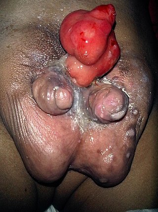
Pediatric urology is a surgical subspecialty of medicine dealing with the disorders of children's genitourinary systems. Pediatric urologists provide care for both boys and girls ranging from birth to early adult age. The most common problems are those involving disorders of urination, reproductive organs and testes.

<span class="mw-page-title-main">Intersex medical interventions</span> Performed to modify atypical or ambiguous genitalia

Intersex medical interventions, also known as intersex genital mutilations (IGM), are surgical, hormonal and other medical interventions performed to modify atypical or ambiguous genitalia and other sex characteristics, primarily for the purposes of making a person's appearance more typical and to reduce the likelihood of future problems. The history of intersex surgery has been characterized by controversy due to reports that surgery can compromise sexual function and sensation, and create lifelong health issues. Timing, evidence, necessity and indications for surgeries in infancy, adolescence or adult age have been controversial, associated with issues of consent.

<span class="mw-page-title-main">Prostatectomy</span> Surgical removal of all or part of the prostate gland

Prostatectomy is the surgical removal of all or part of the prostate gland. This operation is done for benign conditions that cause urinary retention, as well as for prostate cancer and for other cancers of the pelvis.

Nocturia is defined by the International Continence Society (ICS) as “the complaint that the individual has to wake at night one or more times for voiding .” The term is derived from Latin nox, night, and Greek [τα] ούρα, urine. Causes are varied and can be difficult to discern. Although not every patient needs treatment, most people seek treatment for severe nocturia, waking up to void more than 2–3 times per night.

An Indiana pouch is a surgically-created urinary diversion used to create a way for the body to store and eliminate urine for patients who have had their urinary bladders removed as a result of bladder cancer, pelvic exenteration, bladder exstrophy or who are not continent due to a congenital, neurogenic bladder. This particular urinary diversion results in a continent reservoir that the patient must catheterize to empty urine. This concept and technique was developed by Drs. Mike Mitchell, Randall Rowland, and Richard Bihrle at Indiana University.

Urethroplasty is the surgical repair of an injury or defect within the walls of the urethra. Trauma, iatrogenic injury and infections are the most common causes of urethral injury/defect requiring repair. Urethroplasty is regarded as the gold standard treatment for urethral strictures and offers better outcomes in terms of recurrence rates than dilatations and urethrotomies. It is probably the only useful modality of treatment for long and complex strictures though recurrence rates are higher for this difficult treatment group.

<span class="mw-page-title-main">Urachal cancer</span> Medical condition

Urachal cancer is a very rare type of cancer arising from the urachus or its remnants. The disease might arise from metaplastic glandular epithelium or embryonic epithelial remnants originating from the cloaca region.

<span class="mw-page-title-main">Mani Menon</span>

Mani Menon, born 9 July 1948 in Trichur, India, is an American surgeon whose work has helped to lay the foundation for modern Robotic Cancer Surgery. He is the founding director and the Raj and Padma Vattikuti Distinguished Chair of the Vattikuti Urology Institute at the Henry Ford Hospital in Detroit, MI, where he established the first cancer-oriented robotics program in the world. Menon is widely regarded for his role in the development of robotic surgery techniques for the treatment of patients with prostate, kidney, and bladder cancers, as well as for the development of robotic kidney transplantation.
Menon is the recipient of the Gold Cystoscope award, Hugh Hampton Young award, the Keyes Medal, the prestigious B.C. Roy award.

<span class="mw-page-title-main">Diphallia</span> Genital medical condition

Diphallia, penile duplication (PD), diphallic terata, or diphallasparatus, is an extremely rare developmental abnormality in which a male is born with two penises. The first reported case was by Johannes Jacob Wecker in 1609. Its occurrence is 1 in 5.5 million boys in the United States.

<span class="mw-page-title-main">Cystitis cystica</span> Medical condition

Cystitis cystica is an uncommon chronic reactive inflammatory disease that is believed to be brought on by a tumor, calculi, infection, or obstruction of the urothelium. Cystitis glandularis is a proliferative progression of cystitis cystica that is distinguished by urothelial glandular metaplasia.

<span class="mw-page-title-main">Buried penis</span> Male congenital condition

Buried penis, also called hidden penis or retractile penis, is a congenital or acquired condition in which the penis is partially or completely hidden below the surface of the skin. A buried penis can lead to urinary difficulties, poor hygiene, infection, and inhibition of normal sexual function.

<span class="mw-page-title-main">Eosinophilic cystitis</span> Medical condition

Eosinophilic cystitis is a rare type of interstitial cystitis first reported in 1960 by Edwin Brown. Eosinophilic cystitis has been linked to a number of etiological factors, including allergies, bladder tumors, trauma to the bladder, parasitic infections, and chemotherapy drugs, though the exact cause of the condition is still unknown. The antigen-antibody response is most likely the cause of eosinophilic cystitis. This results in the generation of different immunoglobulins, which activate eosinophils and start the inflammatory process.

<span class="mw-page-title-main">Artificial urinary sphincter</span> Medical device

An artificial urinary sphincter (AUS) is an implanted device to treat moderate to severe stress urinary incontinence, most commonly in men. The AUS is designed to supplement the function of the natural urinary sphincter that restricts urine flow out of the bladder.

Male genital examination is a physical examination of the genital in males to detect ailments and to assess sexual development, and is normally a component of an annual physical examination. The examination includes checking the penis, scrotum, and urethral meatus. A comprehensive assessment of the male genitals assesses the pubic hair based on Sexual Maturity Rating and the size of the testicles and penis. The exam can also be conducted to verify a person's age and biological sex. The genitourinary system can also be assessed as part of the male genital examination. During a genital examination, the doctor can detect any of the following: structural abnormalities, urethral opening abnormalities, problems related to not being circumcised, lumps, tumors, redness, excoriation, edema, lesions, swelling, cancer, hair-related issues, and many others. In some instances where a physical examination of the male genitals is not sufficient to diagnose an individual, then an internal genital examination using imaging or ultrasounds will be needed for further evaluation.

Jerry G. Blaivas is an American urologist and senior faculty at the Icahn School of Medicine at Mount Sinai in New York City and adjunct professor of Urology at SUNY Downstate Medical School in Brooklyn, as well as professor of clinical urology at College of Physicians and Surgeons, Columbia University and clinical professor of Urology at Weill Medical College of Cornell University. He has four patents pending, has received four research grants for which he served as the principal investigator, and served as a major in the United States Army assigned to the Walson Army Hospital. He additionally served as president for the Urodynamic Society.

<span class="mw-page-title-main">Vaginal anomalies</span> Congenital defect; abnormal or absent vagina

Vaginal anomalies are abnormal structures that are formed during the prenatal development of the female reproductive system and are rare congenital defects that result in an abnormal or absent vagina.

Penile implants may be employed to treat erectile dysfunction or urinary troubles after a spinal cord injury.

References

  1. Muecke EC: The role of the cloacal membrane in exstrophy: the first successful experimental study. J Urol 1964; 92:659.
  2. Larsen, WJ. (2001). Human embryology (3 ed.). Philadelphia, Pa.: Churchill Livingstone. p. 275. ISBN   0-443-06583-7.
  3. 1 2 Gearhart JP, Mathews R (2011). "Chapter 124: Exstrophy-epispadias complex". In Wein AJ (ed.). Campbell-Walsh Urology (10th ed.). Philadelphia, Pa: Saunders Elsevier.
  4. 1 2 Anand S, Lotfollahzadeh S (2022). "Bladder Exstrophy". National Center for Biotechnology Information, U.S. National Library of Medicine. PMID   33085303 . Retrieved 25 July 2021.
  5. Gearhart JP, Ben-Chaim J, Jeffs RD, Sanders RC (June 1995). "Criteria for the prenatal diagnosis of classic bladder exstrophy". Obstetrics and Gynecology. 85 (6): 961–4. doi:10.1016/0029-7844(95)00069-4. PMID   7770267. S2CID   2096064.
  6. Nelson CP, Dunn RL, Wei JT, Gearhart JP (September 2005). "Surgical repair of bladder exstrophy in the modern era: contemporary practice patterns and the role of hospital case volume". The Journal of Urology. 174 (3): 1099–102. doi:10.1097/01.ju.0000169132.14799.33. PMID   16094068.
  7. Kasprenski M, Benz K, Maruf M, Jayman J, Di Carlo H, Gearhart J (March 2020). "Modern Management of the Failed Bladder Exstrophy Closure: A 50-yr Experience". European Urology Focus. 6 (2): 383–389. doi:10.1016/j.euf.2018.09.008. PMID   30292419. S2CID   52926501.
  8. Gearhart JP, Jeffs RD (1998). "The bladder exstrophy-epispadias complex". In Walsh PC (ed.). Campbell's urology (7th ed.). Philadelphia: WB Saunders. p. 1939.
  9. "Hydronephrosis". The Lecturio Medical Concept Library. Retrieved 25 July 2021.
  10. Grady RW, Mitchell ME (October 1999). "Complete primary repair of exstrophy". The Journal of Urology. 162 (4): 1415–20. doi:10.1016/S0022-5347(05)68327-9. PMID   10492227.
  11. Husmann DA, Gearhart JP (October 2004). "Loss of the penile glans and/or corpora following primary repair of bladder exstrophy using the complete penile disassembly technique". The Journal of Urology. 172 (4 Pt 2): 1696–700, discussion 1700–1. doi:10.1097/01.ju.0000138675.16931.cb. PMID   15371793.
  12. Moliterno D (2013). Therapeutic advances in thrombosis. Chichester, West Sussex: Wiley-Blackwell. pp. 779–798. ISBN   9781405196253.
  13. Oesterling JE, Jeffs RD (February 1987). "The importance of a successful initial bladder closure in the surgical management of classical bladder exstrophy: analysis of 144 patients treated at the Johns Hopkins Hospital between 1975 and 1985". The Journal of Urology. 137 (2): 258–62. doi:10.1016/s0022-5347(17)43972-3. PMID   3806816.
  14. Husmann DA, McLorie GA, Churchill BM (August 1989). "Closure of the exstrophic bladder: an evaluation of the factors leading to its success and its importance on urinary continence". The Journal of Urology. 142 (2 Pt 2): 522–4, discussion 542–3. doi:10.1016/s0022-5347(17)38803-1. PMID   2746771.
  15. Novak TE, Costello JP, Orosco R, Sponseller PD, Mack E, Gearhart JP (August 2010). "Failed exstrophy closure: management and outcome". Journal of Pediatric Urology. 6 (4): 381–4. doi:10.1016/j.jpurol.2009.10.009. PMID   19906564.
  16. Gargollo PC, Borer JG (2007). "Contemporary outcomes in bladder exstrophy". Current Opinion in Urology. 17 (4): 272–80. doi:10.1097/MOU.0b013e3281ddb32f. PMID   17558272. S2CID   6290323.
  17. Lattimer JK, Smith MJK: Exstrophy closure: a follow up on 70 cases. J Urol 1966; 95:356.
  18. 1 2 Shapiro E, Jeffs RD, Gearhart JP, Lepor H (August 1985). "Muscarinic cholinergic receptors in bladder exstrophy: insights into surgical management". The Journal of Urology. 134 (2): 308–10. doi:10.1016/s0022-5347(17)47139-4. PMID   4020984.
  19. Ives E, Coffey R, Carter CO (April 1980). "A family study of bladder exstrophy". Journal of Medical Genetics. 17 (2): 139–41. doi:10.1136/jmg.17.2.139. PMC   1048521 . PMID   7381870.
  20. Lancaster PA (1987). "Epidemiology of bladder exstrophy: a communication from the International Clearinghouse for Birth Defects monitoring systems". Teratology. 36 (2): 221–227. doi:10.1002/tera.1420360210. PMID   3424208.