Related Research Articles
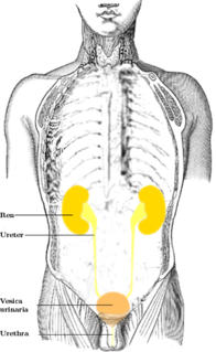
The ureters are tubes made of smooth muscle that propel urine from the kidneys to the urinary bladder. In a human adult, the ureters are usually 20–30 cm (8–12 in) long and around 3–4 mm (0.12–0.16 in) in diameter. The ureter is lined by urothelial cells, a type of transitional epithelium, and has an additional smooth muscle layer in third closest to the bladder that assists with peristalsis.

A ureterocele is a congenital abnormality found in the ureter. In this condition the distal ureter balloons at its opening into the bladder, forming a sac-like pouch. It is most often associated with a duplicated collection system, where two ureters drain their respective kidney instead of one. Simple ureterocele, where the condition involves only a single ureter, represents only twenty percent of cases.
Potter sequence is the atypical physical appearance of a baby due to oligohydramnios experienced when in the uterus. It includes clubbed feet, pulmonary hypoplasia and cranial anomalies related to the oligohydramnios. Oligohydramnios is the decrease in amniotic fluid volume sufficient to cause deformations in morphogenesis of the baby.
The development of the urinary system begins during prenatal development, and relates to the development of the urogenital system – both the organs of the urinary system and the sex organs of the reproductive system. The development continues as a part of sexual differentiation.

The trigone is a smooth triangular region of the internal urinary bladder formed by the two ureteric orifices and the internal urethral orifice.
Hydronephrosis describes hydrostatic dilation of the renal pelvis and calyces as a result of obstruction to urine flow downstream. Alternatively, hydroureter describes the dilation of the ureter, and hydronephroureter describes the dilation of the entire upper urinary tract.
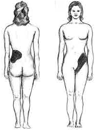
Renal colic is a type of abdominal pain commonly caused by obstruction of ureter from dislodged kidney stones. The most frequent site of obstruction is the vesico-ureteric junction (VUJ), the narrowest point of the upper urinary tract. Acute obstruction and the resultant urinary stasis can distend the ureter (hydroureter) and cause a reflexive peristaltic smooth muscle spasm, which leads to a very intense visceral pain transmitted via the ureteric plexus.

The common iliac arteries are two large arteries that originate from the aortic bifurcation at the level of the fourth lumbar vertebra. They end in front of the sacroiliac joint, one on either side, and each bifurcates into the external and internal iliac arteries.
Congenital vertebral anomalies are a collection of malformations of the spine. Most, around 85%, are not clinically significant, but they can cause compression of the spinal cord by deforming the vertebral canal or causing instability. This condition occurs in the womb. Congenital vertebral anomalies include alterations of the shape and number of vertebrae.

Vesicoureteral reflux (VUR), also known as vesicoureteric reflux, is a condition in which urine flows retrograde, or backward, from the bladder into one or both ureters and then to the renal calyx or kidneys. Urine normally travels in one direction from the kidneys to the bladder via the ureters, with a 1-way valve at the vesicoureteral (ureteral-bladder) junction preventing backflow. The valve is formed by oblique tunneling of the distal ureter through the wall of the bladder, creating a short length of ureter (1–2 cm) that can be compressed as the bladder fills. Reflux occurs if the ureter enters the bladder without sufficient tunneling, i.e., too "end-on".
Tricuspid insufficiency (TI), more commonly called tricuspid regurgitation (TR), is a type of valvular heart disease in which the tricuspid valve of the heart, located between the right atrium and right ventricle, does not close completely when the right ventricle contracts (systole). TR allows the blood to flow backwards from the right ventricle to the right atrium, which increases the volume and pressure of the blood both in the right atrium and the right ventricle, which may increase central venous volume and pressure if the backward flow is sufficiently severe.
Hematocolpos is a medical condition in which the vagina is pooled with menstrual blood due to multiple factors leading to the blockage of menstrual blood flow. The medical definition of hematocolpos is 'an accumulation of blood within the vagina'. It is often caused by the combination of menstruation with an imperforate hymen. It is sometimes seen in Robinow syndrome, uterus didelphys, or other vaginal anomalies.
Reflux nephropathy is kidney damage (nephropathy) due to urine flowing backward (reflux) from the bladder toward the kidneys; the latter is called vesicoureteral reflux (VUR). Longstanding VUR can result in small and scarred kidneys during the first five years of life in affected children. The end results of reflux nephropathy can include high blood pressure, excessive protein loss in the urine, and eventually kidney failure.

An abdominal x-ray is an x-ray of the abdomen. It is sometimes abbreviated to AXR, or KUB.

Ectopic ureter is a medical condition where the ureter, rather than terminating at the urinary bladder, terminates at a different site. In males this site is usually the urethra, in females this is usually the urethra or vagina. It can be associated with renal dysplasia, frequent urinary tract infections, and urinary incontinence. Ectopic ureters are found in 1 of every 2000–4000 patients, and can be difficult to diagnose, but are most often seen on CT scans.
Congenital stenosis of vena cava is a congenital anomaly in which the superior vena cava or inferior vena cava has an aberrant interruption or coarctation.
Bladder outlet obstruction occurs when urine is unable to flow from the kidneys through the ureters and out of the bladder through the urethra. Decreased flow of urine leads to swelling of the urinary tract, called hydronephrosis. This process of decreased flow of urine through the urinary tract can begin as early as during intrauterine life and it prevents normal development of fetal kidneys and fetal urine. Low levels of fetal urine leads to low amniotic fluid levels and incomplete lung maturation. Older children and adults can also experience bladder outlet obstruction; however, this process is usually reversible and isn't associated with as many poor outcomes as in infants with congenital bladder outlet obstruction.

Duplicated ureter or Duplex Collecting System is a congenital condition in which the ureteric bud, the embryological origin of the ureter, splits, resulting in two ureters draining a single kidney. It is the most common renal abnormality, occurring in approximately 1% of the population.

A ureteric balloon catheter is a balloon catheter intended for treating strictures of the ureter. In fact it is a double J stent on which a balloon is mounted. It is connected to a delivery device (pusher) to introduce it from the bladder into the ureter. The system comprises a non-return valve device, and a pusher with a stylet and two ports.

Renal ultrasonography is the examination of one or both kidneys using medical ultrasound.
References
- ↑ "Enlarged ureter". Greifswald Medical School . Archived from the original on 2017-02-07. Retrieved 2017-02-06.CS1 maint: discouraged parameter (link)
- ↑ Page 140 in: Mitchell E. Tublin (2015). Diagnostic Imaging: Genitourinary E-Book. Edition 3. Elsevier Health Sciences. ISBN 9780323400428.
- Bailey and Love's Short Practice of Surgery