Vesicoureteral reflux

Last updated
Vesicoureteral reflux
Ultrasound Scan ND 0119092150 0939241.png
Ultrasound image showing abnormal vesicoureteral junction and dilated distal ureter resulting in primary vesicoureteral reflux (VUR).
Specialty Urology   OOjs UI icon edit-ltr-progressive.svg

Vesicoureteral reflux (VUR), also known as vesicoureteric reflux, is a condition in which urine flows retrograde, or backward, from the bladder into one or both ureters and then to the renal calyx or kidneys. [1] [2] Urine normally travels in one direction (forward, or anterograde) from the kidneys to the bladder via the ureters, with a one-way valve at the vesicoureteral (ureteral-bladder) junction preventing backflow. The valve is formed by oblique tunneling of the distal ureter through the wall of the bladder, creating a short length of ureter (1–2 cm) that can be compressed as the bladder fills. Reflux occurs if the ureter enters the bladder without sufficient tunneling, i.e., too "end-on".

Contents

Signs and symptoms

Most children with vesicoureteral reflux are asymptomatic. Vesicoureteral reflux may be diagnosed as a result of further evaluation of dilation of the kidney or ureters draining urine from the kidney while in utero as well as when a sibling has VUR (though routine testing in either circumstance is controversial). Reflux also increases risk of acute bladder and kidney infections, so testing for reflux may be performed after a child has one or more infections.

In infants, the signs and symptoms of a urinary tract infection may include only fever and lethargy, with poor appetite and sometimes foul-smelling urine, while older children typically present with discomfort or pain with urination and frequent urination.

Causes

In healthy individuals the ureters enter the urinary bladder obliquely and run submucosally for some distance. This, in addition to the ureter's muscular attachments, helps secure and support them posteriorly. Together these features produce a valvelike effect that occludes the ureteric opening during storage and voiding of urine. In people with VUR, failure of this mechanism occurs, with resultant backward (retrograde) flow of urine.

Primary VUR

Insufficient submucosal length of the ureter relative to its diameter causes inadequacy of the valvular mechanism. This is precipitated by a congenital defect or lack of longitudinal muscle of the portion of the ureter within the bladder resulting in an ureterovesicular junction (UVJ) abnormality.

Secondary VUR

In this category the ureters' valvular mechanism is initially intact and healthy but becomes overwhelmed by increased bladder pressures associated with obstruction, which distorts the ureterovesicular junction. The obstructions may be anatomical or functional. Secondary VUR can be further divided into anatomical and functional groups.

Anatomical

Posterior urethral valves; urethral or meatal stenosis. These causes are treated surgically when possible.

Functional

Bladder instability, neurogenic bladder and non-neurogenic bladder. Bladder infections may cause reflux due to the elevated pressures associated with inflammation. [3]

Resolution of functional VUR will usually occur if the precipitating cause is treated and resolved. Medical and/or surgical treatment may be indicated.

Diagnosis

VCUG demonstrating bilateral Grade II (non-dilating) vesicoureteral reflux. Vesicoureteral-reflux-004.jpg
VCUG demonstrating bilateral Grade II (non-dilating) vesicoureteral reflux.

The following procedures may be used to diagnose VUR:

An abdominal ultrasound might suggest the presence of VUR if ureteral dilatation is present; however, in many circumstances of VUR of low to moderate, even high severity, the sonogram may be completely normal, thus providing insufficient utility as a single diagnostic test in the evaluation of children suspected of having VUR, such as those presenting with prenatal hydronephrosis or urinary tract infection (UTI). [4]

VCUG is the method of choice for grading and initial workup, while RNC is preferred for subsequent evaluations as there is less exposure to radiation. A high index of suspicion should be attached to any case where a child presents with a urinary tract infection, and anatomical causes should be excluded. A VCUG and abdominal ultrasound should be performed in these cases

DMSA scintigraphy is used for the evaluation of the parenchymal damage, which is seen as cortical scars. After the first febrile UTI, the diagnostic role of an initial scintigraphy for detecting the damage before the VCUG was investigated and it was suggested that VCUG can be omitted in children who has no cortical scars and urinary tract dilatation. [5] [6]

Early diagnosis in children is crucial as studies have shown that the children with VUR who present with a UTI and associated acute pyelonephritis are more likely to develop permanent renal cortical scarring than those children without VUR, with an odds ratio of 2.8. [7] Thus VUR not only increases the frequency of UTIs, but also the risk of damage to upper urinary structures and end-stage renal disease. [8]

Severity

Vesicoureteral reflux (VUR) is graded according to severity. [1]

The younger the patient and the lower the grade at presentation the higher the chance of spontaneous resolution. Approximately 85% of grade I & II VUR cases will resolve spontaneously. Approximately 50% of grade III cases and a lower percentage of higher grades will also resolve spontaneously.

Treatment

The goal of treatment is to minimize infections, as it is infections that cause renal scarring and not the vesicoureteral reflux. [9] Minimizing infections is primarily done by prophylactic antibiotics in newborns and infants who are not potty trained. However, in children who are older, physicians and parents should focus on bowel and bladder management. Children who hold their bladder or who are constipated have a greater number of infections than children who void on a regular schedule. When medical management fails to prevent recurrent urinary tract infections, or if the kidneys show progressive renal scarring then surgical interventions may be necessary. Medical management is recommended in children with Grade I-III VUR as most cases will resolve spontaneously. A trial of medical treatment is indicated in patients with Grade IV VUR especially in younger patients or those with unilateral disease. Of the patients with Grade V VUR only infants are trialled on a medical approach before surgery is indicated, in older patients surgery is the only option.

Endoscopic injection

Endoscopic injection involves applying a gel around the ureteral opening to create a valve function and stop urine from flowing back up the ureter. The gel consists of two types of sugar-based molecules called dextranomer and hyaluronic acid. Trade names for this combination include Deflux and Zuidex. Both constituents are well known from previous uses in medicine. They are also biocompatible, which means that they do not cause significant reactions within the body. In fact, hyaluronic acid is produced and found naturally within the body.

Medical treatment

Medical treatment entails low dose antibiotic prophylaxis until resolution of VUR occurs. Antibiotics are administered nightly at half the normal therapeutic dose. The specific antibiotics used differ with the age of the patient and include:

After 2 months the following antibiotics are suitable:

Urine cultures are performed 3 monthly to exclude breakthrough infection. Annual radiological investigations are likewise indicated. Good perineal hygiene, and timed and double voiding are also important aspects of medical treatment. Bladder dysfunction is treated with the administration of anticholinergics.

Surgical management

A surgical approach is necessary in cases where a breakthrough infection results despite prophylaxis, or there is non-compliance with the prophylaxis. Similarly if the VUR is severe (Grade IV & V), there are pyelonephritic changes or congenital abnormalities. Other reasons necessitating surgical intervention are failure of renal growth, formation of new scars, renal deterioration and VUR in girls approaching puberty.

There are four types of surgical procedure available for the treatment of VUR: endoscopic (STING/HIT procedures); laparoscopic; robotic-assisted laparoscopic; and open procedures (Cohen procedure, Leadbetter-Politano procedure, Lich-Gregoir technique). Laparoscopic and robotic-assisted laparoscopic procedures are often imitation of classical open procedures in laparoscopic or robotic-assisted laparoscopic environments. [10]

Surveillance

The American Urological Association recommends ongoing monitoring of children with VUR until the abnormality resolves or is no longer clinically significant. The recommendations are for annual evaluation of blood pressure, height, weight, analysis of the urine, and kidney ultrasound. [7]

Epidemiology

The prevalence of VUR is difficult to ascertain at any one time, it differs depending on the population looked at. The prevalence of VUR in healthy children has been estimated 0.4-1.8% However in children with UTI the prevalence is up to 30%. Probably the prevalence in healthy population is significantly higher than the traditional estimates, up to 10% of the population. [11] Younger children are more prone to VUR because of the relative shortness of the submucosal ureters. This susceptibility decreases with age as the length of the ureters increases as the children grow.

Four times as many girls as boys are diagnosed with VUR during childhood. Boys most commonly present during their first year, and girls present more cumulatively throughout childhood. [12]

History

As early as the time of Graeco-Roman physician and anatomist Galen described the urinary tract and noted that there were specific mechanisms to prevent the reflux of urine. [13]

Related Research Articles

<span class="mw-page-title-main">Urology</span> Medical specialty

Urology, also known as genitourinary surgery, is the branch of medicine that focuses on surgical and medical diseases of the urinary-tract system and the reproductive organs. Organs under the domain of urology include the kidneys, adrenal glands, ureters, urinary bladder, urethra, and the male reproductive organs.

<span class="mw-page-title-main">Urinary tract infection</span> Infection that affects part of the urinary tract

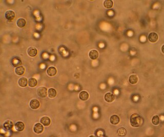
A urinary tract infection (UTI) is an infection that affects a part of the urinary tract. When it affects the lower urinary tract it is known as a bladder infection (cystitis) and when it affects the upper urinary tract it is known as a kidney infection (pyelonephritis). Symptoms from a lower urinary tract infection include pain with urination, frequent urination, and feeling the need to urinate despite having an empty bladder. Symptoms of a kidney infection include fever and flank pain usually in addition to the symptoms of a lower UTI. Rarely the urine may appear bloody. In the very old and the very young, symptoms may be vague or non-specific.

<span class="mw-page-title-main">Prune belly syndrome</span> Medical condition

Prune belly syndrome is a rare, genetic birth defect affecting about 1 in 40,000 births. About 97% of those affected are male. Prune belly syndrome is a congenital disorder of the urinary system, characterized by a triad of symptoms. The syndrome is named for the mass of wrinkled skin that is often present on the abdomen of those with the disorder.

<span class="mw-page-title-main">Cystoscopy</span> Medical procedure; endoscopy of the urinary bladder via the urethra

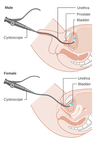
Cystoscopy is endoscopy of the urinary bladder via the urethra. It is carried out with a cystoscope.
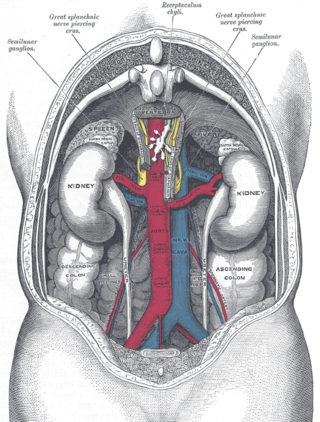
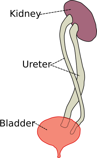
<span class="mw-page-title-main">Ureter</span> Tubes used in the urinary system in most animals

The ureters are tubes made of smooth muscle that propel urine from the kidneys to the urinary bladder. In a human adult, the ureters are usually 20–30 cm (8–12 in) long and around 3–4 mm (0.12–0.16 in) in diameter. The ureter is lined by urothelial cells, a type of transitional epithelium, and has an additional smooth muscle layer that assists with peristalsis in its lowest third.

<span class="mw-page-title-main">Pyelonephritis</span> Medical condition

Pyelonephritis is inflammation of the kidney, typically due to a bacterial infection. Symptoms most often include fever and flank tenderness. Other symptoms may include nausea, burning with urination, and frequent urination. Complications may include pus around the kidney, sepsis, or kidney failure.

<span class="mw-page-title-main">Hydronephrosis</span> Medical condition

Hydronephrosis describes hydrostatic dilation of the renal pelvis and calyces as a result of obstruction to urine flow downstream. Alternatively, hydroureter describes the dilation of the ureter, and hydronephroureter describes the dilation of the entire upper urinary tract.

<span class="mw-page-title-main">Ileal conduit urinary diversion</span> Surgical technique for urinary diversion

An ileal conduit urinary diversion is one of various surgical techniques for urinary diversion. It has sometimes been referred to as the Bricker ileal conduit after its inventor, Eugene M. Bricker. It is a form of incontinent urostomy, and was developed during the 1940s and is still one of the most used techniques for the diversion of urine after a patient has had their bladder removed, due to its low complication rate and high patient satisfaction level. It is usually used in conjunction with radical cystectomy in order to control invasive bladder cancer.

<span class="mw-page-title-main">Posterior urethral valve</span> Medical condition

Posterior urethral valve (PUV) disorder is an obstructive developmental anomaly in the urethra and genitourinary system of male newborns. A posterior urethral valve is an obstructing membrane in the posterior male urethra as a result of abnormal in utero development. It is the most common cause of bladder outlet obstruction in male newborns. The disorder varies in degree, with mild cases presenting late due to milder symptoms. More severe cases can have renal and respiratory failure from lung underdevelopment as result of low amniotic fluid volumes, requiring intensive care and close monitoring. It occurs in about one in 8,000 babies.

In urology, voiding cystourethrography (VCUG) is a frequently performed technique for visualizing a person's urethra and urinary bladder while the person urinates (voids). It is used in the diagnosis of vesicoureteral reflux, among other disorders. The technique consists of catheterizing the person in order to fill the bladder with a radiocontrast agent, typically diatrizoic acid. Under fluoroscopy the radiologist watches the contrast enter the bladder and looks at the anatomy of the patient. If the contrast moves into the ureters and back into the kidneys, the radiologist makes the diagnosis of vesicoureteral reflux, and gives the degree of severity a score. The exam ends when the person voids while the radiologist is watching under fluoroscopy. Consumption of fluid promotes excretion of contrast media after the procedure. It is important to watch the contrast during voiding, because this is when the bladder has the most pressure, and it is most likely this is when reflux will occur. Despite this detailed description of the procedure, at least as of 2016 the technique had not been standardized across practices.

<span class="mw-page-title-main">Ureteral stent</span>

A ureteral stent, or ureteric stent, is a thin tube inserted into the ureter to prevent or treat obstruction of the urine flow from the kidney. The length of the stents used in adult patients varies between 24 and 30 cm. Additionally, stents come in differing diameters or gauges, to fit different size ureters. The stent is usually inserted with the aid of a cystoscope. One or both ends of the stent may be coiled to prevent it from moving out of place; this is called a JJ stent, double J stent or pig-tail stent.

Pyelogram is a form of imaging of the renal pelvis and ureter.

Reflux nephropathy is kidney damage (nephropathy) due to urine flowing backward (reflux) from the bladder toward the kidneys; the latter is called vesicoureteral reflux (VUR). Longstanding VUR can result in small and scarred kidneys during the first five years of life in affected children. The end results of reflux nephropathy can include high blood pressure, excessive protein loss in the urine, and eventually kidney failure.

<span class="mw-page-title-main">Ovarian vein syndrome</span> Medical condition

Ovarian vein syndrome is a rare condition in which a dilated ovarian vein compresses the ureter. This causes chronic or colicky abdominal pain, back pain and/or pelvic pain. The pain can worsen on lying down or between ovulation and menstruation. There can also be an increased tendency towards urinary tract infection or pyelonephritis. The right ovarian vein is most commonly involved, although the disease can be left-sided or affect both sides. It is currently classified as a form of pelvic congestion syndrome.

Urologic diseases or conditions include urinary tract infections, kidney stones, bladder control problems, and prostate problems, among others. Some urologic conditions do not affect a person for that long and some are lifetime conditions. Kidney diseases are normally investigated and treated by nephrologists, while the specialty of urology deals with problems in the other organs. Gynecologists may deal with problems of incontinence in women.

Bladder outlet obstruction occurs when urine is unable to flow from the kidneys through the ureters and out of the bladder through the urethra. Decreased flow of urine leads to swelling of the urinary tract, called hydronephrosis. This process of decreased flow of urine through the urinary tract can begin as early as during intrauterine life and it prevents normal development of fetal kidneys and fetal urine. Low levels of fetal urine leads to low amniotic fluid levels and incomplete lung maturation. Older children and adults can also experience bladder outlet obstruction; however, this process is usually reversible and isn't associated with as many poor outcomes as in infants with congenital bladder outlet obstruction.

<span class="mw-page-title-main">Duplicated ureter</span>

Duplicated ureter or duplex collecting system is a congenital condition in which the ureteric bud, the embryological origin of the ureter, splits, resulting in two ureters draining a single kidney. It is the most common renal abnormality, occurring in approximately 1% of the population.

<span class="mw-page-title-main">Fraley syndrome</span> Medical condition

Fraley syndrome is a condition where the superior infundibulum of the upper calyx of the kidney is obstructed by the crossing renal artery branch, causing distension and dilatation of the calyx and presenting clinically as haematuria and nephralgia. Furthermore, when the renal artery obstructs the proximal collecting system, filling defects can occur anywhere in the calyces, pelvis, or ureter.

<span class="mw-page-title-main">Ureteral cancer</span> Medical condition

Ureteral cancer is cancer of the ureters, muscular tubes that propel urine from the kidneys to the urinary bladder. It is also known as ureter cancer, renal pelvic cancer, and rarely ureteric cancer or uretal cancer. Cancer in this location is rare. Ureteral cancer becomes more likely in older adults, usually ages 70–80, who have previously been diagnosed with bladder cancer.

Ureteric stricture (ureteral stricture) is the pathological narrowing of the ureter which may lead to serious complications such as kidney failure.

References

  1. 1 2 Williams, Gabrielle; Fletcher, Jeffery T.; Alexander, Stephen I.; Craig, Jonathan C. (2008-05-01). "Vesicoureteral Reflux". Journal of the American Society of Nephrology. 19 (5): 847–862. doi: 10.1681/ASN.2007020245 . ISSN   1046-6673. PMID   18322164.
  2. Miyakita, Hideshi; Hayashi, Yutaro; Mitsui, Takahiko; Okawada, Manabu; Kinoshita, Yoshiaki; Kimata, Takahisa; Koikawa, Yasuhiro; Sakai, Kiyohide; Satoh, Hiroyuki; Tokunaga, Masatoshi; Naitoh, Yasuyuki (2020-04-01). "Guidelines for the medical management of pediatric vesicoureteral reflux". International Journal of Urology. 27 (6): 480–490. doi: 10.1111/iju.14223 . ISSN   1442-2042. PMC   7318347 . PMID   32239562.
  3. Institute of Urology & Nephrology, London, UK, The cellular basis of bladder instability UJUS 2009, Retrieved 4-20-2010
  4. Wongbencharat, Kunruedi; Tongpenyai, Yothi; Na-rungsri, Kunyalak (2016-03-01). "Renal ultrasound and DMSA screening for high-grade vesicoureteral reflux". Pediatrics International. 58 (3): 214–218. doi:10.1111/ped.12803. ISSN   1442-200X. PMID   26275163. S2CID   25098104.
  5. Zhang, Xin; Xu, Hong; Zhou, Lijun; Cao, Qi; Shen, Qian; Sun, Li; Fang, Xiaoyan; Guo, Wei; Zhai, Yihui (2014-01-01). "Accuracy of Early DMSA Scan for VUR in Young Children With Febrile UTI". Pediatrics. 133 (1): e30–e38. doi:10.1542/peds.2012-2650. ISSN   0031-4005. PMID   24366989. S2CID   14821315.
  6. Sheu, Ji-Nan; Wu, Kang-Hsi; Chen, Shan-Ming; Tsai, Jeng-Dau; Chao, Yu-Hua; Lue, Ko-Huang (2013). "Acute 99mTc DMSA Scan Predicts Dilating Vesicoureteral Reflux in Young Children With a First Febrile Urinary Tract Infection". Clinical Nuclear Medicine. 38 (3): 163–168. doi:10.1097/rlu.0b013e318279f112. PMID   23354031. S2CID   37633508.
  7. 1 2 Peters CA, Skoog SJ, Arant BS, Copp HL, Elder JS, Hudson RG, Khoury AE, Lorenzo AJ, Pohl HG, Shapiro E, Snodgrass WT, Diaz M (September 2010). "Summary of the AUA Guideline on Management of Primary Vesicoureteral Reflux in Children". The Journal of Urology. 184 (3): 1134–44. doi:10.1016/j.juro.2010.05.065. PMID   20650499.
  8. Brakeman, Paul (2008-07-21). "Vesicoureteral Reflux, Reflux Nephropathy, and End-Stage Renal Disease". Advances in Urology. 2008: 508949. doi: 10.1155/2008/508949 . ISSN   1687-6369. PMC   2478704 . PMID   18670633.
  9. Tekgül, S; Riedmiller, H; Hoebeke, P; Kočvara, R; Nijman, RJ; Radmayr, C; Stein, R; Dogan, HS; European Association of, Urology (September 2012). "EAU guidelines on vesicoureteral reflux in children". European Urology. 62 (3): 534–42. doi:10.1016/j.eururo.2012.05.059. PMID   22698573.
  10. Akhavan, Ardavan; Avery, Daniel; Lendvay, Thomas S. (2014). "Robot-assisted extravesical ureteral reimplantation: Outcomes and conclusions from 78 ureters". Journal of Pediatric Urology. 10 (5): 864–868. doi:10.1016/j.jpurol.2014.01.028. PMID   24642080.
  11. Sargent, M. A. (2000-08-01). "What is the normal prevalence of vesicoureteral reflux?". Pediatric Radiology. 30 (9): 587–593. doi:10.1007/s002470000263. PMID   11009294. S2CID   196420913.
  12. Rink, Richard C.; Mouriquand, Pierre D. E. (2010). Pediatric Urology. Saunders/Elsevier. ISBN   978-1-4160-3204-5.
  13. Nahon, I; Waddington, G; Dorey, G; Adams, R (2011). "The history of urologic surgery: from reeds to robotics". Urologic Nursing. 31 (3): 173–80. doi:10.7257/1053-816X.2011.31.3.173. PMID   21805756.