Related Research Articles
A trisomy is a type of polysomy in which there are three instances of a particular chromosome, instead of the normal two. A trisomy is a type of aneuploidy.

Aneuploidy is the presence of an abnormal number of chromosomes in a cell, for example a human cell having 45 or 47 chromosomes instead of the usual 46. It does not include a difference of one or more complete sets of chromosomes. A cell with any number of complete chromosome sets is called a euploid cell.
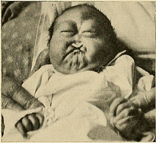
Patau syndrome is a syndrome caused by a chromosomal abnormality, in which some or all of the cells of the body contain extra genetic material from chromosome 13. The extra genetic material disrupts normal development, causing multiple and complex organ defects.

Trisomy 18, also known as Edwards syndrome, is a genetic disorder caused by the presence of a third copy of all or part of chromosome 18. Many parts of the body are affected. Babies are often born small and have heart defects. Other features include a small head, small jaw, clenched fists with overlapping fingers, and severe intellectual disability.

Nondisjunction is the failure of homologous chromosomes or sister chromatids to separate properly during cell division (mitosis/meiosis). There are three forms of nondisjunction: failure of a pair of homologous chromosomes to separate in meiosis I, failure of sister chromatids to separate during meiosis II, and failure of sister chromatids to separate during mitosis. Nondisjunction results in daughter cells with abnormal chromosome numbers (aneuploidy).

Prenatal testing is a tool that can be used to detect some birth defects at various stages prior to birth. Prenatal testing consists of prenatal screening and prenatal diagnosis, which are aspects of prenatal care that focus on detecting problems with the pregnancy as early as possible. These may be anatomic and physiologic problems with the health of the zygote, embryo, or fetus, either before gestation even starts or as early in gestation as practicable. Screening can detect problems such as neural tube defects, chromosome abnormalities, and gene mutations that would lead to genetic disorders and birth defects, such as spina bifida, cleft palate, Down syndrome, trisomy 18, Tay–Sachs disease, sickle cell anemia, thalassemia, cystic fibrosis, muscular dystrophy, and fragile X syndrome. Some tests are designed to discover problems which primarily affect the health of the mother, such as PAPP-A to detect pre-eclampsia or glucose tolerance tests to diagnose gestational diabetes. Screening can also detect anatomical defects such as hydrocephalus, anencephaly, heart defects, and amniotic band syndrome.
Trisomy 8 causes Warkany syndrome 2, a human chromosomal disorder caused by having three copies (trisomy) of chromosome 8. It can appear with or without mosaicism.
Full trisomy 9 is a rare and fatal chromosomal disorder caused by having three copies (trisomy) of chromosome number 9. It can be a viable condition if trisomy affects only part of the cells of the body (mosaicism) or in cases of partial trisomy in which cells have a normal set of two entire chromosomes 9 plus part of a third copy, usually of the short arm of the chromosome.
The Pallister–Killian syndrome (PKS), also termed tetrasomy 12p mosaicism or the Pallister mosaic aneuploidy syndrome, is an extremely rare and severe genetic disorder. PKS is due to the presence of an extra and abnormal chromosome termed a small supernumerary marker chromosome (sSMC). sSMCs contain copies of genetic material from parts of virtually any other chromosome and, depending on the genetic material they carry, can cause various genetic disorders and neoplasms. The sSMC in PKS consists of multiple copies of the short arm of chromosome 12. Consequently, the multiple copies of the genetic material in the sSMC plus the two copies of this genetic material in the two normal chromosome 12's are overexpressed and thereby cause the syndrome. Due to a form of genetic mosaicism, however, individuals with PKS differ in the tissue distributions of their sSMC and therefore show different syndrome-related birth defects and disease severities. For example, individuals with the sSMC in their heart tissue are likely to have cardiac structural abnormalities while those without this sSMC localization have a structurally normal heart.

The palpebral fissure is the elliptic space between the medial and lateral canthi of the two open eyelids. In simple terms, it is the opening between the eyelids. In adult humans, this measures about 10 mm vertically and 30 mm horizontally.
A chromosomal abnormality, chromosomal anomaly, chromosomal aberration, chromosomal mutation, or chromosomal disorder is a missing, extra, or irregular portion of chromosomal DNA. These can occur in the form of numerical abnormalities, where there is an atypical number of chromosomes, or as structural abnormalities, where one or more individual chromosomes are altered. Chromosome mutation was formerly used in a strict sense to mean a change in a chromosomal segment, involving more than one gene. Chromosome anomalies usually occur when there is an error in cell division following meiosis or mitosis. Chromosome abnormalities may be detected or confirmed by comparing an individual's karyotype, or full set of chromosomes, to a typical karyotype for the species via genetic testing.

Micrognathism is a condition where the jaw is undersized. It is also sometimes called mandibular hypoplasia. It is common in infants, but is usually self-corrected during growth, due to the jaws' increasing in size. It may be a cause of abnormal tooth alignment and in severe cases can hamper feeding. It can also, both in adults and children, make intubation difficult, either during anesthesia or in emergency situations.
Klaus Patau was a German-born American geneticist. He received his PhD from the University of Berlin in 1936, worked from 1938 to 1939 in London, and then returned to Germany, where he worked at the Kaiser Wilhelm Institute for Biology until 1947. He emigrated to the United States in 1948 and obtained American citizenship. In 1960 he first reported the extra chromosome in trisomy 13. The syndrome caused by trisomy 13 is often called Patau syndrome. It is also known as Bartholin-Patau syndrome, since the clinical picture associated with trisomy 13 was described by Thomas Bartholin in 1656. At the time, laboratory techniques were unable to distinguish between chromosomes of similar size, so chromosomes were grouped into seven groups by size, lettered A through G. Chromosomes 13 through 15 were in group D, so Patau originally named his eponymous syndrome "trisomy D".

Trisomy 16 is a chromosomal abnormality in which there are 3 copies of chromosome 16 rather than two. It is the most common trisomy leading to miscarriage and the second most common chromosomal cause of it, closely following X-chromosome monosomy. About 6% of miscarriages have trisomy 16. Those mostly occur between 8 and 15 weeks after the last menstrual period.

Aplasia cutis congenita is a rare disorder characterized by congenital absence of skin. Ilona J. Frieden classified ACC in 1986 into 9 groups on the basis of location of the lesions and associated congenital anomalies. The scalp is the most commonly involved area with lesser involvement of trunk and extremities. Frieden classified ACC with fetus papyraceus as type 5. This type presents as truncal ACC with symmetrical absence of skin in stellate or butterfly pattern with or without involvement of proximal limbs. It is the most common congenital cicatricial alopecia, and is a congenital focal absence of epidermis with or without evidence of other layers of the skin.

Pilomatricoma is a benign skin tumor derived from the hair matrix. These neoplasms are relatively uncommon and typically occur on the scalp, face, and upper extremities. Clinically, pilomatricomas present as a subcutaneous nodule or cyst with unremarkable overlying epidermis that can range in size from 0.5 to 3.0 cm, but the largest reported case was 24 cm.

Cutis marmorata is a benign skin condition which, if persistent, occurs in Cornelia de Lange syndrome, trisomy 13 and trisomy 18 syndromes. When a newborn infant is exposed to low environmental temperatures, an evanescent, lacy, reticulated red and/or blue cutaneous vascular pattern appears over most of the body surface. This vascular change represents an accentuated physiologic vasomotor response that disappears with increasing age, although it is sometimes discernible even in older children. It is also seen in cardiogenic shock.

Trisomy X, also known as triple X syndrome and characterized by the karyotype 47,XXX, is a chromosome disorder in which a female has an extra copy of the X chromosome. It is relatively common and occurs in 1 in 1,000 females, but is rarely diagnosed; fewer than 10% of those with the condition know they have it.
References
- ↑ Rapini, Ronald P.; Bolognia, Jean L.; Jorizzo, Joseph L. (2007). Dermatology: 2-Volume Set. St. Louis: Mosby. ISBN 1-4160-2999-0.
| | This cutaneous condition article is a stub. You can help Wikipedia by expanding it. |