Sebaceous hyperplasia

Last updated
Sebaceous hyperplasia
Sebaceous hyperplasia.jpg
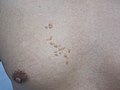
Sebaceous hyperplasia in 55-year-old woman, diagnosis was histologically verified.
Specialty Dermatology

Sebaceous hyperplasia is a disorder of the sebaceous glands in which they become enlarged, producing flesh-colored or yellowish, shiny, often umbilicated bumps. [1] Sebaceous hyperplasia, primarily affecting older patients in high-concentration areas like the face, head, and neck, typically has a 2-4 mm diameter and causes no symptoms. The lesions are often surrounded by telangiectatic blood vessels, also known as "crown vessels," and a central dell, which is in line with the origin of the lesions.

Contents

Sebaceous glands are glands located within the skin and are responsible for secreting an oily substance named sebum. They are commonly associated with hair follicles but they can be found in hairless regions of the skin as well. Their secretion lubricates the skin, protecting it from drying out or becoming irritated. [2]

Murine studies suggest topical irritants and carcinogens may contribute to sebaceous hyperplasia development, with immunosuppression with cyclopsporin A or HIV infection increasing the likelihood.

Sebaceous hyperplasia is a condition that can be diagnosed clinically but requires a biopsy for confirmation. It shares similarities with folliculosebaceous unit architecture but has larger and expanded sebaceous glands. Identifying sebaceous hyperplasia using dermatoscopy can help identify it from other lesions. The dermoscopic characteristics include "crown vessels" clusters of white or yellow nodules, a distinct asymmetrical milky-white structure called the cumulus sign, and a central umbilication called the "bonbon toffee sign."

Sebaceous hyperplasia treatment involves various techniques like cryotherapy, bichloroacetic acid, shave excision, carbon dioxide laser ablation, electrodessication, erbium/yttrium aluminum garnet laser ablation, and pulsed-dye laser photothermolysis.

Signs and symptoms

Sebaceous hyperplasia primarily affects older patients in areas with high concentrations of sebaceous glands, such as the face, head, and neck. [3] It typically manifests as yellowish-colored skin with small papules that are often surrounded by telangiectatic blood vessels, also known as "crown vessels," and a central dell that is in line with the origin of the lesions, which is a dilated central follicular infundibulum. [4] [5] Most lesions have a diameter of 2-4 mm and cause no symptoms. [6]

Causes

Although the etiology is unknown, murine studies have revealed that topical irritants and carcinogens may be involved in the development of sebaceous hyperplasia. [7] [8]

Immunosuppression with cyclopsporin A [9] or HIV infection increases the likelihood of developing sebaceous hyperplasia significantly. [10]

Diagnosis

Although sebaceous hyperplasia can be diagnosed clinically, a biopsy is necessary for confirmation in cases of doubt. [11] The differential diagnosis for sebaceous hyperplasia includes nevus sebaceous, sebaceous carcinoma, sebaceous adenoma, basal cell carcinoma, molluscum contagiosum, and small epidermal inclusion cysts. [6]

Sebaceous hyperplasia shares histopathological similarities with the typical architecture of the folliculosebaceous unit, but with larger and expanded sebaceous glands. [12] [13]

To help identify sebaceous hyperplasia from other lesions, dermatoscopy may be utilized. A cluster of white or yellow nodules encircled by tiny, nonarborizing branching vessels known as "crown vessels" are among the dermoscopic characteristics of sebaceous hyperplasia. [14] [15] On dermatoscopy, a distinct asymmetrical milky-white structure known as the cumulus sign has been identified in cases with sebaceous hyperplasia. Unlike the traditional crown vessels, some writers describe the blood vessels as "multiple tree-like branches." [16] A central umbilication encircled by the cumulus sign was another distinguishing characteristic of dermatoscopy; the authors dubbed this the "bonbon toffee sign." [17]

Treatment

Treatment for sebaceous hyperplasia is carried out for cosemetic reasons. There are numerous techniques that have been reported, including cryotherapy, bichloroacetic acid, shave excision, carbon dioxide laser ablation, electrodessication, erbium or yttrium aluminum garnet laser ablation, and pulsed-dye laser photothermolysis. [18]

Additional photos

See also

Related Research Articles

<span class="mw-page-title-main">Nevus</span> Mole or birthmark; visible, circumscribed, chronic skin lesion

Nevus is a nonspecific medical term for a visible, circumscribed, chronic lesion of the skin or mucosa. The term originates from nævus, which is Latin for "birthmark"; however, a nevus can be either congenital or acquired. Common terms, including mole, birthmark, and beauty mark, are used to describe nevi, but these terms do not distinguish specific types of nevi from one another.

<span class="mw-page-title-main">Seborrheic keratosis</span> Skin disease

A seborrheic keratosis is a non-cancerous (benign) skin tumour that originates from cells, namely keratinocytes, in the outer layer of the skin called the epidermis. Like liver spots, seborrheic keratoses are seen more often as people age.

<span class="mw-page-title-main">Actinic keratosis</span> Skin disorder

Actinic keratosis (AK), sometimes called solar keratosis or senile keratosis, is a pre-cancerous area of thick, scaly, or crusty skin. Actinic keratosis is a disorder of epidermal keratinocytes that is induced by ultraviolet (UV) light exposure. These growths are more common in fair-skinned people and those who are frequently in the sun. They are believed to form when skin gets damaged by UV radiation from the sun or indoor tanning beds, usually over the course of decades. Given their pre-cancerous nature, if left untreated, they may turn into a type of skin cancer called squamous cell carcinoma. Untreated lesions have up to a 20% risk of progression to squamous cell carcinoma, so treatment by a dermatologist is recommended.

Bromoderma is a skin condition characterized by an eruption of papules and pustules on the skin. It is caused by hypersensitivity to bromides, such as those found in certain drugs. There is at least one reported case of bromoderma caused by excessive consumption of a soft drink containing brominated vegetable oil.

<span class="mw-page-title-main">Fordyce spots</span> Sebaceous glands that are present in most individuals

Fordyce spots are harmless and painless visible sebaceous glands typically appearing as white/yellow small bumps or spots on the inside of lips or cheeks, gums, or genitalia. They are common, and are present in around 80% of adults. Treatment is generally not required and attempts to remove them typically result in pain and scarring.

<span class="mw-page-title-main">Dermatoscopy</span> Medical examination of the skin

Dermatoscopy, also known as dermoscopy or epiluminescence microscopy, is the examination of skin lesions with a dermatoscope. It is a tool similar to a camera to allow for inspection of skin lesions unobstructed by skin surface reflections. The dermatoscope consists of a magnifier, a light source, a transparent plate and sometimes a liquid medium between the instrument and the skin. The dermatoscope is often handheld, although there are stationary cameras allowing the capture of whole body images in a single shot. When the images or video clips are digitally captured or processed, the instrument can be referred to as a digital epiluminescence dermatoscope. The image is then analyzed automatically and given a score indicating how dangerous it is. This technique is useful to dermatologists and skin cancer practitioners in distinguishing benign from malignant (cancerous) lesions, especially in the diagnosis of melanoma.

<span class="mw-page-title-main">Adenoma sebaceum</span> Medical condition

Adenoma sebaceum, also known as facial angiofibroma is a misnamed cutaneous disorder consisting of angiofibromas that begin in childhood and appear clinically as red papules on the face especially on the nasolabial folds, cheek and chin, often misidentified as acne not responding to treatment. Adenoma sebaceum may at times be associated with tuberous sclerosis. Gradually the papules become more prominent with time and persist throughout life. Cosmetic removal by argon or pulse dye laser or scalpel is indicated.

<span class="mw-page-title-main">Angiolymphoid hyperplasia with eosinophilia</span> Medical condition

Angiolymphoid hyperplasia with eosinophilia usually presents with pink to red-brown, dome-shaped, dermal papules or nodules of the head or neck, especially about the ears and on the scalp.

Targetoid hemosiderotic hemangioma, also known as a hobnail hemangioma is a skin condition characterized by a central brown or purplish papule that is surrounded by an ecchymotic halo. It may appear similar to melanoma. It was first described by Santa Cruz and Aronberg in 1988.

<span class="mw-page-title-main">Atypical fibroxanthoma</span> Medical condition

Atypical fibroxanthoma (AFX) of the skin is a low-grade malignancy related to malignant fibrous histiocytoma, which it resembles histologically. Atypical fibroxanthoma manifests as a hard, pink or red papule or nodule that grows over the course of several months and may bleed or ulcerate. They typically occur on the head and neck. Atypical fibroxanthoma is usually asymptomatic.

<span class="mw-page-title-main">Cutaneous myxoma</span> Medical condition

A cutaneous myxoma, or superficial angiomyxoma, consists of a multilobulated myxoid mass containing stellate or spindled fibroblasts with pools of mucin forming cleft-like spaces. There is often a proliferation of blood vessels and an inflammatory infiltrate. Staining is positive for vimentin, negative for cytokeratin and desmin, and variable for CD34, Factor VIIIa, SMA, MSA and S-100.

Erosive pustular dermatitis of the scalp presents with pustules, erosions, and crusts on the scalp of primarily older Caucasian females, and on biopsy, has a lymphoplasmacytic infiltrate with or without foreign body giant cells and pilosebaceous atrophy.

Melanonychia is a black or brown pigmentation of a nail, and may be present as a normal finding on many digits in Afro-Caribbeans, as a result of trauma, systemic disease, or medications, or as a postinflammatory event from such localized events as lichen planus or fixed drug eruption.

<span class="mw-page-title-main">Linear IgA bullous dermatosis</span> Medical condition

Linear IgA bullous dermatosis is a rare immune-mediated blistering skin disease frequently associated with medication exposure, especially vancomycin, with men and women being equally affected. It was first described by Tadeusz Chorzelski in 1979 and may be divided into two types:

<span class="mw-page-title-main">Malignant acrospiroma</span> Medical condition

Malignant acrospiroma, also known as nodular hidradenocarcinoma, malignant nodular clear-cell hidradenoma, mucoepidermoid hidradenocarcinoma, clear-cell hidradenocarcinoma, malignant clear-cell acrospiroma, and clear-cell eccrine carcinoma, is a rare and aggressive tumour of the eccrine sweat glands. It was first documented by Keasbey and Hadley in 1954.

<span class="mw-page-title-main">Sebaceous carcinoma</span> Medical condition

Sebaceous carcinoma, also known as sebaceous gland carcinoma (SGc), sebaceous cell carcinoma, and meibomian gland carcinoma is an uncommon malignant cutaneous tumor. Most are typically about 1.4 cm at presentation. SGc originates from sebaceous glands in the skin and, therefore, may originate anywhere in the body where these glands are found. SGc can be divided into 2 types: periocular and extraocular. The periocular region is rich in sebaceous glands making it a common site of origin. The cause of these lesions in the vast majority of cases is unknown. Occasional cases may be associated with Muir-Torre syndrome. SGc accounts for approximately 0.7% of all skin cancers, and the incidence of SGc is highest in Caucasian, Asian, and Indian populations. Due to the rarity of this tumor and variability in clinical and histological presentation, SGc is often misdiagnosed as an inflammatory condition or a more common neoplasm. SGc is commonly treated with wide local excision or Mohs micrographic surgery, and the relative survival rates at 5 and 10 years are 92.72 and 86.98%, respectively.

<span class="mw-page-title-main">Nevus sebaceous</span> Medical condition

Nevus sebaceus or sebaceous nevus is a congenital, hairless plaque that typically occurs on the face or scalp. Such nevi are classified as epidermal nevi and can be present at birth, or early childhood, and affect males and females of all races equally. The condition is named for an overgrowth of sebaceous glands, a relatively uncommon hamartoma, in the area of the nevus. NSJ is first described by Josef Jadassohn in 1895.

<span class="mw-page-title-main">Sebaceous adenoma</span> Medical condition

Sebaceous adenomais a type of adenoma, characterized by a slow-growing tumor usually presenting as a pink, flesh-coloured, or yellow papule or nodule.

<span class="mw-page-title-main">Sebaceoma</span> Medical condition

Sebaceoma, also known as a sebaceous epithelioma, is a cutaneous condition that appears as a yellow or orange papule.

Hair follicle nevus is a cutaneous condition that presents as a small papule from which fine hairs protrude evenly from the surface.

References

  1. James, William D.; Berger, Timothy G.; et al. (2006). Andrews' Diseases of the Skin: Clinical Dermatology. Saunders Elsevier. p. 662. ISBN   978-0-7216-2921-6.
  2. Jo Ann Coers Eurell; Brian L. Frappier (2006). Dellmann's textbook of veterinary histology. Wiley. p. 29. ISBN   9780781741484.
  3. Kumar, P.; Marks, R. (1987). "Sebaceous gland hyperplasia and senile comedones: a prevalence study in elderly hospitalized patients". British Journal of Dermatology. 117 (2): 231–236. doi:10.1111/j.1365-2133.1987.tb04121.x. ISSN   0007-0963. PMID   2958079.
  4. Luderschmidt, Christoph.; Plewig, Gerd. (1978). "Circumscribed Sebaceous Gland Hyperplasia: Autoradiographic and Histoplanimetric Studies". Journal of Investigative Dermatology. 70 (4). Elsevier BV: 207–209. doi:10.1111/1523-1747.ep12541329. ISSN   0022-202X. PMID   641372.
  5. Tetzlaff, Michael T.; North, Jeffrey; Esmaeli, Bita (2019). "Update on sebaceous neoplasia: the morphologic spectrum and molecular genetic drivers of carcinoma". Diagnostic Histopathology. 25 (3). Elsevier BV: 102–109. doi:10.1016/j.mpdhp.2019.01.001. ISSN   1756-2317.
  6. 1 2 Hogan, Daniel; Mohammad, Samya (2011). "Sebaceous hyperplasia". Expert Review of Dermatology. 6 (1): 91–96. doi:10.1586/edm.11.2. ISSN   1746-9872.
  7. Ito, Masaaki; Motoyoshi, Katsuhiro; Suzuki, Megumi; Sato, Yoshio (1985). "Sebaceous Gland Hyperplasia on Rabbit Pinna Induced by Tetradecane". Journal of Investigative Dermatology. 85 (3). Elsevier BV: 249–254. doi:10.1111/1523-1747.ep12276720. ISSN   0022-202X. PMID   2411823.
  8. Rice, Jerry M.; Anderson, Lucy M. (1986). "Sebaceous adenomas with associated epidermal hyperplasia and papilloma formation as a major type of tumor induced in mouse skin by high doses of carcinogens". Cancer Letters. 33 (3). Elsevier BV: 295–306. doi:10.1016/0304-3835(86)90069-8. ISSN   0304-3835. PMID   3100020.
  9. Boschnakow, A.; May, T.; Assaf, C.; Tebbe, B.; Zouboulis, CH.C. (2003). "Ciclosporin A-induced sebaceous gland hyperplasia". British Journal of Dermatology. 149 (1). Oxford University Press (OUP): 198–200. doi:10.1046/j.1365-2133.2003.05397.x. ISSN   0007-0963.
  10. Short, K. A.; Williams, A.; Creamer, D.; Fuller, L. C. (2008). "Sebaceous gland hyperplasia, human immunodeficiency virus and highly active anti-retroviral therapy". Clinical and Experimental Dermatology. 33 (3): 354–355. doi:10.1111/j.1365-2230.2007.02670.x. ISSN   0307-6938. PMID   18346183.
  11. Lim, Geoffrey F.S.; Cusack, Carrie Ann R.; Kist, Joseph M. (2014). "Perioral Lesions and Dermatoses". Dental Clinics of North America. 58 (2). Elsevier BV: 401–435. doi:10.1016/j.cden.2013.12.009. ISSN   0011-8532. PMID   24655530.
  12. Kumar, P.; Barton, S.P.; Marks, R. (1988). "Tissue measurements in senile sebaceous gland hyperplasia". British Journal of Dermatology. 118 (3): 397–402. doi:10.1111/j.1365-2133.1988.tb02434.x. ISSN   0007-0963. PMID   3355781.
  13. Eisen, Daniel B.; Michael, Daniel J. (2009). "Sebaceous lesions and their associated syndromes: Part I". Journal of the American Academy of Dermatology. 61 (4): 549–560. doi:10.1016/j.jaad.2009.04.058. PMID   19751879.
  14. Kim, Nancy H.; Zell, Deborah S.; Kolm, Isabel; Oliviero, Margaret; Rabinovitz, Harold S. (2008-07-01). "The Dermoscopic Differential Diagnosis of Yellow Lobularlike Structures". Archives of Dermatology. 144 (7). American Medical Association (AMA). doi:10.1001/archderm.144.7.962. ISSN   0003-987X. PMID   18645159.
  15. Zaballos, Pedro; Ara, Mariano; Puig, Susana; Malvehy, Josep (2005-06-01). "Dermoscopy of Sebaceous Hyperplasia". Archives of Dermatology. 141 (6). American Medical Association (AMA). doi:10.1001/archderm.141.6.808. ISSN   0003-987X.
  16. Bryden, A. M.; Dawe, R. S.; Fleming, C. (2004). "Dermatoscopic features of benign sebaceous proliferation". Clinical and Experimental Dermatology. 29 (6). Oxford University Press (OUP): 676–677. doi:10.1111/j.1365-2230.2004.1612.x. ISSN   0307-6938. PMID   15550152.
  17. Oztas, P; Polat, M; Oztas, M; Alli, N; Ustun, H (2008-09-10). "Bonbon toffee sign: a new dermatoscopic feature for sebaceous hyperplasia". Journal of the European Academy of Dermatology and Venereology. 22 (10). Wiley: 1200–1202. doi:10.1111/j.1468-3083.2008.02827.x. ISSN   0926-9959. PMID   18540985.
  18. No, David; McClaren, Marla; Chotzen, Vera; Kilmer, Suzanne L. (2004). "Sebaceous Hyperplasia Treated With a 1450-nm Diode Laser". Dermatologic Surgery. 30 (3): 382–384. doi:10.1111/j.1524-4725.2004.30105.x. ISSN   1076-0512. PMID   15008865.

Further reading