Dermatoscopy

Last updated
Dermatoscopy
Dermatoscope1.JPG
Immersion oil dermatoscope.
Specialty dermatology
MeSH D046169
Polarized light dermatoscope. Dermatoscope.jpg
Polarized light dermatoscope.

Dermatoscopy also known as dermoscopy [1] or epiluminescence microscopy, is the examination of skin lesions with a dermatoscope. It is a tool similar to a camera to allow for inspection of skin lesions unobstructed by skin surface reflections. The dermatoscope consists of a magnifier, a light source (polarized or non-polarized), a transparent plate and sometimes a liquid medium between the instrument and the skin. The dermatoscope is often handheld, although there are stationary cameras allowing the capture of whole body images in a single shot. When the images or video clips are digitally captured or processed, the instrument can be referred to as a digital epiluminescence dermatoscope. The image is then analyzed automatically and given a score indicating how dangerous it is. This technique is useful to dermatologists and skin cancer practitioners in distinguishing benign from malignant (cancerous) lesions, especially in the diagnosis of melanoma.

Contents

Types

There are two main types of dermatoscopes, hand held portable and stationary mounted type.

A hand held dermatoscope is composed of a transilluminating light source and a magnifying optic (usually a 10-fold magnification). There are three main modes of dermatoscopy: [2]

Polarized light allows for visualization of deeper skin structures, while non-polarized light provide information about the superficial skin. Most modern dermatoscopes allow the user to toggle between the two modes, which provide complementary information. Others may also allow the user to have different zoom levels and color overlay.

A stationary type allows a full body image to be captured in one snap. It is then transferred into image analysis algorithms that generates a three dimensional model of the person. Lesions on the person are marked and analyzed using Artificial intelligence.

Advantages

With doctors who are experts in dermatoscopy, the diagnostic accuracy for melanoma is significantly better than those who do not have any specialized training. [3] Thus, there is considerable improvement in the sensitivity (detection of melanomas) as well as specificity (percentage of non-melanomas correctly diagnosed as benign), compared with naked eye examination. The accuracy by dermatoscopy was increased up to 20% in the case of sensitivity and up to 10% in the case of specificity, compared with naked eye examination. [4] [5] By using dermatoscopy the specificity is thereby increased, reducing the frequency of unnecessary surgical excisions of benign lesions. [6] [7]

Applications

  1. The typical application of dermatoscopy is early detection of melanoma (see above)
  2. Digital dermatoscopy (videodermatoscopy) is used for monitoring skin lesions suspicious of melanoma. Digital dermatoscopy images are stored and compared to images obtained during the patient's next visit. Suspicious changes in such a lesion are an indication for excision. Skin lesions, which appear unchanged over time are considered benign. [8] [9] Common systems for digital dermoscopy are Fotofinder, Molemax, DermoGenius, Easyscan and HEINE.
  3. Aid in the diagnosis of skin tumors - such as basal cell carcinomas, [10] squamous cell carcinomas, [11] cylindromas, [12] dermatofibromas, angiomas, seborrheic keratosis and many other common skin tumors have classical dermatoscopic findings. [13]
  4. Aid in the diagnosis of scabies and pubic louse. By staining the skin with India ink, a dermatoscope can help identify the location of the mite in the burrow, facilitating scraping of the scabetic burrow. By magnifying pubic louse, it allows for rapid diagnosis of the difficult to see small insects. [14] [15]
  5. Aid in the diagnosis of warts. By allowing a physician to visualize the structure of a wart, to distinguish it from corn, callouses, trauma, or foreign bodies. By examining warts at late stages of treatment, to assure that therapy is not stopped prematurely due to difficult to visualize wart structures.
  6. Aid in the diagnosis of fungal infections. To differentiate "black dot" tinea, or tinea capitis (fungal scalp infection) from alopecia areata. [16]
  7. Aid in the diagnosis of hair and scalp diseases, such as alopecia areata, [17] female androgenic alopecia, [18] monilethrix, [19] Netherton syndrome, [20] and woolly hair syndrome. [21] Dermoscopy of hair and scalp is called trichoscopy. [22] [23]
  8. Determination of surgical margin of hard to define skin cancers. Examples would be Bowen's disease, superficial basal cell carcinomas, and lentigo malignas. These tumors have very indistinct margins. By allowing the surgeon to correctly identify the true extent of the tumor, repeat surgery often is decreased.
  9. Differentiation of tinea nigra from malignant melanoma or junctional melanocytic nevus. [24]

Artificial intelligence

Artificial intelligence is used to automatically distinguish benign from malignant (cancerous) lesions. [25] Modern software technology allows the usage of databases to aid in this process. [26] [27] Patients will consent their lesion pictures to be stored in a database which acts as an archive and allow artificial intelligence programs to compare newly taken ones. The program then compares key features of a new image with known features of benign and malignant lesions. Oftentimes a score is given to a specific lesion, indicating how dangerous and likely it is to be a malignant lesion. It is then flagged for further examination through a dermatologist. This speeds up the diagnosis process.

One limit is that since not many patients get their lesions documented, the sample size is minuscule compared to what an AI needs.

Proposed solutions include generating synthetic images of skin lesions to improve the algorithm. Then, the AI needs to differentiate whether the sample came from the synthetic samples or from real data sets. It needs to minimize the probability that it will predict its outputs as fake while also maximizing its probability to correctly distinguish between real and fake samples. [28]

History

Skin surface microscopy started in 1663 by Johan Christophorous Kolhaus and was improved with the addition of immersion oil in 1878 by Ernst Abbe. [29] The German dermatologist, Johann Saphier, added a built-in light source to the instrument. Leon Goldman was the first dermatologist to coin the term "dermascopy" and to use the dermatoscope to evaluate pigmented cutaneous lesions.

In 1989 dermatologists from the Ludwigs-Maximilian-University of Munich developed a new device for dermoscopy. A team of physicians led by Professor Otto Braun-Falco in collaboration with the medical device manufacturer HEINE Optotechnik developed a dermatoscope, which was hand-held and illuminated by a halogen lamp. It also featured an achromatic lens with a 10-fold magnification. To reduce light reflection the lesion was covered with immersion oil. This dermatoscope helped to diagnose pigmented skin lesions more quickly and easily. The approach was confirmed by Wilhelm Stolz et al. from the Department of Dermatology and Allergology of the University of Munich and published in the "Lancet"(1989). [30]

At the Medical University of Vienna a dermatoscope based on cross-polarization was invented and patented, a methodology further used in digital dermatoscopes such as the MoleMax device or by FotoFinder. Following, in 2001, a California medical device manufacturer, 3Gen, introduced the first polarized handheld dermatoscope, the DermLite. Polarized illumination, coupled with a cross-polarised viewer, reduces (polarised) skin surface reflection, thus allowing visualisation of skin structures (the light from which is depolarised) without using an immersion fluid. Examination of several lesions is thus more convenient because physicians no longer have to stop and apply immersion oil, alcohol, or water to the skin before examining each lesion. With the marketing of polarised dermatoscopes, dermatoscopy increased in popularity among physicians worldwide. Although images produced by polarised light dermatoscopes are slightly different from those produced by a traditional skin contact glass dermatoscope, they have certain advantages, such as vascular patterns not being potentially missed through compression of the skin by a glass contact plate. [31]

Due to the fairly standardised imaging, and limited amount of diagnoses compared to clinical dermatology, dermatoscopic images became one center of interest for automated medical image analysis. While in the past decades computer vision algorithms and hardware-based method were used [32] [33] large standardized public image collections such as HAM10000 [34] enabled application of convolutional neural networks. The latter approach has now shown experimental evidence of human-level accuracy in larger/international, [35] [36] [37] [38] and smaller/local trials, [39] [40] [41] but this application is not without dispute. [42] [43]

Capture procedure

Full-body capture

Related Research Articles

<span class="mw-page-title-main">Melanocytic nevus</span> Skin condition, mole

A melanocytic nevus is usually a noncancerous condition of pigment-producing skin cells. It is a type of melanocytic tumor that contains nevus cells. Some sources equate the term mole with "melanocytic nevus", but there are also sources that equate the term mole with any nevus form.

<span class="mw-page-title-main">Skin cancer</span> Medical condition involving uncontrolled growth of skin cells

Skin cancers are cancers that arise from the skin. They are due to the development of abnormal cells that have the ability to invade or spread to other parts of the body. Skin cancer is the most commonly diagnosed form of cancer in humans. There are three main types of skin cancers: basal-cell skin cancer (BCC), squamous-cell skin cancer (SCC) and melanoma. The first two, along with a number of less common skin cancers, are known as nonmelanoma skin cancer (NMSC). Basal-cell cancer grows slowly and can damage the tissue around it but is unlikely to spread to distant areas or result in death. It often appears as a painless raised area of skin that may be shiny with small blood vessels running over it or may present as a raised area with an ulcer. Squamous-cell skin cancer is more likely to spread. It usually presents as a hard lump with a scaly top but may also form an ulcer. Melanomas are the most aggressive. Signs include a mole that has changed in size, shape, color, has irregular edges, has more than one color, is itchy or bleeds.

<span class="mw-page-title-main">Nevus</span> Mole or birthmark; visible, circumscribed, chronic skin lesion

Nevus is a nonspecific medical term for a visible, circumscribed, chronic lesion of the skin or mucosa. The term originates from nævus, which is Latin for "birthmark"; however, a nevus can be either congenital or acquired. Common terms, including mole, birthmark, and beauty mark, are used to describe nevi, but these terms do not distinguish specific types of nevi from one another.

<span class="mw-page-title-main">Telogen effluvium</span> Medical condition

Telogen effluvium is a scalp disorder characterized by the thinning or shedding of hair resulting from the early entry of hair in the telogen phase. It is in this phase that telogen hairs begin to shed at an increased rate, where normally the approximate rate of hair loss is 125 hairs per day.

<span class="mw-page-title-main">Acral lentiginous melanoma</span> Medical condition

Acral lentiginous melanoma is an aggressive type of skin cancer. Melanoma is a group of serious skin cancers that arise from pigment cells (melanocytes); acral lentiginous melanoma is a kind of lentiginous skin melanoma. Acral lentiginous melanoma is the most common subtype in people with darker skins and is rare in people with lighter skin types. It is not caused by exposure to sunlight or UV radiation, and wearing sunscreen does not protect against it. Acral lentiginous melanoma is commonly found on the palms, soles, under the nails, and in the oral mucosa. It occurs on non-hair-bearing surfaces of the body, which have not necessarily been exposed to sunlight. It is also found on mucous membranes.

<span class="mw-page-title-main">Seborrheic keratosis</span> Skin disease

A seborrheic keratosis is a non-cancerous (benign) skin tumour that originates from cells, namely keratinocytes, in the outer layer of the skin called the epidermis. Like liver spots, seborrheic keratoses are seen more often as people age.

<span class="mw-page-title-main">Tinea capitis</span> Cutaneous fungal infection of the scalp

Tinea capitis is a cutaneous fungal infection (dermatophytosis) of the scalp. The disease is primarily caused by dermatophytes in the genera Trichophyton and Microsporum that invade the hair shaft. The clinical presentation is typically single or multiple patches of hair loss, sometimes with a 'black dot' pattern, that may be accompanied by inflammation, scaling, pustules, and itching. Uncommon in adults, tinea capitis is predominantly seen in pre-pubertal children, more often boys than girls.

<span class="mw-page-title-main">Actinic keratosis</span> Skin disorder

Actinic keratosis (AK), sometimes called solar keratosis or senile keratosis, is a pre-cancerous area of thick, scaly, or crusty skin. Actinic keratosis is a disorder of epidermal keratinocytes that is induced by ultraviolet (UV) light exposure. These growths are more common in fair-skinned people and those who are frequently in the sun. They are believed to form when skin gets damaged by UV radiation from the sun or indoor tanning beds, usually over the course of decades. Given their pre-cancerous nature, if left untreated, they may turn into a type of skin cancer called squamous cell carcinoma. Untreated lesions have up to a 20% risk of progression to squamous cell carcinoma, so treatment by a dermatologist is recommended.

<span class="mw-page-title-main">Dysplastic nevus</span> Medical condition

A dysplastic nevus or atypical mole is a nevus (mole) whose appearance is different from that of common moles. In 1992, the NIH recommended that the term "dysplastic nevus" be avoided in favor of the term "atypical mole". An atypical mole may also be referred to as an atypical melanocytic nevus, atypical nevus, B-K mole, Clark's nevus, dysplastic melanocytic nevus, or nevus with architectural disorder.

<span class="mw-page-title-main">Merkel-cell carcinoma</span> Rare and highly aggressive skin cancer

Merkel-cell carcinoma (MCC) is a rare and aggressive skin cancer occurring in about three people per million members of the population. It is also known as cutaneous APUDoma, primary neuroendocrine carcinoma of the skin, primary small cell carcinoma of the skin, and trabecular carcinoma of the skin. Factors involved in the development of MCC include the Merkel cell polyomavirus, a weakened immune system, and exposure to ultraviolet radiation. Merkel-cell carcinoma usually arises on the head, neck, and extremities, as well as in the perianal region and on the eyelid. It is more common in people over sixty years old, Caucasian people, and males. MCC is less common in children.

<span class="mw-page-title-main">Lentigo maligna</span> Medical condition

Lentigo maligna is where melanocyte cells have become malignant and grow continuously along the stratum basale of the skin, but have not invaded below the epidermis. Lentigo maligna is not the same as lentigo maligna melanoma, as detailed below. It typically progresses very slowly and can remain in a non-invasive form for years.

<span class="mw-page-title-main">Monilethrix</span> Medical condition

Monilethrix is a rare autosomal dominant hair disease that results in short, fragile, broken hair that appears beaded. It comes from the Latin word for necklace (monile) and the Greek word for hair (thrix). Hair becomes brittle, and breaks off at the thinner parts between the beads. It appears as a thinning or baldness of hair and was first described in 1897 by Walter Smith

<span class="mw-page-title-main">Skin biopsy</span> Removal of skin cells for medical examination

Skin biopsy is a biopsy technique in which a skin lesion is removed to be sent to a pathologist to render a microscopic diagnosis. It is usually done under local anesthetic in a physician's office, and results are often available in 4 to 10 days. It is commonly performed by dermatologists. Skin biopsies are also done by family physicians, internists, surgeons, and other specialties. However, performed incorrectly, and without appropriate clinical information, a pathologist's interpretation of a skin biopsy can be severely limited, and therefore doctors and patients may forgo traditional biopsy techniques and instead choose Mohs surgery.

MoleMax was the first digital epiluminescence microscopy (dermatoscopy) system developed in cooperation with medical faculty Department of Dermatology of the Medical University of Vienna. It is currently owned and distributed by DermaMedicalSystems.

Teledermatology is a subspecialty in the medical field of dermatology and probably one of the most common applications of telemedicine and e-health. In teledermatology, telecommunication technologies are used to exchange medical information over a distance using audio, visual, and data communication. Applications comprise health care management such as diagnoses, consultation, and treatment as well as (continuous) education.

Albert Bernard Ackerman, M.D. was an American dermatologist and pathologist who was "a founding figure in the field of dermatopathology."

<span class="mw-page-title-main">Trichoscopy</span>

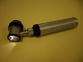
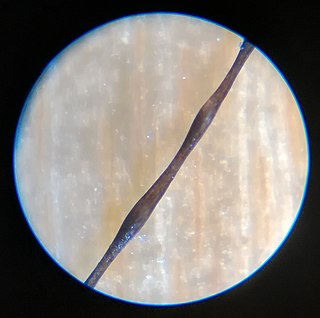
Trichoscopy is a method of hair and scalp evaluation and is used for diagnosing hair and scalp diseases. The method is based on dermoscopy. In trichoscopy hair and scalp structures may be visualized at many-fold magnification. Currently magnifications ranging from 10-fold to 70-fold are most popular in research and clinical practice.

Antonella Tosti is an Italian physician and scientist with major contributions in the field of dermatology, including developing dermoscopy for the diagnosis and care of hair diseases, a world recognized expert in hair disorders. Her contributions to knowledge about nails include research about videodermoscopy of the hyponychium and the nail plate.

<span class="mw-page-title-main">Lidia Rudnicka</span> Polish-American dermatologist

Lidia Rudnicka is a Polish-American dermatologist with contributions to the field of scleroderma research, hair diseases and melanoma prevention.

International Dermoscopy Society (IDS) is a non-governmental organization offering comprehensive promotion of dermoscopy, also known as dermatoscopy. It has over 16,000 international members from over 160 different countries.

References

  1. Berk-Krauss, Juliana; Laird, Mary E. (2017-12-01). "What's in a Name—Dermoscopy vs Dermatoscopy" . JAMA Dermatology. 153 (12): 1235. doi:10.1001/jamadermatol.2017.3905. ISSN   2168-6068. PMID   29238835.
  2. Argenziano, G; Soyer, HP (July 2001). "Dermoscopy of pigmented skin lesions--a valuable tool for early diagnosis of melanoma". The Lancet. Oncology. 2 (7): 443–9. doi:10.1016/s1470-2045(00)00422-8. PMID   11905739.
  3. Lorentzen, H; Weismann, K; Petersen, CS; Larsen, FG; Secher, L; Skødt, V (1999). "Clinical and dermatoscopic diagnosis of malignant melanoma. Assessed by expert and non-expert groups". Acta Dermato-Venereologica. 79 (4): 301–4. doi: 10.1080/000155599750010715 . PMID   10429989.
  4. Vestergaard, ME; Macaskill, P; Holt, PE; Menzies, SW (2008). "Dermoscopy compared with naked eye examination for the diagnosis of primary melanoma: a meta-analysis of studies performed in a clinical setting". British Journal of Dermatology . 159 (3): 669–76. doi:10.1111/j.1365-2133.2008.08713.x. PMID   18616769. S2CID   46404467.Argenziano, G; Fabbrocini, G; Carli, P; De Giorgi, V; Sammarco, E; Delfino, M (1998). "Epiluminescence microscopy for the diagnosis of doubtful melanocytic skin lesions. Comparison of the ABCD rule of dermatoscopy and a new 7-point checklist based on pattern analysis". Archives of Dermatology. 134 (12): 1563–70. doi:10.1001/archderm.134.12.1563. PMID   9875194.
  5. Ascierto, P.A.; Palmieri, G.; Celentano, E.; Parasole, R.; Caraco, C.; Daponte, A.; Chiofalo, M.G.; Melucci, M.T.; Mozzillo, N.; Satriano, R.A.; Castello, G. (2000). "Sensitivity and specificity of epiluminescence microscopy: evaluation on a sample of 2731 excised cutaneous pigmented lesions". British Journal of Dermatology. 142 (5): 893–8. doi:10.1046/j.1365-2133.2000.03468.x. PMID   10809845. S2CID   70893256. http://www.bcbstx.com/provider/pdf/medicalpolicies/medicine/201-023.pdf%5B%5D%5B%5D
  6. Bono, A; Bartoli, C; Cascinelli, N; Lualdi, M; Maurichi, A; Moglia, D; Tragni, G; Tomatis, S; Marchesini, R (2002). "Melanoma detection. A prospective study comparing diagnosis with the naked eye, dermatoscopy and telespectrophotometry". Dermatology. 205 (4): 362–6. doi:10.1159/000066436. PMID   12444332. S2CID   25348202.
  7. "Crutchfield Dermatology". Crutchfield Dermatology. Archived from the original on 2016-10-11. Retrieved 2010-05-12.
  8. Argenziano, G; Mordente, I; Ferrara, G; Sgambato, A; Annese, P; Zalaudek, I (2008). "Dermoscopic monitoring of melanocytic skin lesions: clinical outcome and patient compliance vary according to follow-up protocols". The British Journal of Dermatology. 159 (2): 331–6. doi:10.1111/j.1365-2133.2008.08649.x. PMID   18510663. S2CID   32027237.
  9. Roma, Paolo; Savarese, Imma; Martino, Antonia; Martino, Domenico; Annese, Pietro; Capoluongo, Patrizio; Mordente, Ines; Nicolino, Rachele; Zalaudek, Iris; Argenziano, Giuseppe (2007). "Slow-growing melanoma: Report of five cases". Journal of Dermatological Case Reports. 1 (1): 1–3. doi:10.3315/jdcr.2007.1.1001. PMC   3157767 . PMID   21886697.
  10. Scalvenzi, M; Lembo, S; Francia, MG; Balato, A (2008). "Dermoscopic patterns of superficial basal cell carcinoma". International Journal of Dermatology. 47 (10): 1015–8. doi:10.1111/j.1365-4632.2008.03731.x. PMID   18986346. S2CID   205394964.
  11. Felder, S; Rabinovitz, H; Oliviero, M; Kopf, A (2006). "Dermoscopic differentiation of a superficial basal cell carcinoma and squamous cell carcinoma in situ". Dermatologic Surgery. 32 (3): 423–5. doi:10.1111/j.1524-4725.2006.32085.x. PMID   16640692.
  12. Sicinska, Justyna; Rakowska, Adriana; Czuwara-Ladykowska, Joanna; Mroz, Andrzej; Lipinski, Marcin; Nasierowska-Guttmejer, Anna; Sikorska, Jolanta; Sklinda, Katarzyna; Slowinska, Monika; Kowalska-Oledzka, Elzbieta; Walecka, Irena; Walecki, Jerzy; Rudnicka, Lidia (2007). "Cylindroma transforming into basal cell carcinoma in a patient with Brooke-Spiegler syndrome". Journal of Dermatological Case Reports. 1 (1): 4–9. doi:10.3315/jdcr.2007.1.1002. PMC   3157764 . PMID   21886698.
  13. Campos-Do-Carmo, G; Ramos-E-Silva, M (2008). "Dermoscopy: basic concepts". International Journal of Dermatology. 47 (7): 712–9. doi: 10.1111/j.1365-4632.2008.03556.x . PMID   18613881. S2CID   205394507.
  14. Wu, Ming-Yun; Hu, Shu-Lin; Hsu, Che-Hao (June 2008). "Use of Non-contact Dermatoscopy in the Diagnosis of Scabies" (PDF). Dermatol Sinica: 112–4. Archived from the original (PDF) on 2011-07-09. Retrieved 2009-09-28.
  15. Chuh, A; Lee, A; Wong, W; Ooi, C; Zawar, V (2007). "Diagnosis of Pediculosis pubis: a novel application of digital epiluminescence dermatoscopy". Journal of the European Academy of Dermatology and Venereology. 21 (6): 837–8. doi:10.1111/j.1468-3083.2006.02040.x. PMID   17567326. S2CID   34621172.
  16. Slowinska, M; Rudnicka, L; Schwartz, RA; Kowalska-Oledzka, E; Rakowska, A; Sicinska, J; Lukomska, M; Olszewska, M; Szymanska, E (2008). "Comma hairs: a dermatoscopic marker for tinea capitis: a rapid diagnostic method". Journal of the American Academy of Dermatology. 59 (5 Suppl): S77–9. doi:10.1016/j.jaad.2008.07.009. PMID   19119131.
  17. Inui, S; Nakajima, T; Itami, S (2008). "Significance of dermoscopy in acute diffuse and total alopecia of the female scalp: review of twenty cases". Dermatology. 217 (4): 333–6. doi:10.1159/000155644. PMID   18799878. S2CID   19963257.
  18. Rakowska, A.; et al. (2008). "Trichoscopy criteria for diagnosing female androgenic alopecia". Nature Precedings. doi: 10.1038/npre.2008.1913.1 . hdl:10101/npre.2008.1913.1.
  19. Rakowska, A; Slowinska, M; Czuwara, J; Olszewska, M; Rudnicka, L (2007). "Dermoscopy as a tool for rapid diagnosis of monilethrix". Journal of Drugs in Dermatology. 6 (2): 222–4. PMID   17373184.
  20. Rakowska, A; Kowalska-Oledzka, E; Slowinska, M; Rosinska, D; Rudnicka, L (2009). "Hair shaft videodermoscopy in netherton syndrome". Pediatric Dermatology. 26 (3): 320–2. doi:10.1111/j.1525-1470.2008.00778.x. PMID   19706096. S2CID   42660583.
  21. Rakowska, Adriana; Slowinska, Monika; Kowalska-Oledzka, Elzbieta; Rudnicka, Lidia (2008). "Trichoscopy in genetic hair shaft abnormalities". Journal of Dermatological Case Reports. 2 (2): 14–20. doi:10.3315/jdcr.2008.1009. PMC   3157768 . PMID   21886705.
  22. Rudnicka L, Olszewska M, Rakowska A, Kowalska-Oledzka E, Slowinska M (July 2008). "Trichoscopy: a new method for diagnosing hair loss". Journal of Drugs in Dermatology. 7 (7): 651–4. PMID   18664157.
  23. Rakowska A, Slowinska M, Kowalska-Oledzka E, Rudnicka L (2008). "Trichoscopy (hair and scalp videodermoscopy) in the healthy female. Method standardization and norms for measurable parameters". J Dermatol Case Rep. 3 (1): 14–19. doi:10.3315/jdcr.2008.1021. PMC   3157785 . PMID   21886722.
  24. Guarenti, IM; Almeida HL, Jr; Leitão, AH; Rocha, NM; Silva, RM (2013). "Scanning electron microscopy of tinea nigra". Anais Brasileiros de Dermatologia. 89 (2): 334–6. doi: 10.1590/abd1806-4841.20142780 . PMC   4008070 . PMID   24770516.
  25. Dinnes, Jacqueline; Deeks, Jonathan J.; Chuchu, Naomi; Ferrante di Ruffano, Lavinia; Matin, Rubeta N.; Thomson, David R.; Wong, Kai Yuen; Aldridge, Roger Benjamin; Abbott, Rachel; Fawzy, Monica; Bayliss, Susan E.; Grainge, Matthew J.; Takwoingi, Yemisi; Davenport, Clare; Godfrey, Kathie (2018-12-04). "Dermoscopy, with and without visual inspection, for diagnosing melanoma in adults". The Cochrane Database of Systematic Reviews. 2018 (12): CD011902. doi:10.1002/14651858.CD011902.pub2. ISSN   1469-493X. PMC   6517096 . PMID   30521682.
  26. Kassem, Mohamed A.; Hosny, Khalid M.; Damaševičius, Robertas; Eltoukhy, Mohamed Meselhy (2021-07-31). "Machine Learning and Deep Learning Methods for Skin Lesion Classification and Diagnosis: A Systematic Review". Diagnostics. 11 (8): 1390. doi: 10.3390/diagnostics11081390 . ISSN   2075-4418. PMC   8391467 . PMID   34441324.
  27. Celebi, M. Emre; Mendonça, Teresa; Marques, Jorge S. (March 9, 2018). Dermoscopy Image Analysis (1st ed.). CRC Press.
  28. La Salvia, Marco; Torti, Emanuele; Leon, Raquel; Fabelo, Himar; Ortega, Samuel; Martinez-Vega, Beatriz; Callico, Gustavo M.; Leporati, Francesco (January 2022). "Deep Convolutional Generative Adversarial Networks to Enhance Artificial Intelligence in Healthcare: A Skin Cancer Application". Sensors. 22 (16): 6145. Bibcode:2022Senso..22.6145L. doi: 10.3390/s22166145 . ISSN   1424-8220. PMC   9416026 . PMID   36015906.
  29. Katz, Brian; Rabinovitz, Harold S. (April 2001). "INTRODUCTION TO DERMOSCOPY". Dermatologic Clinics. 19 (2): 221–258. doi:10.1016/S0733-8635(05)70263-1.
  30. Stolz, Wilhelm (1989). "Skin Surface Microscopy". The Lancet. 334 (8667): 864–865. doi:10.1016/s0140-6736(89)93027-4. PMID   2571785. S2CID   5459081.
  31. "Non-contact dermatoscopic device with full polarization control and liquid lens based autofocus function" (PDF). www.dgao-proceedings.de. Retrieved 2019-01-16.
  32. Dreiseitl, S; Binder, M; Hable, K; Kittler, H (June 2009). "Computer versus human diagnosis of melanoma: evaluation of the feasibility of an automated diagnostic system in a prospective clinical trial". Melanoma Research. 19 (3): 180–4. doi: 10.1097/CMR.0b013e32832a1e41 . PMID   19369900. S2CID   3095768.
  33. Dick, V; Sinz, C; Mittlböck, M; Kittler, H; Tschandl, P (19 June 2019). "Accuracy of Computer-Aided Diagnosis of Melanoma: A Meta-analysis". JAMA Dermatology. 155 (11): 1291–1299. doi:10.1001/jamadermatol.2019.1375. PMC   6584889 . PMID   31215969.
  34. Tschandl, P; Rosendahl, C; Kittler, H (14 August 2018). "The HAM10000 dataset, a large collection of multi-source dermatoscopic images of common pigmented skin lesions". Scientific Data. 5: 180161. arXiv: 1803.10417 . Bibcode:2018NatSD...580161T. doi: 10.1038/sdata.2018.161 . PMC   6091241 . PMID   30106392.
  35. Tschandl, P; Codella, N; Akay, BN; Argenziano, G; Braun, RP; Cabo, H; Gutman, D; Halpern, A; Helba, B; Hofmann-Wellenhof, R; Lallas, A; Lapins, J; Longo, C; Malvehy, J; Marchetti, MA; Marghoob, A; Menzies, S; Oakley, A; Paoli, J; Puig, S; Rinner, C; Rosendahl, C; Scope, A; Sinz, C; Soyer, HP; Thomas, L; Zalaudek, I; Kittler, H (July 2019). "Comparison of the accuracy of human readers versus machine-learning algorithms for pigmented skin lesion classification: an open, web-based, international, diagnostic study". The Lancet. Oncology. 20 (7): 938–947. doi:10.1016/S1470-2045(19)30333-X. PMC   8237239 . PMID   31201137.
  36. Tschandl, P; Rosendahl, C; Akay, BN; Argenziano, G; Blum, A; Braun, RP; Cabo, H; Gourhant, JY; Kreusch, J; Lallas, A; Lapins, J; Marghoob, A; Menzies, S; Neuber, NM; Paoli, J; Rabinovitz, HS; Rinner, C; Scope, A; Soyer, HP; Sinz, C; Thomas, L; Zalaudek, I; Kittler, H (1 January 2019). "Expert-Level Diagnosis of Nonpigmented Skin Cancer by Combined Convolutional Neural Networks". JAMA Dermatology. 155 (1): 58–65. doi: 10.1001/jamadermatol.2018.4378 . PMC   6439580 . PMID   30484822.
  37. Marchetti, MA; Codella, NCF; Dusza, SW; Gutman, DA; Helba, B; Kalloo, A; Mishra, N; Carrera, C; Celebi, ME; DeFazio, JL; Jaimes, N; Marghoob, AA; Quigley, E; Scope, A; Yélamos, O; Halpern, AC; International Skin Imaging, Collaboration. (February 2018). "Results of the 2016 International Skin Imaging Collaboration International Symposium on Biomedical Imaging challenge: Comparison of the accuracy of computer algorithms to dermatologists for the diagnosis of melanoma from dermoscopic images". Journal of the American Academy of Dermatology. 78 (2): 270–277.e1. doi: 10.1016/j.jaad.2017.08.016 . PMC   5768444 . PMID   28969863.
  38. Haenssle, HA; Fink, C; Toberer, F; Winkler, J; Stolz, W; Deinlein, T; Hofmann-Wellenhof, R; Lallas, A; Emmert, S; Buhl, T; Zutt, M; Blum, A; Abassi, MS; Thomas, L; Tromme, I; Tschandl, P; Enk, A; Rosenberger, A; Reader Study Level I and Level II, Groups. (January 2020). "Man against machine reloaded: performance of a market-approved convolutional neural network in classifying a broad spectrum of skin lesions in comparison with 96 dermatologists working under less artificial conditions". Annals of Oncology. 31 (1): 137–143. doi: 10.1016/j.annonc.2019.10.013 . PMID   31912788.
  39. Tschandl, P; Kittler, H; Argenziano, G (September 2017). "A pretrained neural network shows similar diagnostic accuracy to medical students in categorizing dermatoscopic images after comparable training conditions". The British Journal of Dermatology. 177 (3): 867–869. doi:10.1111/bjd.15695. PMID   28569993. S2CID   207076965.
  40. Yu, C; Yang, S; Kim, W; Jung, J; Chung, KY; Lee, SW; Oh, B (2018). "Acral melanoma detection using a convolutional neural network for dermoscopy images". PLOS ONE. 13 (3): e0193321. Bibcode:2018PLoSO..1393321Y. doi: 10.1371/journal.pone.0193321 . PMC   5841780 . PMID   29513718.
  41. Esteva, A; Kuprel, B; Novoa, RA; Ko, J; Swetter, SM; Blau, HM; Thrun, S (2 February 2017). "Dermatologist-level classification of skin cancer with deep neural networks". Nature. 542 (7639): 115–118. Bibcode:2017Natur.542..115E. doi:10.1038/nature21056. PMC   8382232 . PMID   28117445.
  42. Lallas, A; Argenziano, G (October 2018). "Artificial intelligence and melanoma diagnosis: ignoring human nature may lead to false predictions". Dermatology Practical & Conceptual. 8 (4): 249–251. doi:10.5826/dpc.0804a01. PMC   6246056 . PMID   30479851.
  43. Navarrete-Dechent, C; Dusza, SW; Liopyris, K; Marghoob, AA; Halpern, AC; Marchetti, MA (October 2018). "Automated Dermatological Diagnosis: Hype or Reality?". The Journal of Investigative Dermatology. 138 (10): 2277–2279. doi: 10.1016/j.jid.2018.04.040 . PMC   7701995 . PMID   29864435.
  44. "VECTRA WB360 Imaging System | Canfield Scientific". www.canfieldsci.com. Retrieved 2022-12-12.
  45. "DermaGraphix | Canfield Scientific". www.canfieldsci.com. Retrieved 2022-12-12.