Related Research Articles
A lesion is any damage or abnormal change in the tissue of an organism, usually caused by injury or diseases. Lesion is derived from the Latin laesio meaning "injury". Lesions may occur in plants as well as animals.

Nevus is a nonspecific medical term for a visible, circumscribed, chronic lesion of the skin or mucosa. The term originates from nævus, which is Latin for "birthmark"; however, a nevus can be either congenital or acquired. Common terms, including mole, birthmark, and beauty mark, are used to describe nevi, but these terms do not distinguish specific types of nevi from one another.

A dysplastic nevus or atypical mole is a nevus (mole) whose appearance is different from that of common moles. In 1992, the NIH recommended that the term "dysplastic nevus" be avoided in favor of the term "atypical mole". An atypical mole may also be referred to as an atypical melanocytic nevus, atypical nevus, B-K mole, Clark's nevus, dysplastic melanocytic nevus, or nevus with architectural disorder.

Dysplastic nevus syndrome, also known as familial atypical multiple mole–melanoma (FAMMM) syndrome, is an inherited cutaneous condition described in certain families, and characterized by unusual nevi and multiple inherited melanomas. First described in 1820, the condition is inherited in an autosomal dominant pattern, and caused by mutations in the CDKN2A gene. In addition to melanoma, individuals with the condition are at increased risk for pancreatic cancer.

Lionel Tarassenko, is a British engineer and academic, who is a leading expert in the application of signal processing and machine learning to healthcare. Tarassenko is President of Reuben College, Oxford.

Dermatoscopy also known as dermoscopy or epiluminescence microscopy, is the examination of skin lesions with a dermatoscope. It is a tool similar to a camera to allow for inspection of skin lesions unobstructed by skin surface reflections. The dermatoscope consists of a magnifier, a light source, a transparent plate and sometimes a liquid medium between the instrument and the skin. The dermatoscope is often handheld, although there are stationary cameras allowing the capture of whole body images in a single shot. When the images or video clips are digitally captured or processed, the instrument can be referred to as a digital epiluminescence dermatoscope. The image is then analyzed automatically and given a score indicating how dangerous it is. This technique is useful to dermatologists and skin cancer practitioners in distinguishing benign from malignant (cancerous) lesions, especially in the diagnosis of melanoma.

Computer-aided detection (CADe), also called computer-aided diagnosis (CADx), are systems that assist doctors in the interpretation of medical images. Imaging techniques in X-ray, MRI, Endoscopy, and ultrasound diagnostics yield a great deal of information that the radiologist or other medical professional has to analyze and evaluate comprehensively in a short time. CAD systems process digital images or videos for typical appearances and to highlight conspicuous sections, such as possible diseases, in order to offer input to support a decision taken by the professional.

Skin biopsy is a biopsy technique in which a skin lesion is removed to be sent to a pathologist to render a microscopic diagnosis. It is usually done under local anesthetic in a physician's office, and results are often available in 4 to 10 days. It is commonly performed by dermatologists. Skin biopsies are also done by family physicians, internists, surgeons, and other specialties. However, performed incorrectly, and without appropriate clinical information, a pathologist's interpretation of a skin biopsy can be severely limited, and therefore doctors and patients may forgo traditional biopsy techniques and instead choose Mohs surgery.
Animal-type melanoma is a rare subtype of melanoma that is characterized by heavily pigmented dermal epithelioid and spindled melanocytes. Animal-type melanoma is also known to be called equine-type melanoma, pigment synthesizing melanoma, and pigmented epithelioid melanocytoma (PEM). While melanoma is known as the most aggressive skin cancer, the mortality for PEM is lower than in other melanoma types. Animal-type melanoma earned its name due to the resemblance of melanocytic tumors in grey horses.

MeVisLab is a cross-platform application framework for medical image processing and scientific visualization. It includes advanced algorithms for image registration, segmentation, and quantitative morphological and functional image analysis. An IDE for graphical programming and rapid user interface prototyping is available.

Artificial intelligence in healthcare is a term used to describe the use of machine-learning algorithms and software, or artificial intelligence (AI), to copy human cognition in the analysis, presentation, and understanding of complex medical and health care data, or to exceed human capabilities by providing new ways to diagnose, treat, or prevent disease. Specifically, AI is the ability of computer algorithms to approximate conclusions based solely on input data.

David Atienza Alonso is a Spanish/Swiss scientist in the disciplines of computer and electrical engineering. His research focuses on hardware‐software co‐design and management for energy‐efficient and thermal-aware computing systems, always starting from a system‐level perspective to the actual electronic design. He is a full professor of electrical and computer engineering at the Swiss Federal Institute of Technology in Lausanne (EPFL) and the head of the Embedded Systems Laboratory (ESL). He is an IEEE Fellow (2016), and an ACM Fellow (2022).
Automated machine learning (AutoML) is the process of automating the tasks of applying machine learning to real-world problems.
Ronald Marc Summers is an American radiologist and senior investigator at the Diagnostic Radiology Department at the NIH Clinical Center in Bethesda, Maryland. He is chief of the Clinical Image Processing Service and directs the Imaging Biomarkers and Computer-Aided Diagnosis (CAD) Laboratory. A researcher in the field of radiology and computer-aided diagnosis, he has co-authored over 500 journal articles and conference proceedings papers and is a coinventor on 12 patents. His lab has conducted research applying artificial intelligence and deep learning to radiology.

Georgia "Gina" D. Tourassi is the Director of the Oak Ridge National Laboratory health data sciences institute and adjunct Professor of radiology at Duke University. She works on biomedical informatics, computer-aided diagnosis and artificial intelligence (AI) in health care.

Aidoc Medical is an Israeli technology company that develops computer-aided simple triage and notification systems. Aidoc has obtained FDA and CE mark approval for its stroke, pulmonary embolism, cervical fracture, intracranial hemorrhage, intra-abdominal free gas, and incidental pulmonary embolism algorithms.

Merative L.P., formerly IBM Watson Health, is an American medical technology company that provides products and services that help clients facilitate medical research, clinical research, real world evidence, and healthcare services, through the use of artificial intelligence, data analytics, cloud computing, and other advanced information technology. Merative is owned by Francisco Partners, an American private equity firm headquartered in San Francisco, California. In 2022, IBM divested and spun-off their Watson Health division into Merative. As of 2023, it remains a standalone company headquartered in Ann Arbor with innovation centers in Hyderabad, Bengaluru, and Chennai.

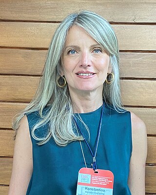
Konstantina "Nantia" Nikita is a Greek electrical and computer engineer and a professor at the School of Electrical and Computer Engineering at the National Technical University of Athens (NTUA), Greece. She is director of the Mobile Radiocommunications Lab and founder and director of the Biomedical Simulations and Imaging Lab, NTUA. Since 2015, she has been an Irene McCulloch Distinguished Adjunct Professor of Biomedical Engineering and Medicine at Keck School of Medicine and Viterbi School of Engineering, University of Southern California.
Pallavi Tiwari is an Indian American biomedical engineer who is a professor at the University of Wisconsin–Madison. Her research considers the development of computer algorithms to accelerate the diagnosis and treatment of disease. She was elected Fellow of the National Academy of Inventors.
References
- ↑ "Deutsch". www.ibmt.fraunhofer.de (in German). Retrieved 2022-09-29.
- ↑ Haenssle, H. A.; Fink, C.; Schneiderbauer, R.; Toberer, F.; Buhl, T.; Blum, A.; Kalloo, A.; Hassen, A. Ben Hadj; Thomas, L.; Enk, A.; Uhlmann, L.; Reader study level-I and level-II Groups; Alt, Christina; Arenbergerova, Monika; Bakos, Renato (2018-08-01). "Man against machine: diagnostic performance of a deep learning convolutional neural network for dermoscopic melanoma recognition in comparison to 58 dermatologists". Annals of Oncology. 29 (8): 1836–1842. doi: 10.1093/annonc/mdy166 . hdl: 11368/2966390 . ISSN 1569-8041. PMID 29846502.
- ↑ humans.txt. "TrichoLAB". tricholab.com. Retrieved 2022-09-29.