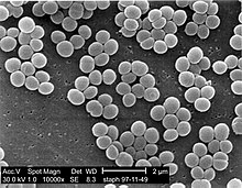
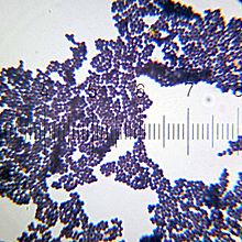
Staphylococcus

Last updated

Staphylococcus
Staphylococcus aureus 01.jpg
Scanning electron micrograph of S. aureus colonies: Note the grape-like clustering common to Staphylococcus species.
Scientific classification OOjs UI icon edit-ltr.svg
Domain: Bacteria
Phylum: Bacillota
Class: Bacilli
Order: Bacillales
Family: Staphylococcaceae
Genus: Staphylococcus
Rosenbach 1884
Species

Staphylococcus is a genus of Gram-positive bacteria in the family Staphylococcaceae from the order Bacillales. Under the microscope, they appear spherical (cocci), and form in grape-like clusters. Staphylococcus species are facultative anaerobic organisms (capable of growth both aerobically and anaerobically).

Contents

The name was coined in 1880 by Scottish surgeon and bacteriologist Alexander Ogston (1844–1929), following the pattern established five years earlier with the naming of Streptococcus . [1] It combines the prefix "staphylo-" (from Ancient Greek : σταφυλή, romanized: staphylē, lit. 'bunch of grapes' [2] ), and suffixed by the New Latin : coccus, lit. 'spherical bacterium' (from Ancient Greek: κόκκος, romanized: kókkos, lit.'grain, seed, berry' [3] ).

Staphylococcus was one of the leading infections in hospitals and many strains of this bacterium have become antibiotic resistant. Despite strong attempts to get rid of them, staph bacteria stay present in hospitals, where they can infect people who are most at risk of infection. [4]

Staphylococcus includes at least 44 species. Of these, nine have two subspecies, one has three subspecies, and one has four subspecies. [5] Many species cannot cause disease and reside normally on the skin and mucous membranes of humans and other animals. Staphylococcus species have been found to be nectar-inhabiting microbes. [6] They are also a small component of the soil microbiome. [7]

Taxonomy

The taxonomy is based on 16s rRNA sequences, [8] and most of the staphylococcal species fall into 11 clusters:

  1. S. aureus group – S. argenteus, S. aureus, S. schweitzeri, S. simiae
  2. S. auricularis group – S. auricularis
  3. S. carnosus group – S. carnosus, S. condimenti, S. debuckii, S. massiliensis, S. piscifermentans, S. simulans
  4. S. epidermidis group – S. capitis, S. caprae, S. epidermidis, S. saccharolyticus
  5. S. haemolyticus group – S. borealis, S. devriesei, S. haemolyticus, S. hominis
  6. S. hyicus-intermedius group – S. agnetis, S. chromogenes, S. cornubiensis, S. felis, S. delphini, S. hyicus, S. intermedius, S. lutrae, S. microti, S. muscae, S. pseudintermedius, S. rostri, S. schleiferi
  7. S. lugdunensis group – S. lugdunensis
  8. S. saprophyticus group – S. arlettae, S. caeli, S. cohnii, S. equorum, S. gallinarum, S. kloosii, S. leei, S. nepalensis, S. saprophyticus, S. succinus, S. xylosus
  9. S. sciuri group – S. fleurettii, S. lentus, S. sciuri, S. stepanovicii, S. vitulinus
  10. S. simulans group – S. simulans
  11. S. warneri group – S. pasteuri, S. warneri

A twelfth group – that of S. caseolyticus – has now been removed to a new genus, Macrococcus , the species of which are currently the closest known relatives of Staphylococcus. [9]

Two species were described in 2015 – Staphylococcus argenteus and Staphylococcus schweitzeri – both of which were previously considered variants of S. aureus. [10]

A new coagulase negative species – Staphylococcus edaphicus – has been isolated from Antarctica. [11] This species is probably a member of the S. saprophyticus group.

Groups

Based on an analysis of orthologous gene content three groups (A, B and C) have been proposed. [12]

Group A includes S. aureus, S. borealis, S. capitis, S. epidermidis, S. haemolyticus, S. hominis, S. lugdunensis, S. pettenkoferi, S. simiae and S. warneri.

Group B includes S. arlettae, S. cohnii, S. equorum, S. saprophyticus and S. xylosus.

Group C includes S. delphini, S. intermedius and S. pseudintermedius.

Notes

The S. saprophyticus and S. sciuri groups are generally novobiocin-resistant, as is S. hominis subsp. novobiosepticus.

Members of the S. sciuri group are oxidase-positive due to their possession of the enzyme cytochrome c oxidase. This group is the only clade within the staphylococci to possess this gene.

The S. sciuri group appears to be the closest relations to the genus Macrococcus .

S. pulvereri has been shown to be a junior synonym of S. vitulinus. [13]

Within these clades, the S. haemolyticus and S. simulans groups appear to be related, as do the S. aureus and S. epidermidis groups. [14]

S. lugdunensis appears to be related to the S. haemolyticus group.

S. petrasii may be related to S. haemolyticus, but this needs to be confirmed.

The taxonomic position of S. lyticans,S. petrasii, and S. pseudolugdunensis has yet to be clarified. The published descriptions of these species do not appear to have been validly published.

Biochemical identification

Assignment of a strain to the genus Staphylococcus requires it to be a Gram-positive coccus [15] that forms clusters, has an appropriate cell wall structure (including peptidoglycan type and teichoic acid presence) and G + C content of DNA in a range of 30–40 mol%.

Staphylococcus species can be differentiated from other aerobic and facultative anaerobic, Gram-positive cocci by several simple tests. [15] Staphylococcus species are facultative anaerobes (capable of growth both aerobically and anaerobically). [15] All species grow in the presence of bile salts.

All species of Staphylococcus aureus were once thought to be coagulase-positive, but this has since been disproven. [16] [17] [18]

Growth can also occur in a 6.5% NaCl solution. [15] On Baird-Parker medium, Staphylococcus species grow fermentatively, except for S. saprophyticus, which grows oxidatively. Staphylococcus species are resistant to bacitracin (0.04 U disc: resistance = < 10 mm zone of inhibition) and susceptible to furazolidone (100 μg disc: resistance = < 15 mm zone of inhibition). Further biochemical testing is needed to identify to the species level.

Coagulase production

One of the most important phenotypical features used in the classification of staphylococci is their ability to produce coagulase, an enzyme that causes blood clot formation.

Seven species are currently recognised as being coagulase-positive: S. aureus, S. delphini, S. hyicus, S. intermedius, S. lutrae, S. pseudintermedius, and S. schleiferi subsp. coagulans. These species belong to two separate groups – the S. aureus (S. aureus alone) group and the S. hyicus-intermedius group (the remaining five).

An eighth species has also been described – Staphylococcus leei – from patients with gastritis. [19]

S. aureus is coagulase-positive, meaning it produces coagulase. However, while the majority of S. aureus strains are coagulase-positive, some may be atypical in that they do not produce coagulase. S. aureus is catalase-positive (meaning that it can produce the enzyme catalase) and able to convert hydrogen peroxide (H2O2) to water and oxygen, which makes the catalase test useful to distinguish staphylococci from enterococci and streptococci.

S. pseudintermedius inhabits and sometimes infects the skin of domestic dogs and cats. This organism, too, can carry the genetic material that imparts multiple bacterial resistance. It is rarely implicated in infections in humans, as a zoonosis.

S. epidermidis , a coagulase-negative species, is a commensal of the skin, but can cause severe infections in immunosuppressed patients and those with central venous catheters. S. saprophyticus , another coagulase-negative species that is part of the normal vaginal flora, is predominantly implicated in genitourinary tract infections in sexually active young women. In recent years, several other Staphylococcus species have been implicated in human infections, notably S. lugdunensis , S. schleiferi , and S. caprae .

Common abbreviations for coagulase-negative staphylococci are CoNS, CNS, or CNST. [20] The American Society for Microbiology abbreviates coagulase-negative staphylococci as "CoNS".

Genomics and molecular biology

The first S. aureus genomes to be sequenced were those of N315 and Mu50, in 2001. Many more complete S. aureus genomes have been submitted to the public databases, making it one of the most extensively sequenced bacteria. The use of genomic data is now widespread and provides a valuable resource for researchers working with S. aureus. Whole genome technologies, such as sequencing projects and microarrays, have shown an enormous variety of S. aureus strains. Each contains different combinations of surface proteins and different toxins. Relating this information to pathogenic behaviour is one of the major areas of staphylococcal research. The development of molecular typing methods has enabled the tracking of different strains of S. aureus. This may lead to better control of outbreak strains. A greater understanding of how the staphylococci evolve, especially due to the acquisition of mobile genetic elements encoding resistance and virulence genes is helping to identify new outbreak strains and may even prevent their emergence. [21]

The widespread incidence of antibiotic resistance across various strains of S. aureus, or across different species of Staphylococcus has been attributed to horizontal gene transfer of genes encoding antibiotic/metal resistance and virulence. A recent study demonstrated the extent of horizontal gene transfer among Staphylococcus to be much greater than previously expected, and encompasses genes with functions beyond antibiotic resistance and virulence, and beyond genes residing within the mobile genetic elements. [22]

Various strains of Staphylococcus are available from biological research centres, such as the National Collection of Type Cultures.

Host range

Unknown variety of Staphylococcus, Gram-stained - numbered ticks on the scale are 11 um apart 20101017 231210 Staphylococcus.jpg
Unknown variety of Staphylococcus, Gram-stained – numbered ticks on the scale are 11  µm apart

Members of the genus Staphylococcus frequently colonize the skin and upper respiratory tracts of mammals and birds and also in marine sponge. [15] Marine sponge associated Staphylococcus species are highly salt tolerant. [15] Some species specificity has been observed in host range, such that the Staphylococcus species observed on some animals appear more rarely on more distantly related host species. [23] Some of the observed host specificity includes:

Populations at risk for Staphylococcus aureus infection

It is said that anyone can develop a staph infection, although certain groups of people are at greater risk, including people with chronic conditions such as diabetes, cancer, vascular disease, eczema, lung disease, and people who inject drugs. In healthcare facilities, the risk of more serious staph infection is higher because many patients have weakened immune systems or have undergone procedures. In healthcare, the risk of more serious staph infection is higher for patients in intensive care units (ICUs), patients who have undergone certain types of surgeries and patients with medical devices inserted in their bodies. [25]

Staphylococcus aureus has emerged as a leading agent of sepsis. It facilitates factors such as tissue adhesion, immune evasion, and host cell injury. In the bloodstream, these factors cause inflammation, impair immune cell function, alter coagulation, and compromise vascular integrity. When left untreated, S. aureus triggers pathophysiologic disturbances that are further amplified by the host inflammatory response, culminating in the severe clinical manifestations of sepsis and septic shock. [26]

Clinical

Staphylococcus can cause a wide variety of diseases in humans and animals through either toxin production or penetration. Staphylococcal toxins are a common cause of food poisoning, for they can be produced by bacteria growing in improperly stored food items. The most common sialadenitis is caused by staphylococci, as bacterial infections. [27] Staphylococci break down leucine into isovaleric acid, the main odor of foot odor. [28]

See also

Related Research Articles

<i>Staphylococcus aureus</i> Species of Gram-positive bacterium

Staphylococcus aureus is a Gram-positive spherically shaped bacterium, a member of the Bacillota, and is a usual member of the microbiota of the body, frequently found in the upper respiratory tract and on the skin. It is often positive for catalase and nitrate reduction and is a facultative anaerobe that can grow without the need for oxygen. Although S. aureus usually acts as a commensal of the human microbiota, it can also become an opportunistic pathogen, being a common cause of skin infections including abscesses, respiratory infections such as sinusitis, and food poisoning. Pathogenic strains often promote infections by producing virulence factors such as potent protein toxins, and the expression of a cell-surface protein that binds and inactivates antibodies. S. aureus is one of the leading pathogens for deaths associated with antimicrobial resistance and the emergence of antibiotic-resistant strains, such as methicillin-resistant S. aureus (MRSA), is a worldwide problem in clinical medicine. Despite much research and development, no vaccine for S. aureus has been approved.

<span class="mw-page-title-main">Coagulase</span> Class of bacterial proteins

Coagulase is a protein enzyme produced by several microorganisms that enables the conversion of fibrinogen to fibrin. In the laboratory, it is used to distinguish between different types of Staphylococcus isolates. Importantly, S. aureus is generally coagulase-positive, meaning that a positive coagulase test would indicate the presence of S. aureus or any of the other 11 coagulase-positive Staphylococci. A negative coagulase test would instead show the presence of coagulase-negative organisms such as S. epidermidis or S. saprophyticus. However, it is now known that not all S. aureus are coagulase-positive. Whereas coagulase-positive Staphylococci are usually pathogenic, coagulase-negative Staphylococci are more often associated with opportunistic infection.

Staphylococcus lugdunensis is a coagulase-negative member of the genus Staphylococcus, consisting of Gram-positive bacteria with spherical cells that appear in clusters.

<i>Staphylococcus haemolyticus</i> Species of bacterium

Staphylococcus haemolyticus is a member of the coagulase-negative staphylococci (CoNS). It is part of the skin flora of humans, and its largest populations are usually found at the axillae, perineum, and inguinal areas. S. haemolyticus also colonizes primates and domestic animals. It is a well-known opportunistic pathogen, and is the second-most frequently isolated CoNS. Infections can be localized or systemic, and are often associated with the insertion of medical devices. The highly antibiotic-resistant phenotype and ability to form biofilms make S. haemolyticus a difficult pathogen to treat. Its most closely related species is Staphylococcus borealis.

<i>Staphylococcus saprophyticus</i> Species of bacterium

Staphylococcus saprophyticus is a Gram-positive coccus belonging to the genus Staphylococcus. S. saprophyticus is a common cause of community-acquired urinary tract infections.

Staphylococcus caprae is a Gram-positive, coccus bacteria and a member of the genus Staphylococcus. S. caprae is coagulase-negative. It was originally isolated from goats, but members of this species have also been isolated from human samples.

Staphylococcus hominis is a coagulase-negative member of the bacterial genus Staphylococcus, consisting of Gram-positive, spherical cells in clusters. It occurs very commonly as a harmless commensal on human and animal skin and is known for producing thioalcohol compounds that contribute to body odour. Like many other coagulase-negative staphylococci, S. hominis may occasionally cause infection in patients whose immune systems are compromised, for example by chemotherapy or predisposing illness.

<i>Staphylococcus xylosus</i> Species of bacterium

Staphylococcus xylosus is a species of bacteria belonging to the genus Staphylococcus. It is a Gram-positive bacterium that forms clusters of cells. Like most staphylococcal species, it is coagulase-negative and exists as a commensal on the skin of humans and animals and in the environment.

Staphylococcus warneri is a member of the bacterial genus Staphylococcus, consisting of Gram-positive bacteria with spherical cells appearing in clusters. It is catalase-positive, oxidase-negative, and coagulase-negative, and is a common commensal organism found as part of the skin flora on humans and animals. Like other coagulase-negative staphylococci, S. warneri rarely causes disease, but may occasionally cause infection in patients whose immune system is compromised.

<i>Staphylococcus epidermidis</i> Species of bacterium

Staphylococcus epidermidis is a Gram-positive bacterium, and one of over 40 species belonging to the genus Staphylococcus. It is part of the normal human microbiota, typically the skin microbiota, and less commonly the mucosal microbiota and also found in marine sponges. It is a facultative anaerobic bacteria. Although S. epidermidis is not usually pathogenic, patients with compromised immune systems are at risk of developing infection. These infections are generally hospital-acquired. S. epidermidis is a particular concern for people with catheters or other surgical implants because it is known to form biofilms that grow on these devices. Being part of the normal skin microbiota, S. epidermidis is a frequent contaminant of specimens sent to the diagnostic laboratory.

Lysostaphin is a Staphylococcus simulans metalloendopeptidase. It can function as a bacteriocin (antimicrobial) against Staphylococcus aureus.

<span class="mw-page-title-main">Staphylococcal infection</span> Medical condition

A staphylococcal infection or staph infection is an infection caused by members of the Staphylococcus genus of bacteria.

mecA is a gene found in bacterial cells which allows them to be resistant to antibiotics such as methicillin, penicillin and other penicillin-like antibiotics.

<i>Staphylococcus capitis</i> Species of bacterium

Staphylococcus capitis is a coagulase-negative species (CoNS) of Staphylococcus. It is part of the normal flora of the skin of the human scalp, face, neck, scrotum, and ears and has been associated with prosthetic valve endocarditis, but is rarely associated with native valve infection.

Staphylococcus delphini is a Gram-positive, coagulase-positive member of the bacterial genus Staphylococcus consisting of single, paired, and clustered cocci. Strains of this species were originally isolated from aquarium-raised dolphins suffering from skin lesions.

<i>Staphylococcus hyicus</i> Species of bacterium

Staphylococcus hyicus is a Gram-positive, facultatively anaerobic bacterium in the genus Staphylococcus. It consists of clustered cocci and forms white circular colonies when grown on blood agar. S. hyicus is a known animal pathogen. It causes disease in poultry, cattle, horses, and pigs. Most notably, it is the agent that causes porcine exudative epidermitis, also known as greasy pig disease, in piglets. S. hyicus is generally considered to not be zoonotic, however it has been shown to be able to cause bacteremia and sepsis in humans.

Staphylococcus intermedius is a Gram-positive, catalase positive member of the bacterial genus Staphylococcus consisting of clustered cocci. Strains of this species were originally isolated from the anterior nares of pigeons, dogs, cats, mink, and horses. Many of the isolated strains show coagulase activity. Clinical tests for detection of methicillin-resistant S. aureus may produce false positives by detecting S. intermedius, as this species shares some phenotypic traits with methicillin-resistant S. aureus strains. It has been theorized that S. intermedius has previously been misidentified as S. aureus in human dog bite wound infections, which is why molecular technologies such as MALDI-TOF and PCR are preferred in modern veterinary clinical microbiology laboratories for their more accurate identifications over biochemical tests. S. intermedius is largely phenotypically indiscriminate from Staphylococcus pseudintermedius and Staphylococcus delphini, and therefore the three organisms are considered to be included in the more general 'Staphylococcus intermedius group'.

Staphylococcus schleiferi is a Gram-positive, cocci-shaped bacterium of the family Staphylococcaceae. It is facultatively anaerobic, coagulase-variable, and can be readily cultured on blood agar where the bacterium tends to form opaque, non-pigmented colonies and beta (β) hemolysis. There exists two subspecies under the species S. schleiferi: Staphylococcus schleiferi subsp. schleiferi and Staphylococcus schleiferi subsp. coagulans.

Staphylococcus pseudintermedius is a gram positive coccus bacteria of the genus Staphylococcus found worldwide. It is primarily a pathogen for domestic animals, but has been known to affect humans as well. S. pseudintermedius is an opportunistic pathogen that secretes immune modulating virulence factors, has many adhesion factors, and the potential to create biofilms, all of which help to determine the pathogenicity of the bacterium. Diagnoses of Staphylococcus pseudintermedius have traditionally been made using cytology, plating, and biochemical tests. More recently, molecular technologies like MALDI-TOF, DNA hybridization and PCR have become preferred over biochemical tests for their more rapid and accurate identifications. This includes the identification and diagnosis of antibiotic resistant strains.

Staphylococcus borealis is a bacterial species, member of the genus Staphylococcus, closely related to Staphylococcus haemolyticus and described in 2020. Its cells are Gram positive, coccoid in shape, with a diameter of 0.65 to 1.23 µm and form clusters. Additionally, they are facultative anaerobic, coagulase negative and catalase positive. The type strain was isolated from human blood culture at the University Hospital of North Norway, in 1997. Four additional strains included in the description were isolated from skin swabs, from healthy volunteers. The genome sequence of the type strain is deposited in DNA Data Bank of Japan, European Nucleotide Archive and GenBank under the accession number JABVEJ000000000.

References

  1. "staphylococcus | Origin and meaning of staphylococcus by Online Etymology Dictionary". www.etymonline.com. Retrieved 2018-07-25.
  2. stafulh/  in Liddell, Henry George ; Scott, Robert (1940) A Greek–English Lexicon, revised and augmented throughout by Jones, Sir Henry Stuart , with the assistance of McKenzie, Roderick. Oxford: Clarendon Press. In the Perseus Digital Library , Tufts University.
  3. ko)kkos  in Liddell and Scott
  4. "Staph infections". mayoclinic.org. Retrieved 2022-11-27.
  5. Harris LG, Foster SJ, Richards RG (December 2002). "An introduction to Staphylococcus aureus, and techniques for identifying and quantifying S. aureus adhesins in relation to adhesion to biomaterials: review". European Cells & Materials. 4: 39–60. doi: 10.22203/ecm.v004a04 . PMID   14562246.
  6. Jacquemyn H, Lenaerts M, Brys R, Willems K, Honnay O, Lievens B (2013). "Among-population variation in microbial community structure in the floral nectar of the bee-pollinated forest herb Pulmonaria officinalis L." PLOS ONE. 8 (3): e56917. Bibcode:2013PLoSO...856917J. doi: 10.1371/journal.pone.0056917 . PMC   3594240 . PMID   23536759.
  7. Madigan M, Martinko J, eds. (2005). Brock Biomlogy of Microorganisms (11th ed.). Prentice Hall. ISBN   978-0-13-144329-7.[ page needed ]
  8. Takahashi T, Satoh I, Kikuchi N (April 1999). "Phylogenetic relationships of 38 taxa of the genus Staphylococcus based on 16S rRNA gene sequence analysis". International Journal of Systematic Bacteriology. 49 (2): 725–8. doi: 10.1099/00207713-49-2-725 . PMID   10319495.
  9. Kloos WE, Ballard DN, George CG, Webster JA, Hubner RJ, Ludwig W, Schleifer KH, Fiedler F, Schubert K (July 1998). "Delimiting the genus Staphylococcus through description of Macrococcus caseolyticus gen. nov., comb. nov. and Macrococcus equipercicus sp. nov., and Macrococcus bovicus sp. no. and Macrococcus carouselicus sp. nov". International Journal of Systematic Bacteriology. 48 (3): 859–77. doi: 10.1099/00207713-48-3-859 . PMID   9734040.
  10. Tong SY, Schaumburg F, Ellington MJ, Corander J, Pichon B, Leendertz F, Bentley SD, Parkhill J, Holt DC, Peters G, Giffard PM (January 2015). "Novel staphylococcal species that form part of a Staphylococcus aureus-related complex: the non-pigmented Staphylococcus argenteus sp. nov. and the non-human primate-associated Staphylococcus schweitzeri sp. nov". International Journal of Systematic and Evolutionary Microbiology. 65 (Pt 1): 15–22. doi:10.1099/ijs.0.062752-0. PMC   4298100 . PMID   25269845.
  11. Pantůček R, Sedláček I, Indráková A, Vrbovská V, Mašlaňová I, Kovařovic V, Švec P, Králová S, Krištofová L, Kekláková J, Petráš P, Doškař J (October 2017). "mecCgene and genomic islands with suspected role in adaptation to extreme environment". Applied and Environmental Microbiology. 84 (2): e01746–17. doi:10.1128/AEM.01746-17. PMC   5752872 . PMID   29079617.
  12. Coates-Brown R, Moran JC, Pongchaikul P, Darby AC and MJ Horsburgh MJ (2018) "Comparative genomics of Staphylococcus reveals determinants of speciation and diversification of antimicrobial defense". Front Microbiol
  13. Svec P, Vancanneyt M, Sedlácek I, Engelbeen K, Stetina V, Swings J, Petrás P (November 2004). "Reclassification of Staphylococcus pulvereri Zakrzewska-Czerwinska et al. 1995 as a later synonym of Staphylococcus vitulinus Webster et al. 1994". International Journal of Systematic and Evolutionary Microbiology. 54 (Pt 6): 2213–5. doi: 10.1099/ijs.0.63080-0 . PMID   15545460.
  14. Ghebremedhin B, Layer F, König W, König B (March 2008). "Genetic classification and distinguishing of Staphylococcus species based on different partial gap, 16S rRNA, hsp60, rpoB, sodA, and tuf gene sequences". Journal of Clinical Microbiology. 46 (3): 1019–25. doi:10.1128/JCM.02058-07. PMC   2268370 . PMID   18174295.
  15. 1 2 3 4 5 6 7 8 Paul, Sulav Indra; Rahman, Md. Mahbubur; Salam, Mohammad Abdus; Khan, Md. Arifur Rahman; Islam, Md. Tofazzal (2021-12-15). "Identification of marine sponge-associated bacteria of the Saint Martin's island of the Bay of Bengal emphasizing on the prevention of motile Aeromonas septicemia in Labeo rohita". Aquaculture. 545: 737156. doi:10.1016/j.aquaculture.2021.737156. ISSN   0044-8486.
  16. Ryan KJ, Ray CG, eds. (2004). Sherris Medical Microbiology (4th ed.). McGraw Hill. ISBN   978-0-8385-8529-0.[ page needed ]
  17. PreTest, Surgery, 12th ed., p. 88
  18. Matthews KR, Roberson J, Gillespie BE, Luther DA, Oliver SP (1997). "Identification and Differentiation of Coagulase-Negative Staphylococcus aureus by Polymerase Chain Reaction". Journal of Food Protection. 60 (6): 686–8. doi: 10.4315/0362-028X-60.6.686 . PMID   31195568.
  19. Jin M, Rosario W, Watler E, Calhoun DH (March 2004). "Development of a large-scale HPLC-based purification for the urease from Staphylococcus leei and determination of subunit structure". Protein Expression and Purification. 34 (1): 111–7. doi:10.1016/j.pep.2003.10.012. PMID   14766306.
  20. Becker K, Heilmann C, Peters G (October 2014). "Coagulase-negative staphylococci". Clinical Microbiology Reviews. 27 (4): 870–926. doi:10.1128/CMR.00109-13. PMC   4187637 . PMID   25278577.
  21. Lindsay J, ed. (2008). Staphylococcus: Molecular Genetics. Caister Academic Press. ISBN   978-1-904455-29-5.[ page needed ]
  22. Chan CX, Beiko RG, Ragan MA (August 2011). "Lateral transfer of genes and gene fragments in Staphylococcus extends beyond mobile elements". Journal of Bacteriology. 193 (15): 3964–77. doi:10.1128/JB.01524-10. PMC   3147504 . PMID   21622749.
  23. Kloos WE (1980). "Natural populations of the genus Staphylococcus". Annual Review of Microbiology. 34: 559–92. doi:10.1146/annurev.mi.34.100180.003015. PMID   7002032.
  24. "Symptoms and Treatments of Staph Infection (Cellulitis)". Just-Health.net. December 14, 2013.
  25. "Staphylococcus aureus in Healthcare Settings | HAI". CDC. 2020-12-10. Retrieved 2022-04-23.
  26. Powers, Michael E.; Wardenburg, Juliane Bubeck (13 February 2014). "Igniting the Fire: Staphylococcus aureus Virulence Factors in the Pathogenesis of Sepsis". PLOS Pathogens. 10 (2): e1003871. doi: 10.1371/journal.ppat.1003871 . ISSN   1553-7374. PMC   3923759 . PMID   24550724.
  27. "Sialoadenitis: inflammation of the salivary glands". The Medical Consumer's Advocate. 2001-01-04. Retrieved 2011-01-04.
  28. Stevens D, Cornmell R, Taylor D, Grimshaw SG, Riazanskaia S, Arnold DS, Fernstad SJ, Smith AM, Heaney LM, Reynolds JC, Thomas CL, Harker M. Spatial variations in the microbial community structure and diversity of the human foot is associated with the production of odorous volatiles. FEMS Microbiol Ecol. 2015 Jan;91(1):1-11. doi: 10.1093/femsec/fiu018. Epub 2014 Dec 8. PMID: 25764539.