Glycerophospholipid

Last updated
Membrane structures. Top, an archaeal phospholipid: 1, isoprene chains; 2, ether linkages; 3, L-glycerol moiety; 4, phosphate group. Middle, a bacterial or eukaryotic phospholipid: 5, fatty acid chains; 6, ester linkages; 7, D-glycerol moiety; 8, phosphate group. Bottom: 9, lipid bilayer of bacteria and eukaryotes; 10, lipid monolayer of some archaea. Archaea membrane.svg
Membrane structures. Top, an archaeal phospholipid: 1, isoprene chains; 2, ether linkages; 3,  L-glycerol moiety; 4, phosphate group. Middle, a bacterial or eukaryotic phospholipid: 5, fatty acid chains; 6, ester linkages; 7,  D-glycerol moiety; 8, phosphate group. Bottom: 9, lipid bilayer of bacteria and eukaryotes; 10, lipid monolayer of some archaea.

Glycerophospholipids or phosphoglycerides are glycerol-based phospholipids. They are the main component of biological membranes in eukaryotic cells. They are a type of lipid, of which its composition affects membrane structure and properties. [1] Two major classes are known: those for bacteria and eukaryotes and a separate family for archaea. [2]

Contents

Structures

Glycerophospholipids are derived from glycerol-3-phosphate in a de novo pathway. [3] The term glycerophospholipid signifies any derivative of glycerophosphoric acid that contains at least one O-acyl, or O-alkyl, or O-alk-1'-enyl residue attached to the glycerol moiety. [4] The phosphate group forms an ester linkage to the glycerol. The long-chained hydrocarbons are typically attached through ester linkages in bacteria/eukaryotes and by ether linkages in archaea. In bacteria and procaryotes, the lipids consist of diesters commonly of C16 or C18 fatty acids. These acids are straight-chained and, especially for the C18 members, can be unsaturated. For archaea, the hydrocarbon chains have chain lengths of C10, C15, C20 etc. since they are derived from isoprene units. These chains are branched, with one methyl substituent per C5 subunit. These chains are linked to the glycerol phosphate by ether linkages. [2] The two hydrocarbon chains attached to the glycerol are hydrophobic while the polar head, which mainly consists of the phosphate group attached to the third carbon of the glycerol backbone, is hydrophilic. [5] This dual characteristic leads to the amphipathic nature of glycerophospholipids.

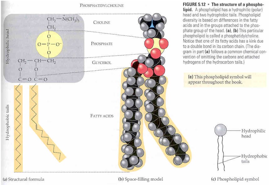
They are usually organized into a bilayer in membranes with the polar hydrophilic heads sticking outwards to the aqueous environment and the non-polar hydrophobic tails pointing inwards. [6] Glycerophospholipids consist of various diverse species which usually differ slightly in structure. The most basic structure is a phosphatidate. This species is an important intermediate in the synthesis of many phosphoglycerides. The presence of an additional group attached to the phosphate allows for many different phosphoglycerides.

By convention, structures of these compounds show the 3 glycerol carbon atoms vertically with the phosphate attached to carbon atom number three (at the bottom). Plasmalogens and phosphatidates are examples. [7]

Nomenclature and stereochemistry

In general, glycerophospholipids use an "sn" notation, which stands for stereospecific numbering. [8] When the letters "sn" appear in the nomenclature, by convention the hydroxyl group of the second carbon of glycerol (2-sn) is on the left on a Fischer projection. The numbering follows the one of Fischer's projections, being 1-sn the carbon at the top and 3-sn the one at the bottom. [9]

The advantage of this particular notation is that the spatial configuration (D or L) of the glycero-molecule is determined intuitively by the residues on the positions sn-1 and sn-3.

For example sn-glycero-3-phosphoric acid and sn-glycero-1-phosphoric acid are enantiomers.

Most vegetable oils have unsaturated fatty acids in the sn-2 position, with saturated fatty acids in the 1-sn and/or 3-sn position. [8] Animal fats more often have saturated fatty acids in the 2-sn, with unsaturated fatty acids in the 1-sn and/or 3-sn position. [8]

Examples

Plasmalogens

Plasmalogens are a type of phosphoglyceride. The first carbon of glycerol has a hydrocarbon chain attached via an ether, not ester, linkage. The linkages are more resistant to chemical attack than ester linkages are. The second (central) carbon atom has a fatty acid linked by an ester. The third carbon links to an ethanolamine or choline by means of a phosphate ester. These compounds are key components of the membranes of muscles and nerves.

Phosphatidates

Phosphatidates are lipids in which the first two carbon atoms of the glycerol are fatty acid esters, and the 3 is a phosphate ester. The phosphate serves as a link to another alcohol-usually ethanolamine, choline, serine, or a carbohydrate. The identity of the alcohol determines the subcategory of the phosphatidate. There is a negative charge on the phosphate and, in the case of choline or serine, a positive quaternary ammonium ion. (Serine also has a negative carboxylate group.) The presence of charges give a "head" with an overall charge. The phosphate ester portion ("head") is hydrophilic, whereas the remainder of the molecule, the fatty acid "tail", is hydrophobic. These are important components for the formation of lipid bilayers.

Phosphatidylethanolamines, phosphatidylcholines, and other phospholipids are examples of phosphatidates.

Phosphatidylcholines

Phosphatidylcholines are lecithins. Choline is the alcohol, with a positively charged quaternary ammonium, bound to the phosphate, with a negative charge. Lecithins are present in all living organisms. An egg yolk has a high concentration of lecithins, which are commercially important as an emulsifying agent in products such as mayonnaise. Lecithins are also present in brain and nerve tissue.

Phosphatidylinositol

Phosphatidylinositol makes up a small component of the cytosol in eukaryotic cell membranes and gives molecules a negative charge. Its importance relies in its role in activating sensory receptors that correlate with taste functions.

Phosphatidylserine

Phosphatidylserine is important in cell signaling, specifically apoptosis. Cells will use this phosphatidylserine to enter cells via apoptotic mimicry. The structure of this lipid differs in plants and animals, regarding fatty acid composition. In addition, phosphatidylserine plays an important role in the human brain content, as it makes up 13–15% of the phospholipids in the human cerebral cortex. This lipid is found in a wide range of places. For example, in the human diet, about 130 mg are derived from phosphatidylserine. This has been said to have a positive impact on the brain, as it helps with reduced stress and improved memory. [10]

Sphingomyelin

Sphingomyelin is a type of sphingolipid, which contains a backbone of sphingoid bases. It can be found in the myelin sheath of nerve cell axons in animal cell membranes. Sphingomyelin can be found in eggs or bovine brain. This sphingolipid is synthesized at the endoplasmic reticulum and is enriched at the plasma membrane with a larger concentration on the outside. [11]

Other phospholipids

There are many other phospholipids, some of which are glycolipids. The glycolipids include phosphatidyl sugars where the alcohol functional group is part of a carbohydrate. Phosphatidyl sugars are present in plants and certain microorganisms. A carbohydrate is very hydrophilic due to the large number of hydroxyl groups present.

Uses

Functions and use in membranes

Glycerophospholipids are the main structural component of biological membranes. Their amphipathic nature drives the formation of the lipid bilayer structure of membranes. The cell membrane seen under the electron microscope consists of two identifiable layers, or "leaflets", each of which is made up of an ordered row of glycerophospholipid molecules. The composition of each layer can vary widely depending on the type of cell.

Each glycerophospholipid molecule consists of a small polar head group and two long hydrophobic chains. In the cell membrane, the two layers of phospholipids are arranged as follows:

Apart from their function in cell membranes, they function in other cellular processes such as signal induction and transport. In regards to signaling, they provide the precursors for prostanglandins and other leukotrienes. [12] It is their specific distribution and catabolism that enables them carry out the biological response processes listed above. [13] Their roles as storage centers for secondary messengers in the membrane is also a contributing factor to their ability to act as transporters. [13] They also influence protein function. For example, they are important constituents of lipoproteins (soluble proteins that transport fat in the blood) hence affect their metabolism and function. [6]

Use in emulsification

Glycerophospholipids can also act as an emulsifying agent to promote dispersal of one substance into another. This is sometimes used in candy making and ice-cream making.

Presence in the brain

Neural membranes contain several classes of glycerophospholipids which turnover at different rates with respect to their structure and localization in different cells and membranes. There are three major classes namely; 1-alkyl-2-acyl glycerophospholipid, 1,2-diacyl glycerophospholipid and plasmalogen. The main function of these classes of glycerophospholipids in the neural membranes is to provide stability, permeability and fluidity through specific alterations in their compositions. [13] The glycerophospholipid composition of neural membranes greatly alters their functional efficacy. The length of glycerophospholipid acyl chain and the degree of saturation are important determinants of many membrane characteristics including the formation of lateral domains that are rich in polyunsaturated fatty acids. Receptor-mediated degradation of glycerophospholipids by phospholipases A(l), A(2), C, and D results in generation of second messengers, such as prostaglandins, eicosanoids, platelet activating factor and diacylglycerol. Thus, neural membrane phospholipids are a reservoir for second messengers. They are also involved in apoptosis, modulation of activities of transporters, and membrane-bound enzymes. Marked alterations in neural membrane glycerophospholipid composition have been reported to occur in neurological disorders. These alterations result in changes in membrane fluidity and permeability. These processes along with the accumulation of lipid peroxides and compromised energy metabolism may be responsible for the neurodegeneration observed in neurological disorders. [14]

Metabolism

The metabolism of glycerophospholipids is different in eukaryotes, tumor cells, [15] and prokaryotes. Synthesis in prokaryotes involves the synthesis of glycerophospholipids phosphatidic acid and polar head groups. Phosphatidic acid synthesis in eukaryotes is different, there are two routes, one to the other toward phosphatidylcholine and phosphatidylethanolamine. Glycerophospholipids are generally metabolized in several steps with different intermediates. The very first step in this metabolism involves the addition or transfer of the fatty acid chains to the glycerol backbone to form the first intermediate, lysophosphatidic acid (LPA). LPA then becomes acylated to form the next intermediate phosphatidic acid (PA). PA can be dephosphorylated leading to the formation of diacylglycerol which is essential in the synthesis of phosphatidylcholine (PC). [6] PC is one of the many species of glycerophospholipids. In a pathway called the Kennedy pathway, the polar heads are added to complete the formation of the entire structure consisting of the polar head regions, the two fatty acid chains and the phosphate group attached to the glycerol backbone. In this Kennedy pathway, Choline is converted to CDP-Choline which drives the transfer of the polar head groups to complete the formation of PC. PC can then be further converted to other species of glycerophospholipids such as phosphatidylserine (PS) and phosphatidylethanolamine (PE). [6]

See also

Related Research Articles

<span class="mw-page-title-main">Lipid</span> Substance of biological origin that is soluble in nonpolar solvents

Lipids are a broad group of organic compounds which include fats, waxes, sterols, fat-soluble vitamins, monoglycerides, diglycerides, phospholipids, and others. The functions of lipids include storing energy, signaling, and acting as structural components of cell membranes. Lipids have applications in the cosmetic and food industries, and in nanotechnology.

<span class="mw-page-title-main">Phospholipid</span> Class of lipids

Phospholipids are a class of lipids whose molecule has a hydrophilic "head" containing a phosphate group and two hydrophobic "tails" derived from fatty acids, joined by an alcohol residue. Marine phospholipids typically have omega-3 fatty acids EPA and DHA integrated as part of the phospholipid molecule. The phosphate group can be modified with simple organic molecules such as choline, ethanolamine or serine.

<span class="mw-page-title-main">Choline</span> Chemical compound and essential nutrient

Choline is a cation with the chemical formula [(CH3)3NCH2CH2OH]+. Choline forms various salts, for example choline chloride and choline bitartrate.

<span class="mw-page-title-main">Phosphatidylcholine</span> Class of phospholipids

Phosphatidylcholines (PC) are a class of phospholipids that incorporate choline as a headgroup. They are a major component of biological membranes and can be easily obtained from a variety of readily available sources, such as egg yolk or soybeans, from which they are mechanically or chemically extracted using hexane. They are also a member of the lecithin group of yellow-brownish fatty substances occurring in animal and plant tissues. Dipalmitoylphosphatidylcholine (lecithin) is a major component of the pulmonary surfactant, and is often used in the lecithin–sphingomyelin ratio to calculate fetal lung maturity. While phosphatidylcholines are found in all plant and animal cells, they are absent in the membranes of most bacteria, including Escherichia coli. Purified phosphatidylcholine is produced commercially.

<span class="mw-page-title-main">Sphingomyelin</span> Class of chemical compounds

Sphingomyelin is a type of sphingolipid found in animal cell membranes, especially in the membranous myelin sheath that surrounds some nerve cell axons. It usually consists of phosphocholine and ceramide, or a phosphoethanolamine head group; therefore, sphingomyelins can also be classified as sphingophospholipids. In humans, SPH represents ~85% of all sphingolipids, and typically make up 10–20 mol % of plasma membrane lipids.

<span class="mw-page-title-main">Phosphatidylinositol</span> Chemical compound

Phosphatidylinositol consists of a family of lipids made of a phosphate group, two fatty acid chains, and one inositol molecule. They represent a class of the phosphatidylglycerides. Typically phosphatidylinositols form a minor component on the cytosolic side of eukaryotic cell membranes. The phosphate group gives the molecules a negative charge at physiological pH.

Phosphatidic acids are anionic phospholipids important to cell signaling and direct activation of lipid-gated ion channels. Hydrolysis of phosphatidic acid gives rise to one molecule each of glycerol and phosphoric acid and two molecules of fatty acids. They constitute about 0.25% of phospholipids in the bilayer.

<span class="mw-page-title-main">Plasmalogen</span> Subclass of Glycerophospholipids

Glycerophospholipids of biochemical relevance are divided into three subclasses based on the substitution present at the sn-1 position of the glycerol backbone: acyl, alkyl and alkenyl. Of these, the alkyl and alkenyl moiety in each case form an ether bond, which makes for two types of ether phospholipids, plasmanyl, and plasmenyl. Plasmalogens are plasmenyls with an ester linked lipid at the sn-2 position of the glycerol backbone, chemically designated 1-0(1Z-alkenyl)-2-acyl-glycerophospholipids. The lipid attached to the vinyl ether at sn-1 can be C16:0, C18:0, or C18:1, and the lipid attached to the acyl group at sn-2 can be C22:6 ω-3 or C20:4 ω-6, . Plasmalogens are classified according to their head group, mainly as PC plasmalogens (plasmenylcholines) and PE plasmalogens (plasmenylethalomines) Plasmalogens should not be confused with plasmanyls.

<span class="mw-page-title-main">Ether lipid</span>

In biochemistry, an ether lipid refers to any lipid in which the lipid "tail" group is attached to the glycerol backbone via an ether bond at any position. In contrast, conventional glycerophospholipids and triglycerides are triesters. Structural types include:

<span class="mw-page-title-main">Phosphatidylserine</span> Chemical compound

Phosphatidylserine is a phospholipid and is a component of the cell membrane. It plays a key role in cell cycle signaling, specifically in relation to apoptosis. It is a key pathway for viruses to enter cells via apoptotic mimicry. Its exposure on the outer surface of a membrane marks the cell for destruction via apoptosis.

<span class="mw-page-title-main">Lipid signaling</span> Biological signaling using lipid molecules

Lipid signaling, broadly defined, refers to any biological cell signaling event involving a lipid messenger that binds a protein target, such as a receptor, kinase or phosphatase, which in turn mediate the effects of these lipids on specific cellular responses. Lipid signaling is thought to be qualitatively different from other classical signaling paradigms because lipids can freely diffuse through membranes. One consequence of this is that lipid messengers cannot be stored in vesicles prior to release and so are often biosynthesized "on demand" at their intended site of action. As such, many lipid signaling molecules cannot circulate freely in solution but, rather, exist bound to special carrier proteins in serum.

Lipid metabolism is the synthesis and degradation of lipids in cells, involving the breakdown and storage of fats for energy and the synthesis of structural and functional lipids, such as those involved in the construction of cell membranes. In animals, these fats are obtained from food and are synthesized by the liver. Lipogenesis is the process of synthesizing these fats. The majority of lipids found in the human body from ingesting food are triglycerides and cholesterol. Other types of lipids found in the body are fatty acids and membrane lipids. Lipid metabolism is often considered as the digestion and absorption process of dietary fat; however, there are two sources of fats that organisms can use to obtain energy: from consumed dietary fats and from stored fat. Vertebrates use both sources of fat to produce energy for organs such as the heart to function. Since lipids are hydrophobic molecules, they need to be solubilized before their metabolism can begin. Lipid metabolism often begins with hydrolysis, which occurs with the help of various enzymes in the digestive system. Lipid metabolism also occurs in plants, though the processes differ in some ways when compared to animals. The second step after the hydrolysis is the absorption of the fatty acids into the epithelial cells of the intestinal wall. In the epithelial cells, fatty acids are packaged and transported to the rest of the body.

<span class="mw-page-title-main">Phosphatidylethanolamine</span> Group of chemical compounds

Phosphatidylethanolamine (PE) is a class of phospholipids found in biological membranes. They are synthesized by the addition of cytidine diphosphate-ethanolamine to diglycerides, releasing cytidine monophosphate. S-Adenosyl methionine can subsequently methylate the amine of phosphatidylethanolamines to yield phosphatidylcholines.

<span class="mw-page-title-main">Membrane lipid</span> Lipid molecules on cell membrane

Membrane lipids are a group of compounds which form the lipid bilayer of the cell membrane. The three major classes of membrane lipids are phospholipids, glycolipids, and cholesterol. Lipids are amphiphilic: they have one end that is soluble in water ('polar') and an ending that is soluble in fat ('nonpolar'). By forming a double layer with the polar ends pointing outwards and the nonpolar ends pointing inwards membrane lipids can form a 'lipid bilayer' which keeps the watery interior of the cell separate from the watery exterior. The arrangements of lipids and various proteins, acting as receptors and channel pores in the membrane, control the entry and exit of other molecules and ions as part of the cell's metabolism. In order to perform physiological functions, membrane proteins are facilitated to rotate and diffuse laterally in two dimensional expanse of lipid bilayer by the presence of a shell of lipids closely attached to protein surface, called annular lipid shell.

<span class="mw-page-title-main">Phosphatidylglycerol</span> Lipid

Phosphatidylglycerol is a glycerophospholipid found in pulmonary surfactant and in the plasma membrane where it directly activates lipid-gated ion channels.

<span class="mw-page-title-main">Phosphatidate phosphatase</span>

The enzyme phosphatidate phosphatase (PAP, EC 3.1.3.4) is a key regulatory enzyme in lipid metabolism, catalyzing the conversion of phosphatidate to diacylglycerol:

In enzymology, a 1-acylglycerol-3-phosphate O-acyltransferase is an enzyme that catalyzes the chemical reaction

<span class="mw-page-title-main">Diacylglycerol cholinephosphotransferase</span>

In enzymology, a diacylglycerol cholinephosphotransferase is an enzyme that catalyzes the chemical reaction

Archaeol is composed of two phytanyl chains linked to the sn-2 and sn-3 positions of glycerol. As its phosphate ester, it is a common component of the membranes of archaea.

<span class="mw-page-title-main">1-Lysophosphatidylcholine</span>

2-acyl-sn-glycero-3-phosphocholines are a class of phospholipids that are intermediates in the metabolism of lipids. Because they result from the hydrolysis of an acyl group from the sn-1 position of phosphatidylcholine, they are also called 1-lysophosphatidylcholine. The synthesis of phosphatidylcholines with specific fatty acids occurs through the synthesis of 1-lysoPC. The formation of various other lipids generates 1-lysoPC as a by-product.

References

  1. Harayama, Takeshi; Riezman, Howard (May 2018). "Understanding the diversity of membrane lipid composition". Nature Reviews Molecular Cell Biology. 19 (5): 281–296. doi:10.1038/nrm.2017.138. ISSN   1471-0080.
  2. 1 2 Caforio, Antonella; Driessen, Arnold J.M. (2017). "Archaeal phospholipids: Structural properties and biosynthesis" (PDF). Biochimica et Biophysica Acta (BBA) - Molecular and Cell Biology of Lipids. 1862 (11): 1325–1339. doi:10.1016/j.bbalip.2016.12.006. PMID   28007654.
  3. Hishikawa, Daisuke; Hashidate, Tomomi; Shimizu, Takao; Shindou, Hideo (May 2014). "Diversity and function of membrane glycerophospholipids generated by the remodeling pathway in mammalian cells". Journal of Lipid Research. 55 (5): 799–807. doi: 10.1194/jlr.R046094 . ISSN   0022-2275. PMC   3995458 . PMID   24646950.
  4. IUPAC , Compendium of Chemical Terminology , 2nd ed. (the "Gold Book") (1997). Online corrected version: (2006) " glycerophospholipid ". doi : 10.1351/goldbook.G02648
  5. Montealegre, Cristina; Verardo, Vito; Luisa Marina, María; Caboni, Maria Fiorenza (March 2014). "Analysis of glycerophosphate- and sphingolipids by CE". Electrophoresis. 35 (6): 779–792. doi:10.1002/elps.201300534. PMID   24301713. S2CID   205804071.
  6. 1 2 3 4 Ecker, Josef; Liebisch, Gerhard (April 2014). "Application of stable isotopes to investigate the metabolism of fatty acids, glycerophospholipid and sphingolipid species". Progress in Lipid Research. 54: 14–31. doi:10.1016/j.plipres.2014.01.002. PMID   24462586.
  7. Yong-Mei Zhang and Charles O. Rock (2008). "Thematic Review Series: Glycerolipids. Acyltransferases in bacterial glycerophospholipid synthesis". J Lipid Res. 49 (9): 1867–1874. doi: 10.1194/jlr.R800005-JLR200 . PMC   2515527 . PMID   18369234.
  8. 1 2 3 Alfieri A, Imperlini E, Nigro E, Vitucci D, Orrù S, Daniele A, Buono P, Mancini A (2017). "Effects of Plant Oil Interesterified Triacylglycerols on Lipemia and Human Health". International Journal of Molecular Sciences . 19 (1): E104. doi: 10.3390/ijms19010104 . PMC   5796054 . PMID   29301208.
  9. Moss G.P. (www version) (1976). "Nomenclature of Lipids, Recommendations 1976". IUPAC-IUB Commission on Biochemical Nomenclature (CBN). Part I. Fatty Acid, Neutral Fats, Long-Chain Alcohols and Long-Chain Bases § Lip-1.13. Retrieved 27 Sep 2023.
  10. "Scientific Opinion on the substantiation of health claims related to phosphatidyl serine (ID 552, 711, 734, 1632, 1927) pursuant to Article 13(1) of Regulation (EC) No 1924/2006 | EFSA". www.efsa.europa.eu. 2010-10-19. Retrieved 2023-11-29.
  11. Testi, Roberto (1996-12-01). "Sphingomyelin breakdown and cell fate". Trends in Biochemical Sciences. 21 (12): 468–471. doi:10.1016/S0968-0004(96)10056-6. ISSN   0968-0004.
  12. Hermansson, Martin; Hokynar, Kati; Somerharju, Pentti (July 2011). "Mechanisms of glycerophospholipid homeostasis in mammalian cells". Progress in Lipid Research. 50 (3): 240–257. doi:10.1016/j.plipres.2011.02.004. PMID   21382416.
  13. 1 2 3 Farooqui, AA; Horrocks, LA; Farooqui, T (June 2000). "Glycerophospholipids in brain: their metabolism, incorporation into membranes, functions, and involvement in neurological disorders". Chemistry and Physics of Lipids. 106 (1): 1–29. doi:10.1016/s0009-3084(00)00128-6. PMID   10878232.
  14. Garcia, Christina (2011-06-30). "Metabolism of glycerophospholipids". We Sapiens.org. Archived from the original on 2012-03-23.
  15. Dolce V, Cappello AR, Lappano R, Maggiolini M (November 2011). "Glycerophospholipid synthesis as a novel drug target against cancer". Current Molecular Pharmacology. 4 (3): 167–175. doi:10.2174/1874467211104030167. PMID   21222647.