Platelet-activating factor

Last updated
Platelet-activating factor
Platelet-activating factor.svg
Names
Systematic IUPAC name
(2R)-2-(Acetyloxy)-3-(hexadecyloxy)propyl 2-(trimethylazaniumyl)ethyl phosphate
Identifiers
3D model (JSmol)
ChEBI
ChemSpider
MeSH Platelet+Activating+Factor
PubChem CID
UNII
  • InChI=1S/C26H54NO7P/c1-6-7-8-9-10-11-12-13-14-15-16-17-18-19-21-31-23-26(34-25(2)28)24-33-35(29,30)32-22-20-27(3,4)5/h26H,6-24H2,1-5H3/t26-/m1/s1 X mark.svgN
    Key: HVAUUPRFYPCOCA-AREMUKBSSA-N X mark.svgN
  • InChI=1/C26H54NO7P/c1-6-7-8-9-10-11-12-13-14-15-16-17-18-19-21-31-23-26(34-25(2)28)24-33-35(29,30)32-22-20-27(3,4)5/h26H,6-24H2,1-5H3/t26-/m1/s1
    Key: HVAUUPRFYPCOCA-AREMUKBSBE
  • CCCCCCCCCCCCCCCCOC[C@H](COP(=O)([O-])OCC[N+](C)(C)C)OC(=O)C
Properties
C26H54NO7P
Molar mass 523.683
Except where otherwise noted, data are given for materials in their standard state (at 25 °C [77 °F], 100 kPa).
X mark.svgN  verify  (what is  Yes check.svgYX mark.svgN ?)

Platelet-activating factor, also known as PAF, PAF-acether or AGEPC (acetyl-glyceryl-ether-phosphorylcholine), is a potent phospholipid activator and mediator of many leukocyte functions, platelet aggregation and degranulation, inflammation, and anaphylaxis. It is also involved in changes to vascular permeability, the oxidative burst, chemotaxis of leukocytes, as well as augmentation of arachidonic acid metabolism in phagocytes.

Contents

PAF is produced by a variety of cells, but especially those involved in host defense, such as platelets, endothelial cells, neutrophils, monocytes, and macrophages. PAF is continuously produced by these cells but in low quantities and production is controlled by the activity of PAF acetylhydrolases. It is produced in larger quantities by inflammatory cells in response to specific stimuli. [1]

History

PAF was discovered by French immunologist Jacques Benveniste in the early 1970s. [2] [3] PAF was the first phospholipid known to have messenger functions. Benveniste made significant contributions in the role and characteristics of PAF and its importance in inflammatory response and mediation. Using lab rats and mice, he found that ionophore A23187 (a mobile ion carrier that allows the passage of Mn2+, Ca2+ and Mg2+ and has antibiotic properties against bacteria and fungi) caused the release of PAF. These developments led to the finding that macrophages produce PAF and that macrophages play an important function in aggregation of platelets and liberation of their inflammatory and vasoactive substances.[ citation needed ]

Further studies on PAF were conducted by Constantinos A. Demopoulos in 1979. [4] Demopoulos found that PAF plays a crucial role in heart disease and strokes. His experiment’s data found that atherosclerosis (the accumulation of lipid-rich lesions in the endothelium of the arteries) can be attributed to PAF and PAF-like lipids, and identified biologically active compounds in the polar lipid fractions of olive oil, honey, milk and yoghurt, mackerel, and wine that have PAF-antagonistic properties and inhibit the development of atherosclerosis in animal models. [5] During the course of his studies, he also determined the chemical structure of the compound.

Evolution

PAF can be found in protozoans, yeasts, plants, bacteria, and mammals. PAF has regulatory role in protozoans. The regulatory role is thought to diverge from that point and be maintained as living organisms started to evolve. During evolution, functions of PAF in the cell have been changing and enlarging.[ citation needed ]

PAF has been found in plants but its function has not yet been determined.[ citation needed ]

Fungal PAF

The antifungal protein PAF from Penicillium chrysogenum exhibits growth-inhibitory activity against a broad range of filamentous fungi. Evidence suggests that disruption of Ca2+ signaling/homeostasis plays an important role in the mechanistic basis of PAF as a growth inhibitor. [6]

PAF also elicits hyperpolarization of the plasma membrane and the activation of ion channels, followed by an increase in reactive oxygen species in the cell and the induction of an apoptosis-like phenotype [7]

Cumulative evidence reveals that diabetes is a condition in which cell Ca2+ homeostasis is impaired. Defects in cell Ca2+ regulation were found in erythrocytes, cardiac muscle, platelets, skeletal muscle, kidney, aorta, adipocytes, liver, osteoblasts, arteries, lens, peripheral nerves, brain synaptosomes, retinal tissue, and pancreatic beta cells, confirming that this defect in cell Ca2+ metabolism is a basic pathology associated with the diabetic state. [8]

The defects identified in the mechanical activity of the hearts from type 1 diabetic animals include alteration of Ca2+ signaling via changes in critical processes. [9]

Function

PAF is used to transmit signals between neighboring cells and acts as a hormone, cytokines, and other signaling molecules. The PAF signaling system can trigger inflammatory and thrombotic cascades, amplify these cascades when acting with other mediators, and mediate molecular and cellular interactions (cross talk) between inflammation and thrombosis. [10] Unregulated PAF signaling can cause pathological inflammation and has been found to be a cause in sepsis, shock, and traumatic injury. PAF can be used as a local signaling molecule and travel over very short distances or it can be circulated throughout the body and act via endocrine.

PAF initiates an inflammatory response in allergic reactions. [11] This has been demonstrated in the skin of humans and in the paws and skin of lab rabbits and rodents. The inflammatory response is enhanced by the use of vasodilators, including prostaglandin E1 (PGE,) and PGE2 and inhibited by vasoconstrictors. [12]

PAF also induces apoptosis in a different way that is independent of the PAF receptor. The pathway to apoptosis can be inhibited by negative feedback from PAF acetylhydrolase (PAF-AH), an enzyme that catabolizes platelet-activating factor.

It is an important mediator of bronchoconstriction.

It causes platelets to aggregate and blood vessels to dilate. Thus, it is important to the process of hemostasis. At a concentration of 10−12 mol/L, PAF causes life-threatening inflammation of the airways to induce asthma like symptoms.

Toxins such as fragments of destroyed bacteria induce the synthesis of PAF, which causes a drop in blood pressure and reduced volume of blood pumped by the heart, which leads to shock and possibly death.

Structure

Several molecular species of platelet-activating factor that vary in the length of the O-alkyl side-chain have been identified.

Studies found that PAF could not be modified without losing its biological activity. Thus, small changes in the structure of PAF could render its signaling abilities inert. [13] Investigation led to the understanding that platelet and blood pressure response were dependent on the sn-2 propionyl analog. If the sn-1 was removed then PAF lacked any sort of biological activity. Finally, the sn-3 position of PAF was experimented with by removing methyl groups sequentially. As more and more methyl groups were removed, biological activity diminished until it was eventually inactive.

Biochemistry

Biosynthesis

PAF is produced by stimulated basophils, monocytes, polymorphonuclear neutrophils, platelets, and endothelial cells primarily through lipid remodeling. A variety of stimuli can initiate the synthesis of PAF. These stimuli could be macrophages going through phagocytosis or endothelium cells uptake of thrombin.

There are two different pathways in which PAF can be synthesized: de novo pathway and remodeling. The remodeling pathway is activated by inflammatory agents and it is thought to be the primary source of PAF under pathological conditions. The de novo pathway is used to maintain PAF levels during normal cellular function.

The most common pathway taken to produce PAF is remodeling. The precursor to the remodeling pathway is a phospholipid, which is typically phosphatidylcholine (PC). The fatty acid is removed from the sn-2 position of the three-carbon backbone of the phospholipid by phospholipase A2 (PLA2) to produce the intermediate lyso-PC (LPC). An acetyl group is then added by LPC acetyltransferase (LPCAT) to produce PAF.

Using the de novo pathway, PAF is produced from 1-O-alkyl-2-acetyl-sn-glycerol (AAG). Fatty acids are joined on the sn-1 position with 1-O-hexadecyl being the best for PAF activity. Phosphocholine is then added to the sn-3 site on AAG creating PAF.

Regulation

The concentration of PAF is controlled by the synthesis of the compound and by the actions of PAF acetylhydrolases (PAF-AH). PAF-AH are a family of enzymes that have the ability to catabolize and degrade PAF and turn it into an inactive compound. The enzymes within this family are lipoprotein-associated phospholipase A2, cytoplasmic platelet-activating factor acetylhydrolase 2, and platelet-activating factor acetylhydrolase 1b.

Cations are one form of regulation in the production of PAF. Calcium plays a large role in the inhibition of enzymes that produce PAF in the denovo pathway of PAF biosynthesis.

The regulation of PAF is still not completely understood. Enzymes that are associated with the production of PAF are controlled by metal ions, thiol compounds, fatty acids, pH, compartmentalization, and phosphorylation and dephosphorylation. These controls on these PAF producing enzymes are believed to work in conjunction to control it, but the overall pathway and reasoning is not well understood.

Pharmacology

Inhibition

PAF antagonists do not provoke an inflammatory response upon binding, but block or lessen the effect of PAF. Examples of PAF antagonists are: [14]

Clinical significance

High PAF levels are associated with a variety of medical conditions. Some of these conditions include:

•Allergic reactions
•Stroke
•Sepsis
•Myocardial infarction
•Colitis, inflammation of the large intestine
•Multiple sclerosis

While the effects that PAF has on inflammatory response and cardiovascular conditions are well understood, PAF is still a subject for discussion. Over the past 23 years, papers written on PAF have almost doubled from approximately 7,500 in 1997 to 14,500 in 2020.PubMed (June 2020). "Platelet-activating factor search results and historical activity metrics". PubMed. Research into PAF is ongoing.

See also

Related Research Articles

<span class="mw-page-title-main">Arachidonic acid</span> Fatty acid used metabolically in many organisms

Arachidonic acid is a polyunsaturated omega-6 fatty acid 20:4(ω-6), or 20:4(5,8,11,14). It is structurally related to the saturated arachidic acid found in cupuaçu butter. Its name derives from the Neo-Latin word arachis (peanut), but peanut oil does not contain any arachidonic acid.

<span class="mw-page-title-main">Lipoxin</span> Acronym for lipoxygenase interaction product

A lipoxin (LX or Lx), an acronym for lipoxygenase interaction product, is a bioactive autacoid metabolite of arachidonic acid made by various cell types. They are categorized as nonclassic eicosanoids and members of the specialized pro-resolving mediators (SPMs) family of polyunsaturated fatty acid (PUFA) metabolites. Like other SPMs, LXs form during, and then act to resolve, inflammatory responses. Initially, two lipoxins were identified, lipoxin A4 (LXA4) and LXB4, but more recent studies have identified epimers of these two LXs: the epi-lipoxins, 15-epi-LXA4 and 15-epi-LXB4 respectively.

<span class="mw-page-title-main">Phospholipase A2</span> Peripheral membrane protein

The enzyme phospholipase A2 (EC 3.1.1.4, PLA2, systematic name phosphatidylcholine 2-acylhydrolase) catalyse the cleavage of fatty acids in position 2 of phospholipids, hydrolyzing the bond between the second fatty acid “tail” and the glycerol molecule:

<span class="mw-page-title-main">Ether lipid</span>

In an organic chemistry general sense, an ether lipid implies an ether bridge between an alkyl group and an unspecified alkyl or aryl group, not necessarily glycerol. If glycerol is involved, the compound is called a glyceryl ether, which may take the form of an alkylglycerol, an alkyl acyl glycerol, or in combination with a phosphatide group, a phospholipid.

<span class="mw-page-title-main">Phospholipid scramblase</span> Protein

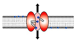
Scramblase is a protein responsible for the translocation of phospholipids between the two monolayers of a lipid bilayer of a cell membrane. In humans, phospholipid scramblases (PLSCRs) constitute a family of five homologous proteins that are named as hPLSCR1–hPLSCR5. Scramblases are not members of the general family of transmembrane lipid transporters known as flippases. Scramblases are distinct from flippases and floppases. Scramblases, flippases, and floppases are three different types of enzymatic groups of phospholipid transportation enzymes. The inner-leaflet, facing the inside of the cell, contains negatively charged amino-phospholipids and phosphatidylethanolamine. The outer-leaflet, facing the outside environment, contains phosphatidylcholine and sphingomyelin. Scramblase is an enzyme, present in the cell membrane, that can transport (scramble) the negatively charged phospholipids from the inner-leaflet to the outer-leaflet, and vice versa.

<span class="mw-page-title-main">Lipid signaling</span> Biological signaling using lipid molecules

Lipid signaling, broadly defined, refers to any biological signaling event involving a lipid messenger that binds a protein target, such as a receptor, kinase or phosphatase, which in turn mediate the effects of these lipids on specific cellular responses. Lipid signaling is thought to be qualitatively different from other classical signaling paradigms because lipids can freely diffuse through membranes. One consequence of this is that lipid messengers cannot be stored in vesicles prior to release and so are often biosynthesized "on demand" at their intended site of action. As such, many lipid signaling molecules cannot circulate freely in solution but, rather, exist bound to special carrier proteins in serum.

Arachidonate 5-lipoxygenase, also known as ALOX5, 5-lipoxygenase, 5-LOX, or 5-LO, is a non-heme iron-containing enzyme that in humans is encoded by the ALOX5 gene. Arachidonate 5-lipoxygenase is a member of the lipoxygenase family of enzymes. It transforms essential fatty acids (EFA) substrates into leukotrienes as well as a wide range of other biologically active products. ALOX5 is a current target for pharmaceutical intervention in a number of diseases.

<span class="mw-page-title-main">PAFAH1B1</span> Protein-coding gene in the species Homo sapiens

Platelet-activating factor acetylhydrolase IB subunit alpha is an enzyme that in humans is encoded by the PAFAH1B1 gene. The protein is often referred to as Lis1 and plays an important role in regulating the motor protein Dynein.

<span class="mw-page-title-main">Platelet-activating factor receptor</span> Protein-coding gene in the species Homo sapiens

The platelet-activating factor receptor(PAF-R) is a G-protein coupled receptor which binds platelet-activating factor. It is encoded in the human by the PTAFR gene.

The enzyme 1-alkyl-2-acetylglycerophosphocholine esterase (EC 3.1.1.47) catalyzes the reaction

<span class="mw-page-title-main">Prostacyclin receptor</span> Mammalian protein found in Homo sapiens

The Prostacyclin receptor, also termed the prostaglandin I2 receptor or just IP, is a receptor belonging to the prostaglandin (PG) group of receptors. IP binds to and mediates the biological actions of prostacyclin (also termed Prostaglandin I2, PGI2, or when used as a drug, epoprostenol). IP is encoded in humans by the PTGIR gene. While possessing many functions as defined in animal model studies, the major clinical relevancy of IP is as a powerful vasodilator: stimulators of IP are used to treat severe and even life-threatening diseases involving pathological vasoconstriction.

<span class="mw-page-title-main">5-Hydroxyeicosatetraenoic acid</span> Chemical compound

5-Hydroxyeicosatetraenoic acid (5-HETE, 5(S)-HETE, or 5S-HETE) is an eicosanoid, i.e. a metabolite of arachidonic acid. It is produced by diverse cell types in humans and other animal species. These cells may then metabolize the formed 5(S)-HETE to 5-oxo-eicosatetraenoic acid (5-oxo-ETE), 5(S),15(S)-dihydroxyeicosatetraenoic acid (5(S),15(S)-diHETE), or 5-oxo-15-hydroxyeicosatetraenoic acid (5-oxo-15(S)-HETE).

<span class="mw-page-title-main">Phospholipase C</span> Class of enzymes

Phospholipase C (PLC) is a class of membrane-associated enzymes that cleave phospholipids just before the phosphate group (see figure). It is most commonly taken to be synonymous with the human forms of this enzyme, which play an important role in eukaryotic cell physiology, in particular signal transduction pathways. Phospholipase C's role in signal transduction is its cleavage of phosphatidylinositol 4,5-bisphosphate (PIP2) into diacyl glycerol (DAG) and inositol 1,4,5-trisphosphate (IP3), which serve as second messengers. Activators of each PLC vary, but typically include heterotrimeric G protein subunits, protein tyrosine kinases, small G proteins, Ca2+, and phospholipids.

<span class="mw-page-title-main">PAFAH2</span> Protein-coding gene in the species Homo sapiens

Platelet-activating factor acetylhydrolase 2, cytoplasmic is an enzyme that in humans is encoded by the PAFAH2 gene. It is one of several PAF acetylhydrolases.

<span class="mw-page-title-main">Lipoprotein-associated phospholipase A2</span> Protein-coding gene in the species Homo sapiens

Lipoprotein-associated phospholipase A2 (Lp-PLA2) also known as platelet-activating factor acetylhydrolase (PAF-AH) is a phospholipase A2 enzyme that in humans is encoded by the PLA2G7 gene. Lp-PLA2 is a 45-kDa protein of 441 amino acids. It is one of several PAF acetylhydrolases.

<span class="mw-page-title-main">12-Hydroxyeicosatetraenoic acid</span> Chemical compound

12-Hydroxyeicosatetraenoic acid (12-HETE) is a derivative of the 20 carbon polyunsaturated fatty acid, arachidonic acid, containing a hydroxyl residue at carbon 12 and a 5Z,8Z,10E,14Z Cis–trans isomerism configuration (Z=cis, E=trans) in its four double bonds. It was first found as a product of arachidonic acid metabolism made by human and bovine platelets through their 12S-lipoxygenase (i.e. ALOX12) enzyme(s). However, the term 12-HETE is ambiguous in that it has been used to indicate not only the initially detected "S" stereoisomer, 12S-hydroxy-5Z,8Z,10E,14Z-eicosatetraenoic acid (12(S)-HETE or 12S-HETE), made by platelets, but also the later detected "R" stereoisomer, 12(R)-hydroxy-5Z,8Z,10E,14Z-eicosatetraenoic acid (also termed 12(R)-HETE or 12R-HETE) made by other tissues through their 12R-lipoxygenase enzyme, ALOX12B. The two isomers, either directly or after being further metabolized, have been suggested to be involved in a variety of human physiological and pathological reactions. Unlike hormones which are secreted by cells, travel in the circulation to alter the behavior of distant cells, and thereby act as Endocrine signalling agents, these arachidonic acid metabolites act locally as Autocrine signalling and/or Paracrine signaling agents to regulate the behavior of their cells of origin or of nearby cells, respectively. In these roles, they may amplify or dampen, expand or contract cellular and tissue responses to disturbances.

<span class="mw-page-title-main">Nicolas Bazan</span>

Nicolas G. Bazan is a neuroscientist, eye researcher, and author. His research focuses on neurodegenerative diseases, neuroinflammation, and cell survival using cellular, molecular, and disease models including lipidomics. He also operates "Nicholas Bazan Wines" with Mark Wahle.

<span class="mw-page-title-main">15-Hydroxyeicosatetraenoic acid</span> Chemical compound

15-Hydroxyeicosatetraenoic acid (also termed 15-HETE, 15(S)-HETE, and 15S-HETE) is an eicosanoid, i.e. a metabolite of arachidonic acid. Various cell types metabolize arachidonic acid to 15(S)-hydroperoxyeicosatetraenoic acid (15(S)-HpETE). This initial hydroperoxide product is extremely short-lived in cells: if not otherwise metabolized, it is rapidly reduced to 15(S)-HETE. Both of these metabolites, depending on the cell type which forms them, can be further metabolized to 15-oxo-eicosatetraenoic acid (15-oxo-ETE), 5S,15S-dihydroxy-eicosatetraenoic acid (5(S),15(S)-diHETE), 5-oxo-15(S)-hydroxyeicosatetraenoic acid (5-oxo-15(S)-HETE, a subset of specialized pro-resolving mediators viz., the lipoxins, a class of pro-inflammatory mediators, the eoxins, and other products that have less well-defined activities and functions. Thus, 15(S)-HETE and 15(S)-HpETE, in addition to having intrinsic biological activities, are key precursors to numerous biologically active derivatives.

<span class="mw-page-title-main">5-Oxo-eicosatetraenoic acid</span> Chemical compound

5-Oxo-eicosatetraenoic acid is a Nonclassic eicosanoid metabolite of arachidonic acid and the most potent naturally occurring member of the 5-HETE family of cell signaling agents. Like other cell signaling agents, 5-oxo-ETE is made by a cell and then feeds back to stimulate its parent cell and/or exits this cell to stimulate nearby cells. 5-Oxo-ETE can stimulate various cell types particularly human leukocytes but possesses its highest potency and power in stimulating the human eosinophil type of leukocyte. It is therefore suggested to be formed during and to be an important contributor to the formation and progression of eosinophil-based allergic reactions; it is also suggested that 5-oxo-ETE contributes to the development of inflammation, cancer cell growth, and other pathological and physiological events.

Specialized pro-resolving mediators are a large and growing class of cell signaling molecules formed in cells by the metabolism of polyunsaturated fatty acids (PUFA) by one or a combination of lipoxygenase, cyclooxygenase, and cytochrome P450 monooxygenase enzymes. Pre-clinical studies, primarily in animal models and human tissues, implicate SPM in orchestrating the resolution of inflammation. Prominent members include the resolvins and protectins.

References

  1. Zimmerman GA, McIntyre TM, Prescott SM, Stafforini DM (May 2002). "The platelet-activating factor signaling system and its regulators in syndromes of inflammation and thrombosis". Critical Care Medicine. 30 (5 Suppl): S294–301. doi:10.1097/00003246-200205001-00020. PMID   12004251. S2CID   648041.
  2. Benveniste J, Henson PM, Cochrane CG (Dec 1972). "Leukocyte-dependent histamine release from rabbit platelets. The role of IgE, basophils, and a platelet-activating factor". The Journal of Experimental Medicine. 136 (6): 1356–77. doi:10.1084/jem.136.6.1356. PMC   2139324 . PMID   4118412.
  3. Benveniste J (Jun 1974). "Platelet-activating factor, a new mediator of anaphylaxis and immune complex deposition from rabbit and human basophils". Nature. 249 (457): 581–2. Bibcode:1974Natur.249..581B. doi:10.1038/249581a0. PMID   4275800. S2CID   4180118.
  4. Demopoulos CA, Pinckard RN, Hanahan DJ (Oct 1979). "Platelet-activating factor. Evidence for 1-O-alkyl-2-acetyl-sn-glyceryl-3-phosphorylcholine as the active component (a new class of lipid chemical mediators)" (PDF). The Journal of Biological Chemistry. 254 (19): 9355–8. doi: 10.1016/S0021-9258(19)83523-8 . PMID   489536.
  5. Lordan R, Tsoupras A, Zabetakis I, Demopoulos CA (December 3, 2019). "Forty Years Since the Structural Elucidation of Platelet-Activating Factor (PAF): Historical, Current, and Future Research Perspectives". Molecules. 24 (23): 4414. doi: 10.3390/molecules24234414 . PMC   6930554 . PMID   31816871.
  6. Binder U, Chu M, Read ND, Marx F (Sep 2010). "The antifungal activity of the Penicillium chrysogenum protein PAF disrupts calcium homeostasis in Neurospora crassa". Eukaryotic Cell. 9 (9): 1374–82. doi:10.1128/EC.00050-10. PMC   2937333 . PMID   20622001.
  7. Marx F, Binder U, Leiter E, Pócsi I (Feb 2008). "The Penicillium chrysogenum antifungal protein PAF, a promising tool for the development of new antifungal therapies and fungal cell biology studies". Cellular and Molecular Life Sciences. 65 (3): 445–54. doi:10.1007/s00018-007-7364-8. PMID   17965829. S2CID   35186093.
  8. Levy J (Feb 1999). "Abnormal cell calcium homeostasis in type 2 diabetes mellitus: a new look on old disease". Endocrine. 10 (1): 1–6. doi:10.1385/ENDO:10:1:1. PMID   10403564. S2CID   43840751.
  9. Yaras N, Ugur M, Ozdemir S, Gurdal H, Purali N, Lacampagne A, Vassort G, Turan B (Nov 2005). "Effects of diabetes on ryanodine receptor Ca release channel (RyR2) and Ca2+ homeostasis in rat heart". Diabetes. 54 (11): 3082–8. doi: 10.2337/diabetes.54.11.3082 . PMID   16249429.
  10. Prescott SM, Zimmerman GA, Stafforini DM, McIntyre TM (2000). "Platelet-activating factor and related lipid mediators". Annual Review of Biochemistry. 69: 419–45. doi:10.1146/annurev.biochem.69.1.419. PMID   10966465.
  11. McIntyre TM, Prescott SM, Stafforini DM (Apr 2009). "The emerging roles of PAF acetylhydrolase". Journal of Lipid Research. 50 Suppl (Suppl): S255–9. doi:10.1194/jlr.R800024-JLR200. PMC   2674695 . PMID   18838739.
  12. Morley J, Page CP, Paul W (Nov 1983). "Inflammatory actions of platelet activating factor (Pafacether) in guinea-pig skin". British Journal of Pharmacology. 80 (3): 503–9. doi:10.1111/j.1476-5381.1983.tb10722.x. PMC   2045011 . PMID   6685552.
  13. Snyder F (Feb 1989). "Biochemistry of platelet-activating factor: a unique class of biologically active phospholipids". Proceedings of the Society for Experimental Biology and Medicine. 190 (2): 125–35. doi:10.3181/00379727-190-42839. PMID   2536942. S2CID   21006141.
  14. Camussi G, Tetta C, Bussolino F, Baglioni C (Oct 1988). "Synthesis and release of platelet-activating factor is inhibited by plasma alpha 1-proteinase inhibitor or alpha 1-antichymotrypsin and is stimulated by proteinases". The Journal of Experimental Medicine. 168 (4): 1293–306. doi:10.1084/jem.168.4.1293. PMC   2189082 . PMID   3049910.
  15. Hayashi J, Hiromura K, Koizumi R, Shimizu Y, Maezawa A, Nojima Y, Naruse T (Mar 2001). "Platelet-activating factor antagonist, SM-12502, attenuates experimental glomerular thrombosis in rats". Nephron. 87 (3): 274–8. doi:10.1159/000045926. PMID   11287764. S2CID   12221065.
  16. "Etizolam".
  17. CID 65889 from PubChem