Sampling of human stratum corneum using a tape-stripping method.
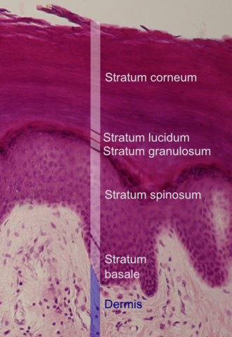
The stratum corneum (Latin for 'horned/horny layer') is the outermost layer of the epidermis of the skin. Consisting of dead tissue, it protects underlying tissue from infection, dehydration, chemicals, and mechanical stress. It is composed of 15 to 20 layers of flattened cells with no nuclei or cell organelles.
Among its properties are mechanical shear, impact resistance, water flux and hydration regulation, microbial proliferation and invasion regulation, initiation of inflammation through cytokine activation and dendritic cell activity, and selective permeability to exclude toxins, irritants, and allergens.[2] The cytoplasm of corneocytes, its cells, shows filamentous keratin. These corneocytes are embedded in a lipid matrix composed of ceramides, cholesterol, and fatty acids.[3]
Desquamation is the process of cell shedding from the surface of the stratum corneum, balancing proliferating keratinocytes that form in the stratum basale. These cells migrate through the epidermis towards the surface in a journey that takes approximately fourteen days.[4]
Structure
The human stratum corneum comprises several levels of flattened corneocytes that are divided into two layers: the stratum disjunctum and stratum compactum. The stratum disjunctum is the uppermost and loosest layer of skin. The skin's protective acid mantle and lipid barrier sit on top of the stratum disjunctum.[5] The stratum compactum is the comparatively deeper, more compacted and more cohesive part of the stratum corneum.[6] The corneocytes of the stratum disjunctum are larger, more rigid and more hydrophobic than those of the stratum compactum.[7]
Research on osmotic permeability suggests the stratum compactum consists of two layers. The stratum disjunctum above these layers can swell, as can the lowest layer of the stratum disjunctum up to two-fold. However, the first layer in the stratum compactum between them has limited swelling capacity and provides the stratum corneum's barrier.[8]
Function
During cornification, the process whereby living keratinocytes are transformed into non-living corneocytes, the cell membrane is replaced by a layer of ceramides which become covalently linked to an envelope of structural proteins (the cornified envelope).[4] This complex surrounds cells in the stratum corneum and contributes to the skin's barrier function. Corneodesmosomes (modified desmosomes) facilitate cellular adhesion by linking adjacent cells within this epidermal layer. These complexes are degraded by proteases, eventually permitting cells to be shed at the surface. Desquamation and formation of the cornified envelope are both required for the maintenance of skin homeostasis. A failure to correctly regulate these processes leads to skin disorders.[4]
Cells of the stratum corneum contain a dense network of keratin, a protein that helps keep the skin hydrated by preventing water evaporation. These cells can also absorb water, further aiding in hydration. In addition, this layer is responsible for the "spring back" or stretchy properties of skin. A weak glutenous protein bond pulls the skin back to its natural shape.
The thickness of the stratum corneum varies throughout the body. In the palms of the hands and the soles of the feet (sometimes knees, elbows,[9] and knuckles) this layer is stabilized and built by the stratum lucidum (clear phase) which allows the cells to concentrate keratin and toughen them before they rise into a typically thicker, more cohesive stratum corneum. The mechanical stress of heavy structural strain causes this stratum lucidum phase in these regions which require additional protection in order to grasp objects, resist abrasion or impact, and avoid injury. In general, the stratum corneum contains 15 to 20 layers of corneocytes. The stratum corneum has a thickness of between 10 and 40 μm.
In reptiles, the stratum corneum is permanent, and is replaced only during times of rapid growth, in a process called ecdysis or moulting. This is conferred by the presence of beta-keratin, which provides a much more rigid skin layer.
In the human forearm, about 1,300 cells per cm2 per hour are shed.[10] The stratum corneum protects the internal structures of the body from external injury and bacterial invasion.
An inability to correctly maintain the skin barrier function due to the dysregulation of epidermal components can lead to skin disorders. For example, a failure to modulate the activity of kallikreins via the disruption of the protease inhibitorLEKTI causes the debilitating disorder Netherton syndrome.[11]
Hyperkeratosis is an increased thickness of the stratum corneum, and is an unspecific finding, seen in many skin conditions.
↑Mitra, Ashim K.; Kwatra, Deep; Vadlapudi, Aswani Dutt (2015). Drug Delivery. Burlington, MA: Jones & Bartlett Learning. pp.285–286. ISBN978-1-284-02568-2.
123Ovaere P; Lippens S; Vandenabeele P; Declercq W. (2009). "The emerging roles of serine protease cascades in the epidermis". Trends in Biochemical Sciences. 34 (9): 453–463. doi:10.1016/j.tibs.2009.08.001. PMID19726197.
↑Murphrey, Morgan B.; Miao, Julia H.; Zito, Patrick M. (2021), "Histology, Stratum Corneum", StatPearls, Treasure Island (FL): StatPearls Publishing, PMID30020671, retrieved 2021-07-18
↑Descargues P, Deraison C, Bonnart C, Kreft M, Kishibe M, Ishida-Yamamoto A, Elias P, Barrandon Y, Zambruno G, Sonnenberg A, Hovnanian A (Jan 2005). "Spink5-deficient mice mimic Netherton syndrome through degradation of desmoglein 1 by epidermal protease hyperactivity". Nat Genet. 37 (1): 56–65. doi:10.1038/ng1493. PMID15619623. S2CID11404025.
This page is based on this Wikipedia article Text is available under the CC BY-SA 4.0 license; additional terms may apply. Images, videos and audio are available under their respective licenses.