Skin is the layer of usually soft, flexible outer tissue covering the body of a vertebrate animal, with three main functions: protection, regulation, and sensation.

In botany, a seed is a plant embryo and food reserve enclosed in a protective outer covering called a seed coat (testa). More generally, the term "seed" means anything that can be sown, which may include seed and husk or tuber. Seeds are the product of the ripened ovule, after the embryo sac is fertilized by sperm from pollen, forming a zygote. The embryo within a seed develops from the zygote and grows within the mother plant to a certain size before growth is halted.
Sheath pronounced as, may refer to:

Melanocytes are melanin-producing neural crest-derived cells located in the bottom layer of the skin's epidermis, the middle layer of the eye, the inner ear, vaginal epithelium, meninges, bones, and heart. Melanin is a dark pigment primarily responsible for skin color. Once synthesized, melanin is contained in special organelles called melanosomes which can be transported to nearby keratinocytes to induce pigmentation. Thus darker skin tones have more melanosomes present than lighter skin tones. Functionally, melanin serves as protection against UV radiation. Melanocytes also have a role in the immune system.

The integumentary system is the set of organs forming the outermost layer of an animal's body. It comprises the skin and its appendages, which act as a physical barrier between the external environment and the internal environment that it serves to protect and maintain the body of the animal. Mainly it is the body's outer skin.

The hair follicle is an organ found in mammalian skin. It resides in the dermal layer of the skin and is made up of 20 different cell types, each with distinct functions. The hair follicle regulates hair growth via a complex interaction between hormones, neuropeptides, and immune cells. This complex interaction induces the hair follicle to produce different types of hair as seen on different parts of the body. For example, terminal hairs grow on the scalp and lanugo hairs are seen covering the bodies of fetuses in the uterus and in some newborn babies. The process of hair growth occurs in distinct sequential stages: anagen is the active growth phase, catagen is the regression of the hair follicle phase, telogen is the resting stage, exogen is the active shedding of hair phase and kenogen is the phase between the empty hair follicle and the growth of new hair.

In anatomy and zoology, the cortex is the outermost layer of an organ. Organs with well-defined cortical layers include kidneys, adrenal glands, ovaries, the thymus, and portions of the brain, including the cerebral cortex, the best-known of all cortices.

Tooth development or odontogenesis is the complex process by which teeth form from embryonic cells, grow, and erupt into the mouth. For human teeth to have a healthy oral environment, all parts of the tooth must develop during appropriate stages of fetal development. Primary (baby) teeth start to form between the sixth and eighth week of prenatal development, and permanent teeth begin to form in the twentieth week. If teeth do not start to develop at or near these times, they will not develop at all, resulting in hypodontia or anodontia.

The cervical loop is the location on an enamel organ in a developing tooth where the outer enamel epithelium and the inner enamel epithelium join. The cervical loop is a histologic term indicating a specific epithelial structure at the apical side of the tooth germ, consisting of loosely aggregated stellate reticulum in the center surrounded by stratum intermedium. These tissues are enveloped by a basal layer of epithelium known on the outside of the tooth as outer enamel epithelium and on the inside as inner enamel epithelium. During root formation the inner layers of epithelium disappear and only the basal layers are left creating Hertwig's epithelial root sheath (HERS). At this point it is usually referred to as HERS instead of the cervical loop to indicate the structural difference.
Cementogenesis is the formation of cementum, one of the three mineralized substances of a tooth. Cementum covers the roots of teeth and serves to anchor gingival and periodontal fibers of the periodontal ligament by the fibers to the alveolar bone.
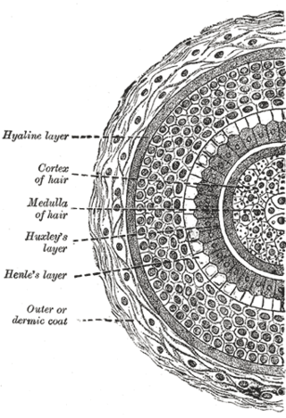
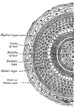
Huxley's layer is the second layer of the inner root sheath of the hair and consists of one or two layers of horny, flattened, nucleated cells. It lies between Henle's layer and the cuticle.
Henle's layer is the third and the outermost layer of the inner root sheath of the hair follicle, consisting of a single layer of cubical cells with clear flattened nuclei. It is named after German physician, pathologist and anatomist Friedrich Gustav Jakob Henle.

The human skin is the outer covering of the body and is the largest organ of the integumentary system. The skin has up to seven layers of ectodermal tissue guarding muscles, bones, ligaments and internal organs. Human skin is similar to most of the other mammals' skin, and it is very similar to pig skin. Though nearly all human skin is covered with hair follicles, it can appear hairless. There are two general types of skin, hairy and glabrous skin (hairless). The adjective cutaneous literally means "of the skin".
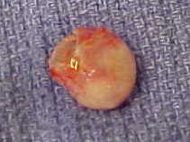
A trichilemmal cyst is a common cyst that forms from a hair follicle, most often on the scalp, and is smooth, mobile, and filled with keratin, a protein component found in hair, nails, skin, and horns. Trichilemmal cysts are clinically and histologically distinct from trichilemmal horns, hard tissue that is much rarer and not limited to the scalp. Rarely, these cysts may grow more extensively and form rapidly multiplying trichilemmal tumors, also called proliferating trichilemmal cysts, which are benign, but may grow aggressively at the cyst site. Very rarely, trichilemmal cysts can become cancerous.
In mammals, trichocytes are the specialized epithelial cells from which the highly mechanically resilient tissues hair and nails are formed. They can be identified by the fact that they express "hard", "trichocyte" or "hair" keratin proteins. These are modified keratins containing large amounts of the amino acid cysteine, which facilitates chemical cross-linking of these proteins to form the tough material from which hair and nail is composed. These cells give rise to non-hair non-keratinized IRSC as well.
The inner root sheath or internal root sheath of the hair follicle is located between the outer root sheath and the hair shaft. It is made of three layers: Henle's layer, Huxley's layer, and the cuticle.
The outer root sheath or external root sheath of the hair follicle encloses the inner root sheath and hair shaft. It is continuous with the basal layer of the interfollicular epidermis (skin).

Loose anagen syndrome, also known as loose anagen hair syndrome, is a hair disorder related to dermatology. It is characterised by the easy and pain free detachment of anagen staged hairs from the scalp. This hair condition can be spontaneous or genetically inherited.

Trichohyalin is a protein that in mammals is encoded by the TCHH gene.
Root sheath may refer to any of these biological structures: