Glutamate receptor-interacting protein

Last updated

Glutamate receptor-interacting protein (GRIP) refers to either a family of proteins that bind to the glutamate receptor or specifically to the GRIP1 protein within this family. Proteins in the glutamate receptor-interacting protein (GRIP) family have been shown to interact with GluR2, a common subunit in the AMPA receptor. [1] This subunit also interacts with other proteins such as protein interacting with C-kinase1 (PICK1) and N-ethylmaleimide-sensitive fusion protein (NSF). Studies have begun to elucidate its function; however, much is still to be learned about these proteins.

Contents

Discovery and history of GRIP 1

Binding of GRIP1 to AMPA Receptors Binding of GRIP1 to AMPA Receptors.jpg
Binding of GRIP1 to AMPA Receptors

The discovery of the Glutamate Receptor Interacting Protein (GRIP-1) came as a result of the observation that Glutamate Receptors, such as the NMDA receptor, cluster at synapses. [2] Shortly after this observation, researchers identified a region on the C-terminal region of NMDA receptors called the tSXV motif that has the ability to bind to the PDZ domain of the PSD-95 protein. [3]

Research on NMDA receptor localization paved the way for research on non-NMDA receptors such as AMPA receptors. Similar to NMDA receptors, it was discovered that AMPA receptors localize in the synaptic terminal of neurons in the central nervous system. [4] By using GFP (green fluorescent protein) antibodies that correspond to the GRIP protein, researchers were able to use fluorescence to determine the location of GRIP in hippocampal neurons. Another GFP antibody was then used to label the GluR2 subunit of AMPA receptors. [4] By using immunocytochemistry and comparing the location of GRIP and AMPA receptors it was determined that GRIP and AMPA receptors experience colocalization in hippocampal neurons. [4] These findings confirmed the initial hypothesis that the GRIP protein plays an important role in binding AMPA receptors to excitatory synapses.

The structure of GRIP contains seven PDZ domains and binds to the C-terminus of the GluR2 subunit of AMPA receptors. [4] Although the number of PDZ domains is different for the proteins PSD-95 and GRIP, the PDZ domain is a common structural motif in proteins that help mediate protein-protein interactions. [5] The AMPA receptor amino acid sequence that the GRIP protein binds to is ESVKI. The conserved serine amino acid in the C- terminus of both AMPA and NMDA receptors suggests that it plays an important role in facilitating the interaction for GRIP and PSD-95. [6]

Role of GRIP in AMPAR cycling

AMPA receptors are constantly being transported between the cell membrane and intracellular space and it was originally thought that GRIP may be responsible for the clustering of AMPA receptors at the excitatory synapse. [1] Although it is still unclear the exact role of GRIP in this trafficking, It appears that PICK1 is more directly responsible for the clustering of AMPA receptors at the surface and that GRIP is involved in the stabilization of AMPA receptors intracellularly. [7] One study showed that when the interaction between GluR2 and GRIP is disrupted, there are no changes in the surface expression of AMPA receptors or the constitutive internalization of AMPA receptors. [8] There is, however, a reduced amount of receptors that remain internalized when receptor cycling is modified by application of AMPA-1. The ratio returns to normal when constitutive recycling is allowed to happen, suggesting that the stabilization of intracellular receptors is critical only under AMPA-induced internalization. [8]

Illustration of roles of GRIP1a and GRIP1b in AMPAR cycling Illustration of roles of GRIP1a and GRIP1b in AMPA cycling.png
Illustration of roles of GRIP1a and GRIP1b in AMPAR cycling

In later studies, two proteins, GRIP-1 (often reduced to GRIP) and ABP-L (also named GRIP-2), were found to be expressed by two separate genes and their respective contributions to AMPA receptor cycling have since been well studied. Each of these proteins have different isoforms due to differential RNA splicing. [9] [10] The isoforms of GRIP-1 are named GRIP-1a and GRIP-1b while those of ABP-L are distinguished as ABP-L and pABP-L. The apparent difference in both cases is that one isoform (GRIP1b and pABP-L respectively) is capable of being conjugated with Palmitic acid, an action called Palmitoylation.

Whereas GRIP initially was thought to be involved in the stabilization of AMPA receptors either at the cell surface or intracellularly when internalization was triggered by AMPA stimulation, it now appears that the GRIP-1 isoforms are involved differentially with the stabilization of AMPA receptors after being internalized due to NMDA stimulation. [11] GRIP-1a has been shown to reduce the expected intracellular levels of AMPA receptors after NMDA stimulation. Conversely, GRIP-1b increases intracellular levels of AMPA receptors under the same conditions.

ABP-L, like GRIP-1b, associates with intracellular stores of AMPA receptors. pABP-L, however, associates with AMPA receptors as the surface membrane. [12] It has not yet been shown under what conditions these interactions are significant in the cycling of AMPAR.

Role of GRIP1 in Fraser syndrome

This diagram depicts the role GRIP1 plays in localizing extra-cellular matrix proteins Fras1 and Frem2 at the dermo-epidermal junction. GRIP1.png
This diagram depicts the role GRIP1 plays in localizing extra-cellular matrix proteins Fras1 and Frem2 at the dermo-epidermal junction.

Mutations to GRIP1 play a role in less than 10% of confirmed cases of the group of congenital defects known as Fraser syndrome. [13] Using immunofluorescence, it has been shown that GRIP1 is found in several kinds of embryonic tissues, including the GI tract, ureter buds, skin and oral and nasal cavities. [14] GRIP1 is also essential for proper function and structure of the dermo-epidermal junction. [15] In mouse models, knocking out GRIP1 protein leads to several deformities that begin in embryo. These deformities include subepidermal hemorrhagic blistering, renal agenesis, syndactylism, polydactylism and cryptopthalmos. [14] One study has shown that complete knock-out of GRIP1 leads to the absence of kidneys. [14] Another study shows blistering of embryonic tissue that GRIP1 is expressed in by day 12 of embryonic life in mice. [15]

The mechanism of GRIP1 in Fraser syndrome is found in the interaction GRIP1 has with the proteins Fras1 and Frem2. [16] Fras1 and Frem2 are extracellular membrane proteins necessary for proper basement membrane function as well as morphogenesis. [16] GRIP1 plays a vital role in localizing Fras1 to the basal surface of epidermal cells as well as localizing Frem2. [16] Knocking out the GRIP1 protein or mutating it leads to poor expression of Fras1 and Frem2. [16] GRIP1 specifically binds with Fras1 through a PDZ motif located on Fras1. [16] Frem2 also has a PDZ domain, although the interaction between GRIP1 and Frem2 is unclear. [16] In one case of Fraser syndrome, GRIP1 lacked PDZ domains 6 and 7. Only the first four PDZ domains of the seven PDZ domains GRIP1 has are required for binding with Fras1, indicating additional mechanisms and proteins GRIP1 interacts with that could lead to Fraser syndrome when mutated. [17] Other mutations in GRIP1 that lead to Fraser syndrome include nonsense mutations, frameshift mutations, splice site mutations, a genome deletion and a deletion of exon 18 of the GRIP1 gene. [13]

Role of GRIP1 in neuron morphology and cargo transport

Neuron morphology, development, and maintenance are dependent on the expression of GRIP1 in the cell. [18] It is vitally important in initial development as knock out experiments in murine models result in skin blisters and embryonic lethality. [15] In developed murine models, disabling mutations like transfecton or dominant negatives in GRIP1 can cause up to 75% loss in “primary, secondary, and higher-order” dendrites in developing neurons. [19] Disabling GRIP1 in live healthy neurons in a dish will cause a 20% reduction in the thickest part of the neuron and up to 70% reduction in the branches. [18]

Defects in neuron morphology due to GRIP1 malfunction can be reserved. One way is to overexpress GRIP1. This leads to increased, but not complete recovery of branching. [18] Another protein, EphB2, which interacts with GRIP1, can be mutated such that a 70-90% recovery of branching is possible. However, overexpression of the wild type leads to a decrease in neuron count. [18]

Motor proteins such as Kinesin (KIF5) are bound to adapter molecules like GRIP1 to move cargo from the Golgi to the extremities of a neuron cell. GRIP1 and KIF5 are very commonly found together due to a good binding affinity (Kd range from 10-20nM [20] ). As for how cargo gets to the right place, there has been a hypothesis called the “smart motor”. [21] It is currently thought that the “smart motor” recognizes the difference between axonal (coated with KLC protein) and dendritic (coated with KHC) proteins. [21] The destination is chosen accordingly. Unfortunately, details about the intermediate transporting steps are unknown. However, at the destination the binding of protein 14-3-3 disrupts the interaction between KIF5 and GRIP1. [19] This releases the cargo.

See also

Related Research Articles

<span class="mw-page-title-main">AMPA receptor</span> Transmembrane protein family

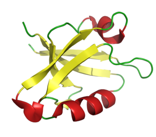
The α-amino-3-hydroxy-5-methyl-4-isoxazolepropionic acid receptor is an ionotropic transmembrane receptor for glutamate (iGluR) that mediates fast synaptic transmission in the central nervous system (CNS). It has been traditionally classified as a non-NMDA-type receptor, along with the kainate receptor. Its name is derived from its ability to be activated by the artificial glutamate analog AMPA. The receptor was first named the "quisqualate receptor" by Watkins and colleagues after a naturally occurring agonist quisqualate and was only later given the label "AMPA receptor" after the selective agonist developed by Tage Honore and colleagues at the Royal Danish School of Pharmacy in Copenhagen. The GRIA2-encoded AMPA receptor ligand binding core was the first glutamate receptor ion channel domain to be crystallized.

<span class="mw-page-title-main">Excitatory synapse</span> Sort of synapse

An excitatory synapse is a synapse in which an action potential in a presynaptic neuron increases the probability of an action potential occurring in a postsynaptic cell. Neurons form networks through which nerve impulses travels, each neuron often making numerous connections with other cells of neurons. These electrical signals may be excitatory or inhibitory, and, if the total of excitatory influences exceeds that of the inhibitory influences, the neuron will generate a new action potential at its axon hillock, thus transmitting the information to yet another cell.

<span class="mw-page-title-main">PDZ domain</span>

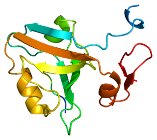
The PDZ domain is a common structural domain of 80-90 amino-acids found in the signaling proteins of bacteria, yeast, plants, viruses and animals. Proteins containing PDZ domains play a key role in anchoring receptor proteins in the membrane to cytoskeletal components. Proteins with these domains help hold together and organize signaling complexes at cellular membranes. These domains play a key role in the formation and function of signal transduction complexes. PDZ domains also play a highly significant role in the anchoring of cell surface receptors to the actin cytoskeleton via mediators like NHERF and ezrin.

<span class="mw-page-title-main">GRIA3</span> Protein-coding gene in humans

Glutamate receptor 3 is a protein that in humans is encoded by the GRIA3 gene.

<span class="mw-page-title-main">DLG4</span> Mammalian protein found in Homo sapiens

PSD-95 also known as SAP-90 is a protein that in humans is encoded by the DLG4 gene.

<span class="mw-page-title-main">DLG1</span> Protein-coding gene in the species Homo sapiens

Discs large homolog 1 (DLG1), also known as synapse-associated protein 97 or SAP97, is a scaffold protein that in humans is encoded by the SAP97 gene.

<span class="mw-page-title-main">PICK1</span> Protein-coding gene in the species Homo sapiens

Protein Interacting with C Kinase - 1 is a protein that in humans is encoded by the PICK1 gene.

<span class="mw-page-title-main">Syntenin-1</span> Protein found in humans

Syntenin-1 is a protein that in humans is encoded by the SDCBP gene.

<span class="mw-page-title-main">GRIA2</span> Mammalian protein found in Homo sapiens

Glutamate ionotropic receptor AMPA type subunit 2 is a protein that in humans is encoded by the GRIA2 gene and it is a subunit found in the AMPA receptors.

<span class="mw-page-title-main">GRIK1</span> Protein-coding gene in the species Homo sapiens

Glutamate receptor, ionotropic, kainate 1, also known as GRIK1, is a protein that in humans is encoded by the GRIK1 gene.

<span class="mw-page-title-main">GRIA4</span>

Glutamate receptor 4 is a protein that in humans is encoded by the GRIA4 gene.

<span class="mw-page-title-main">MPDZ</span> Protein-coding gene in the species Homo sapiens

Multiple PDZ domain protein is a protein that in humans is encoded by the MPDZ gene.

<span class="mw-page-title-main">GRIP1 (gene)</span> Protein-coding gene in the species Homo sapiens

Glutamate receptor-interacting protein 1 is a protein that in humans is encoded by the GRIP1 gene.

<span class="mw-page-title-main">GRIP2</span> Protein-coding gene in the species Homo sapiens

Glutamate receptor-interacting protein 2 is a protein that in humans is encoded by the GRIP2 gene.

<span class="mw-page-title-main">GRIPAP1</span> Protein-coding gene in the species Homo sapiens

GRIP1-associated protein 1 is a protein that in humans is encoded by the GRIPAP1 gene.

<span class="mw-page-title-main">GRID2</span> Protein-coding gene in the species Homo sapiens

Glutamate receptor, ionotropic, delta 2, also known as GluD2, GluRδ2, or δ2, is a protein that in humans is encoded by the GRID2 gene. This protein together with GluD1 belongs to the delta receptor subtype of ionotropic glutamate receptors. They possess 14–24% sequence homology with AMPA, kainate, and NMDA subunits, but, despite their name, do not actually bind glutamate or various other glutamate agonists.

Long-term potentiation (LTP), thought to be the cellular basis for learning and memory, involves a specific signal transmission process that underlies synaptic plasticity. Among the many mechanisms responsible for the maintenance of synaptic plasticity is the cadherin–catenin complex. By forming complexes with intracellular catenin proteins, neural cadherins (N-cadherins) serve as a link between synaptic activity and synaptic plasticity, and play important roles in the processes of learning and memory.

David S. Bredt is an American molecular neuroscientist.

<span class="mw-page-title-main">Synaptic stabilization</span> Modifying synaptic strength via cell adhesion molecules

Synaptic stabilization is crucial in the developing and adult nervous systems and is considered a result of the late phase of long-term potentiation (LTP). The mechanism involves strengthening and maintaining active synapses through increased expression of cytoskeletal and extracellular matrix elements and postsynaptic scaffold proteins, while pruning less active ones. For example, cell adhesion molecules (CAMs) play a large role in synaptic maintenance and stabilization. Gerald Edelman discovered CAMs and studied their function during development, which showed CAMs are required for cell migration and the formation of the entire nervous system. In the adult nervous system, CAMs play an integral role in synaptic plasticity relating to learning and memory.

<span class="mw-page-title-main">Willardiine</span> Chemical compound

Willardiine (correctly spelled with two successive i's) or (S)-1-(2-amino-2-carboxyethyl)pyrimidine-2,4-dione is a chemical compound that occurs naturally in the seeds of Mariosousa willardiana and Acacia sensu lato. The seedlings of these plants contain enzymes capable of complex chemical substitutions that result in the formation of free amino acids (See: #Synthesis). Willardiine is frequently studied for its function in higher level plants. Additionally, many derivates of willardiine are researched for their potential in pharmaceutical development. Willardiine was first discovered in 1959 by R. Gmelin, when he isolated several free, non-protein amino acids from Acacia willardiana (another name for Mariosousa willardiana) when he was studying how these families of plants synthesize uracilyalanines. A related compound, Isowillardiine, was concurrently isolated by a different group, and it was discovered that the two compounds had different structural and functional properties. Subsequent research on willardiine has focused on the functional significance of different substitutions at the nitrogen group and the development of analogs of willardiine with different pharmacokinetic properties. In general, Willardiine is the one of the first compounds studied in which slight changes to molecular structure result in compounds with significantly different pharmacokinetic properties.

References

  1. 1 2 Dong, H; O'Brien, R; Fung, E; Lanahan, A; Worley, P; Hunganir, R (March 1997). "GRIP: A synaptic PDZ domain-containing protein that interacts with AMPA receptors". Nature. 386 (6622): 279–84. Bibcode:1997Natur.386..279D. doi:10.1038/386279a0. PMID   9069286. S2CID   4361791.
  2. Kornau, Hans-Christian; Schenker, Leslie T.; Kennedy, Mary B.; Seeburg, Peter H. (1995). "Domain interaction between NMDA receptor subunits and the postsynaptic density protein PSD-95". Science. 269 (5231): 1737–1740. Bibcode:1995Sci...269.1737K. doi:10.1126/science.7569905. PMID   7569905.
  3. Ehlers, M. D.; Mammen, A. L.; Lau, L.; Huganir, R. L. (1996). "Synaptic targeting of glutamate receptors". Current Opinion in Cell Biology. 8 (4): 484–489. doi: 10.1016/S0955-0674(96)80024-X . PMID   8791455.
  4. 1 2 3 4 Dong, H; O'Brien, R; Fung, E; Lanahan, A; Worley, P; Hunganir, R (1997). "GRIP: A synaptic PDZ domain-containing protein that interacts with AMPA receptors". Nature. 386 (6622): 279–84. Bibcode:1997Natur.386..279D. doi:10.1038/386279a0. PMID   9069286. S2CID   4361791.
  5. Lee, HJ; Zheng, JJ (2010). "PDZ domains and their binding partners: structure, specificity, and modification". Cell Commun Signal. 8: 8. doi: 10.1186/1478-811X-8-8 . PMC   2891790 . PMID   20509869.
  6. Ehlers, M.; Mammen, A.; Lau, L.; Huganir, R. (1998). "Synaptic targeting of glutamate receptors". Current Opinion in Cell Biology. 8 (4): 484–489. doi: 10.1016/S0955-0674(96)80024-X . PMID   8791455.
  7. Xia, J; Zhang, X; Staudinger, J; Huganir, R (January 1999). "Clustering of AMPA receptors by the synaptic PDZ domain-containing protein PICK1". Neuron. 22 (1): 179–87. doi: 10.1016/s0896-6273(00)80689-3 . PMID   10027300.
  8. 1 2 Braithwaite, S; Xia, H; Malenka, R (2002). "Differential roles for NSF and GRIP/ABP in AMPA receptor cycling". Proceedings of the National Academy of Sciences. 99 (10): 7096–101. Bibcode:2002PNAS...99.7096B. doi: 10.1073/pnas.102156099 . PMC   124534 . PMID   12011465.
  9. Dong, H; Zhang, P; Song, I; Petralia, R; Liao, D; Hunganir, R (15 August 1999). "Characterization of the glutamate receptor-interacting proteins GRIP1 and GRIP2". Journal of Neuroscience. 19 (16): 6930–41. doi:10.1523/JNEUROSCI.19-16-06930.1999. PMC   6782851 . PMID   10436050.
  10. Wyszynski, M; Valtschanoff, J; Naisbitt, S; Dunah, A; Kim, E; Standaert, D; et al. (1 August 1999). "Association of AMPA receptors with a subset of glutamate receptor-interacting protein in vivo". Journal of Neuroscience. 19 (15): 6528–37. doi:10.1523/JNEUROSCI.19-15-06528.1999. PMC   6782830 . PMID   10414981.
  11. Hanley, L; Henley, J (2010). "Differential roles of GRIP1a and GRIP1b in AMPA receptor trafficking". Neuroscience Letters. 485 (3): 167–72. doi:10.1016/j.neulet.2010.09.003. PMC   3310156 . PMID   20837103.
  12. DeSouza, S; Fu, J; States, B; Ziff, E (2002). "Differential palmitoylation directs the AMPA receptor-binding protein ABP to spines or to intracellular clusters". Journal of Neuroscience. 22 (9): 3493–503. doi:10.1523/JNEUROSCI.22-09-03493.2002. PMC   6758378 . PMID   11978826.
  13. 1 2 Schanze, D (2014). "Fraser syndrome due to mutations in GRIP1--clinical phenotype in two families and expansion of the mutation spectrum". American Journal of Medical Genetics. 164 (3): 837–840. doi:10.1002/ajmg.a.36343. PMID   24357607. S2CID   11034897.
  14. 1 2 3 Takamiya, K.; Kostourou, V.; Adams, S.; Jadeja, S.; Chalepakis, G.; Scambler, P. J.; Adams, R. H. (2004). "A direct functional link between the multi-PDZ domain protein GRIP1 and the fraser syndrome protein Fras1". Nature Genetics. 36 (2): 172–7. doi: 10.1038/ng1292 . PMID   14730302.
  15. 1 2 3 Bladt, F (2002). "Epidermolysis bullosa and embryonic lethality in mice lacking the multi-PDZ domain protein GRIP1". Proceedings of the National Academy of Sciences of the United States of America. 99 (10): 6816–6821. Bibcode:2002PNAS...99.6816B. doi: 10.1073/pnas.092130099 . PMC   124486 . PMID   11983858.
  16. 1 2 3 4 5 6 Kiyozumi, D (2006). "Breakdown of the reciprocal stabilization of QBRICK/Frem1, Fras1, and Frem2 at the basement membrane provokes fraser syndrome-like defects". Proceedings of the National Academy of Sciences of the United States of America. 103 (32): 11981–11986. Bibcode:2006PNAS..10311981K. doi: 10.1073/pnas.0601011103 . PMC   1567684 . PMID   16880404.
  17. Vogel, M., Zon, P., Brueton, L., Gijzen, M., Tuil, M., Cox, P., ... Haelst, M. (2012). Mutations in GRIP1 cause Fraser syndrome. Journal of Medical Genetics, 303-306.
  18. 1 2 3 4 Hoogenraad, Casper (Jun 19, 2005). "GRIP1 controls dendrite morphogenesis by regulating EphB receptor trafficking". Nature Neuroscience. 8 (7): 906–916. doi:10.1038/nn1487. PMID   15965473. S2CID   23686585.
  19. 1 2 Geiger, Julia (Feb 24, 2014). "The GRIP1/14-3-3 Pathway Coordinates Cargo Trafficking and Dendrite Development". Developmental Cell. 28 (4): 381–393. doi: 10.1016/j.devcel.2014.01.018 . PMID   24576423.
  20. Skoufias, Dimitrios (Jan 14, 1994). "The Carboxyl-terminal Domain of Kinesin Heavy Chain is important for membrane binding". Journal of Biological Chemistry. 269 (2): 1477–1485. doi: 10.1016/S0021-9258(17)42281-2 . PMID   8288613.
  21. 1 2 Setou, Mitsutoshi (May 2, 2002). "Glutamate-receptor-interacting protein GRIP1 directly steers kinesin to dendrites". Letters to Nature. 417 (6884): 83–87. Bibcode:2002Natur.417...83S. doi:10.1038/nature743. PMID   11986669. S2CID   4400494.