Megakaryocyte

Last updated
Megakaryocyte
WVSOM Megakaryocytes arrows.jpg
Two megakaryocytes in bone marrow, marked with arrows.
Details
Location Bone marrow
Function Platelet production
Identifiers
Latin megakaryocytus
MeSH D008533
TH H2.00.04.3.05003
FMA 83555
Anatomical terms of microanatomy

A megakaryocyte ( mega- + karyo- + -cyte , "large-nucleus cell") is a large bone marrow cell with a lobated nucleus that produces blood platelets (thrombocytes), which are necessary for normal clotting. In humans, megakaryocytes usually account for 1 out of 10,000 bone marrow cells, but can increase in number nearly 10-fold during the course of certain diseases. [1] Owing to variations in combining forms and spelling, synonyms include megalokaryocyte and megacaryocyte.

Contents

Structure

In general, megakaryocytes are 10 to 15 times larger than a typical red blood cell, averaging 50–100 μm in diameter. During its maturation, the megakaryocyte grows in size and replicates its DNA without cytokinesis in a process called endomitosis. As a result, the nucleus of the megakaryocyte can become very large and lobulated, which, under a light microscope, can give the false impression that there are several nuclei. In some cases, the nucleus may contain up to 64N DNA, or 32 copies of the normal complement of DNA in a human cell.

The cytoplasm, just as the platelets that bud off from it, contains α-granules and dense bodies.

Development

Blood cell lineage Illu blood cell lineage.jpg
Blood cell lineage

Megakaryocytes are derived from hematopoietic stem cell precursor cells in the bone marrow. They are produced primarily by the liver, kidney, spleen, and bone marrow. These multipotent stem cells live in the marrow sinusoids and are capable of producing all types of blood cells depending on the signals they receive. The primary signal for megakaryocyte production is thrombopoietin or TPO. TPO is sufficient but not absolutely necessary [2] for inducing differentiation of progenitor cells in the bone marrow towards a final megakaryocyte phenotype. Other molecular signals for megakaryocyte differentiation include GM-CSF, IL-3, IL-6, IL-11, chemokines (SDF-1, FGF-4), [3] and erythropoietin. [4] The megakaryocyte develops through the following lineage:

CFU-Me (pluripotential hemopoietic stem cell or hemocytoblast) → megakaryoblastpromegakaryocyte → megakaryocyte.

The cell eventually reaches megakaryocyte stage and loses its ability to divide. However, it is still able to replicate its DNA and continue development, becoming polyploid. [4] The cytoplasm continues to expand and the DNA amount can increase up to 64n in humans and 256n in mice. Many of the morphological features of megakaryocyte differentiation can be recapitulated in non-hematopoietic cells by the expression of Class VI β-tubulin (β6) and they provide a mechanistic basis for understanding these changes. [5]

Function

Platelet release

Once the cell has completed differentiation and become a mature megakaryocyte, it begins the process of producing platelets. The maturation process occurs via endomitotic synchronous replication whereby the cytoplasmic volume enlarges as the number of chromosomes multiplies without cellular division. The cell ceases its growth at 4N, 8N or 16N, becomes granular, and begins to produce platelets. [6] Thrombopoietin plays a role in inducing the megakaryocyte to form small proto-platelet processes. Platelets are held within these internal membranes within the cytoplasm of megakaryocytes. There are two proposed mechanisms for platelet release. In one scenario, these proto-platelet processes break up explosively to become platelets. [7] It is possible to visualize the spontaneous release of platelets using holotomographic live-cell imaging. Alternatively, the cell may form platelet ribbons into blood vessels. The ribbons are formed via pseudopodia and they are able to continuously emit platelets into circulation. In either scenario, each of these proto-platelet processes can give rise to 2000–5000 new platelets upon breakup. Overall, 2/3 of these newly produced platelets will remain in circulation while 1/3 will be sequestered by the spleen.[ medical citation needed ]

Example of platelets release in mature megakaryocytes. This footage shows the formation and spontaneous release of platelets (small round-shaped blood cells), imaged with a live-cell imaging microscope. Platelets release in mature megakaryocytes .gif
Example of platelets release in mature megakaryocytes. This footage shows the formation and spontaneous release of platelets (small round-shaped blood cells), imaged with a live-cell imaging microscope.

Thrombopoietin (TPO) is a 353-amino acid protein encoded on chromosome 3p27. TPO is primarily synthesized in the liver [8] but can be made by kidneys, testes, brain, and even bone marrow stromal cells. It has high homology with erythropoietin. It is essential for the formation of an adequate quantity of platelets.

After budding off platelets, what remains is mainly the cell nucleus. This crosses the bone marrow barrier to the blood and is consumed in the lung by alveolar macrophages.

Effects of cytokines

Cytokines are signals used in the immune system for intercellular communication. There are many cytokines that affect megakaryocytes. Certain cytokines such as IL-3, IL-6, IL-11, LIF, erythropoietin, and thrombopoietin all stimulate the maturation of megakaryocytic progenitor cells. [9] Other signals such as PF4, CXCL5, CXCL7, and CCL5 inhibit platelet formation. [10]

Clinical significance

Megakaryocytes are directly responsible for producing platelets, which are needed for the formation of a thrombus, or blood clot. There are several diseases that are directly attributable to abnormal megakaryocyte function or abnormal platelet function. [11]

Essential thrombocythemia

Essential thrombocythemia (ET) is a disorder characterized by elevated numbers of circulating platelets. The disease occurs in 1–2 per 100,000 people. The 2016 WHO requirements for diagnosis include > 450,000 platelets/μL of blood (normal 150,000–400,000) and characteristic findings in a bone marrow biopsy. Some of the consequences of having such high numbers of platelets include thrombosis or clots in blood vessels. Thrombi form more frequently in arteries than veins. It seems ironic that having platelet counts above 1,000,000 platelets/μL can lead to hemorrhagic events. [12] Approximately half ET cases are due to a mutation in the JAK2 protein, a member of the JAK-STAT signaling pathway. [13] This mutation induces an unregulated proliferative signal from the thrombopoietin (TPO) receptor in the absence of TPO causing clonal expansion of bone marrow cells, especially megakaryocytes. There is a low risk of transformation to leukemia with this disorder. The primary treatment consists of anagrelide or hydroxyurea to lower platelet levels.

Congenital amegakaryocytic thrombocytopenia

Congenital amegakaryocytic thrombocytopenia (CAMT) is a rare inherited disorder. The primary manifestations are thrombocytopenia and megakaryocytopenia, i.e. low numbers of platelets and megakaryocytes. There is an absence of megakaryocytes in the bone marrow with no associated physical abnormalities. [14] The cause for this disorder appears to be a mutation in the gene for the TPO receptor, c-mpl, despite high levels of serum TPO. [15] [16] In addition, there may be abnormalities with the central nervous system including the cerebrum and cerebellum that could cause symptoms. [15] The primary treatment for CAMT is bone marrow transplantation.

Bone marrow/stem cell transplant is the only remedy for this genetic disease. Frequent platelet transfusions are required to keep the patient from bleeding to death until transplant has been completed, although this is not always the case.

There appears to be no generic resource for CAMT patients on the web and this is potentially due to the rarity of the disease.

History

In 1906, James Homer Wright provided evidence that megakaryocytes give rise to blood platelets. [17]

Kelemen first coined the term "thrombopoietin" to describe the humoral substance responsible for the production of platelets. [18]

See also

Related Research Articles

<span class="mw-page-title-main">Haematopoiesis</span> Formation of blood cellular components

Haematopoiesis is the formation of blood cellular components. All cellular blood components are derived from haematopoietic stem cells. In a healthy adult human, roughly ten billion to a hundred billion new blood cells are produced per day, in order to maintain steady state levels in the peripheral circulation.

<span class="mw-page-title-main">Myelodysplastic syndrome</span> Diverse collection of blood-related cancers

A myelodysplastic syndrome (MDS) is one of a group of cancers in which immature blood cells in the bone marrow do not mature, and as a result, do not develop into healthy blood cells. Early on, no symptoms typically are seen. Later, symptoms may include fatigue, shortness of breath, bleeding disorders, anemia, or frequent infections. Some types may develop into acute myeloid leukemia.

<span class="mw-page-title-main">Fanconi anemia</span> Medical condition

Fanconi anemia (FA) is a rare, AR, genetic disease resulting in impaired response to DNA damage in the FA/BRCA pathway. Although it is a very rare disorder, study of this and other bone marrow failure syndromes has improved scientific understanding of the mechanisms of normal bone marrow function and development of cancer. Among those affected, the majority develop cancer, most often acute myelogenous leukemia (AML), MDS, and liver tumors. 90% develop aplastic anemia by age 40. About 60–75% have congenital defects, commonly short stature, abnormalities of the skin, arms, head, eyes, kidneys, and ears, and developmental disabilities. Around 75% have some form of endocrine problem, with varying degrees of severity. 60% of FA is FANC-A, 16q24.3, which has later onset bone marrow failure.

<span class="mw-page-title-main">Thrombocytopenia</span> Abnormally low levels of platelets in the blood

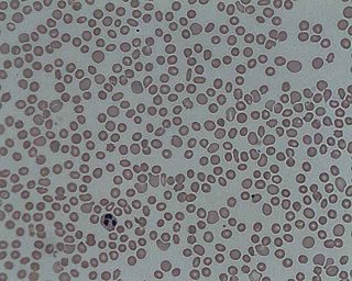
In hematology, thrombocytopenia is a condition characterized by abnormally low levels of platelets in the blood. Low levels of platelets in turn may lead to prolonged or excessive bleeding. It is the most common coagulation disorder among intensive care patients and is seen in a fifth of medical patients and a third of surgical patients.

<span class="mw-page-title-main">Thrombopoietin</span> Mammalian protein found in Homo sapiens

Thrombopoietin (THPO) also known as megakaryocyte growth and development factor (MGDF) is a protein that in humans is encoded by the THPO gene.

<span class="mw-page-title-main">Wiskott–Aldrich syndrome</span> Medical condition

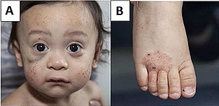
Wiskott–Aldrich syndrome (WAS) is a rare X-linked recessive disease characterized by eczema, thrombocytopenia, immune deficiency, and bloody diarrhea. It is also sometimes called the eczema-thrombocytopenia-immunodeficiency syndrome in keeping with Aldrich's original description in 1954. The WAS-related disorders of X-linked thrombocytopenia (XLT) and X-linked congenital neutropenia (XLN) may present with similar but less severe symptoms and are caused by mutations of the same gene.

<span class="mw-page-title-main">Hematopoietic stem cell</span> Stem cells that give rise to other blood cells

Hematopoietic stem cells (HSCs) are the stem cells that give rise to other blood cells. This process is called haematopoiesis. In vertebrates, the very first definitive HSCs arise from the ventral endothelial wall of the embryonic aorta within the (midgestational) aorta-gonad-mesonephros region, through a process known as endothelial-to-hematopoietic transition. In adults, haematopoiesis occurs in the red bone marrow, in the core of most bones. The red bone marrow is derived from the layer of the embryo called the mesoderm.

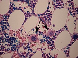
Primary myelofibrosis (PMF) is a rare bone marrow blood cancer. It is classified by the World Health Organization (WHO) as a type of myeloproliferative neoplasm, a group of cancers in which there is activation and growth of mutated cells in the bone marrow. This is most often associated with a somatic mutation in the JAK2, CALR, or MPL genes. In PMF, the bony aspects of bone marrow are remodeled in a process called osteosclerosis; in addition, fibroblast secrete collagen and reticulin proteins that are collectively referred to as (fibrosis). These two pathological processes compromise the normal function of bone marrow resulting in decreased production of blood cells such as erythrocytes, granulocytes and megakaryocytes, the latter cells responsible for the production of platelets.

<span class="mw-page-title-main">Myeloproliferative neoplasm</span> Overproduction of blood cells in the bone marrow

Myeloproliferative neoplasms (MPNs) are a group of rare blood cancers in which excess red blood cells, white blood cells or platelets are produced in the bone marrow. Myelo refers to the bone marrow, proliferative describes the rapid growth of blood cells and neoplasm describes that growth as abnormal and uncontrolled.

Myelokathexis is a congenital disorder of the white blood cells that causes severe, chronic leukopenia and neutropenia. The disorder is believed to be inherited in an autosomal dominant manner. Myelokathexis refers to retention (kathexis) of neutrophils in the bone marrow (myelo). The disorder shows prominent neutrophil morphologic abnormalities.

Mean platelet volume (MPV) is a machine-calculated measurement of the average size of platelets found in blood and is typically included in blood tests as part of the CBC. Since the average platelet size is larger when the body is producing increased numbers of platelets, the MPV test results can be used to make inferences about platelet production in bone marrow or platelet destruction problems.

Hemopoietic growth factors regulate the differentiation and proliferation of particular progenitor cells. Made available through recombinant DNA technology, they hold tremendous potential for medical uses when a person's natural ability to form blood cells is diminished or defective. Recombinant erythropoietin (EPO) is very effective in treating the diminished red blood cell production that accompanies end-stage kidney disease. Erythropoietin is a sialoglycoprotein hormone produced by peritubular cells of kidney.

<span class="mw-page-title-main">GATA1</span> Protein-coding gene in humans

GATA-binding factor 1 or GATA-1 is the founding member of the GATA family of transcription factors. This protein is widely expressed throughout vertebrate species. In humans and mice, it is encoded by the GATA1 and Gata1 genes, respectively. These genes are located on the X chromosome in both species.

<span class="mw-page-title-main">Gray platelet syndrome</span> Medical condition

Gray platelet syndrome (GPS), or platelet alpha-granule deficiency, is a rare congenital autosomal recessive bleeding disorder caused by a reduction or absence of alpha-granules in blood platelets, and the release of proteins normally contained in these granules into the marrow, causing myelofibrosis. The name derives from the initial observation of gray appearance of platelets with a paucity of granules on blood films from a patient with a lifelong bleeding disorder.

The Harrington–Hollingsworth experiment was an experiment that established the autoimmune nature of the blood disorder immune thrombocytopenic purpura. It was performed in 1950 by the academic staff of Barnes-Jewish Hospital in St. Louis, Missouri.

<span class="mw-page-title-main">Thrombopoietin receptor</span> Protein-coding gene in the species Homo sapiens

The thrombopoietin receptor also known as the myeloproliferative leukemia protein or CD110 is a protein that in humans is encoded by the MPL oncogene.

<span class="mw-page-title-main">CFU-GEMM</span>

CFU-GEMM is a colony forming unit that generates myeloid cells. CFU-GEMM cells are the oligopotential progenitor cells for myeloid cells; they are thus also called common myeloid progenitor cells or myeloid stem cells. "GEMM" stands for granulocyte, erythrocyte, monocyte, megakaryocyte.

<span class="mw-page-title-main">Congenital amegakaryocytic thrombocytopenia</span> Medical condition

Congenital amegakaryocytic thrombocytopenia (CAMT) is a rare autosomal recessive bone marrow failure syndrome characterized by severe thrombocytopenia, which can progress to aplastic anemia and leukemia. CAMT usually manifests as thrombocytopenia in the initial month of life or in the fetal phase. Typically CAMPT presents with petechiae, cerebral bleeds, recurrent rectal bleeding, or pulmonary hemorrhage.

<span class="mw-page-title-main">Thrombopoiesis</span>

Thrombopoiesis is the formation of thrombocytes in the bone marrow. Thrombopoietin is the main regulator of thrombopoiesis. Thrombopoietin affects most aspects of the production of platelets. This includes self-renewal and expansion of hematopoietic stem cells, stimulating the increase of megakaryocyte progenitor cells, and supporting these cells so they mature to become platelet-producing cells. The process of Thrombopoiesis is caused by the breakdown of proplatelets. During the process almost all of the membranes, organelles, granules, and soluble macromolecules in the cytoplasm are being consumed. Apoptosis also plays a role in the final stages of thrombopoiesis by letting proplatelet processes to occur from the cytoskeleton of actin.

<span class="mw-page-title-main">William Vainchenker</span>

William Vainchenker, born on 16 December 1947, is a French medical doctor and researcher. He is considered a specialist in hematopoiesis.

References

  1. Branehog I, Ridell B, Swolin B, Weinfeld A (1975). "Megakaryocyte quantifications in relation to thrombokinetics in primary thrombocythaemia and allied diseases". Scand. J. Haematol. 15 (5): 321–32. doi:10.1111/j.1600-0609.1975.tb01087.x. PMID   1060175.
  2. Bunting S, Widmer R, Lipari T, Rangell L, Steinmetz H, Carver-Moore K, Moore MW, Keller GA, de Sauvage FJ (November 1997). "Normal platelets and megakaryocytes are produced in vivo in the absence of thrombopoietin". Blood. 90 (9): 3423–29. doi: 10.1182/blood.V90.9.3423 . PMID   9345025.
  3. Avecilla ST, Hattori K, Heissig B, Tejada R, Liao F, Shido K, Jin DK, Dias S, Zhang F, Hartman TE, Hackett NR, Crystal RG, Witte L, Hicklin DJ, Bohlen P, Eaton D, Lyden D, de Sauvage F, Rafii S (January 2004). "Chemokine-mediated interaction of hematopoietic progenitors with the bone marrow vascular niche is required for thrombopoiesis". Nat Med. 10 (1): 64–71. doi:10.1038/nm973. PMID   14702636. S2CID   21760081.
  4. 1 2 Deutsch VR, Torner A (September 2006). "Megakaryocyte development and platelet production". Br. J. Haematol. 134 (5): 453–66. doi:10.1111/j.1365-2141.2006.06215.x. PMID   16856888. S2CID   23424418.
  5. Yang, H; Ganguly, A; Cabral, F (2012). "Megakaryocyte Lineage Specific Class VI β-Tubulin Suppresses Microtubule Dynamics, Fragments Microtubules, and Blocks Cell Division". Cytoskeleton. 68 (3): 175–87. doi:10.1002/cm.20503. PMC   3082363 . PMID   21309084.
  6. Hoffbrand, A. V.; Pettit, J. E.; Vyas, Paresh (2010). Megakaryocyte and Platelet Production. ISBN   978-0-323-04453-0. Archived from the original on 6 June 2013.
  7. Choi ES, Nichol JL, Hokom MM, et al. (1995). "Platelets generated in vitro from proplatelet-displaying human megakaryocytes are functional". Blood. 85 (2): 402–13. doi: 10.1182/blood.V85.2.402.402 . PMID   7529062.
  8. Jelkmann W (2001). "The role of the liver in the production of thrombopoietin compared with erythropoietin". Eur. J. Gastroenterol. Hepatol. 13 (7): 791–801. doi:10.1097/00042737-200107000-00006. PMID   11474308.
  9. Gordon MS, Hoffman R (1992). "Growth factors affecting human thrombocytopoiesis: potential agents for the treatment of thrombocytopenia". Blood. 80 (2): 302–07. doi: 10.1182/blood.V80.2.302.302 . PMID   1627792.
  10. Pang L, Weiss MJ, Poncz M (2005). "Megakaryocyte biology and related disorders". J. Clin. Invest. 115 (12): 3332–38. doi:10.1172/JCI26720. PMC   1297258 . PMID   16322777.
  11. Nurden AT (2005). "Qualitative disorders of platelets and megakaryocytes". Journal of Thrombosis and Haemostasis. 3 (8): 1773–82. doi: 10.1111/j.1538-7836.2005.01428.x . PMID   16102044. S2CID   22268111.
  12. Michiels JJ, Berneman ZN, Schroyens W, Van Vliet HH (2004). "Pathophysiology and treatment of platelet-mediated microvascular disturbances, major thrombosis and bleeding complications in essential thrombocythaemia and polycythaemia vera". Platelets. 15 (2): 67–84. doi:10.1080/09537100310001646969. PMID   15154599. S2CID   218867974.
  13. Kralovics R, Passamonti F, Buser AS, Teo SS, et al. (2005-04-28). "A gain-of-function mutation of JAK2 in myeloproliferative disorders". N Engl J Med. 352 (17): 1779–90. doi: 10.1056/NEJMoa051113 . PMID   15858187.
  14. Freedman MH, Estrov Z (1990). "Congenital amegakaryocytic thrombocytopenia: an intrinsic hematopoietic stem cell defect". Am. J. Pediatr. Hematol. Oncol. 12 (2): 225–30. doi:10.1097/00043426-199022000-00020. PMID   2378417. S2CID   23164119.
  15. 1 2 Ihara K, Ishii E, Eguchi M, Takada H, Suminoe A, Good RA, Hara T (1999). "Identification of mutations in the c-mpl gene in congenital amegakaryocytic thrombocytopenia". Proc. Natl. Acad. Sci. 96 (6): 3133–36. Bibcode:1999PNAS...96.3132I. doi: 10.1073/pnas.96.6.3132 . PMC   15907 . PMID   10077649.
  16. Ballmaier M, Germeshausen M, Schulze H, Cherkaoui K, Lang S, Gaudig A, Krukemeier S, Eilers M, Strauss G, Welte K (2001). "C-mpl mutations are the cause of congenital amegakaryocytic thrombocytopenia". Blood. 97 (1): 139–46. doi: 10.1182/blood.V97.1.139 . PMID   11133753.
  17. Wright, JH (June 7, 1906). "The origin and nature of the blood plates". Boston Medical and Surgical Journal . CLIV (23): 643–645. doi:10.1056/NEJM190606071542301.
  18. Kelemen, E; Cserhati, I; Tanos, B (December 1958). "Demonstration and some properties of human thrombopoietin in thrombocythaemic sera". Acta Haematologica . 20 (6): 350–355. doi:10.1159/000205503. PMID   13616931.