Avidin

Last updated
Avidin family
Tetramer.png
Tetrameric structure of streptavidin with 2 bound biotins
Identifiers
SymbolAvidin
Pfam PF01382
InterPro IPR005468
PROSITE PDOC00499
CATH 1slf
SCOP2 1slf / SCOPe / SUPFAM
Available protein structures:
Pfam   structures / ECOD  
PDB RCSB PDB; PDBe; PDBj
PDBsum structure summary
Biotin - Avidin can bind up to four molecules of biotin simultaneously with a high degree of affinity and specificity Biotin structure.svg
Biotin - Avidin can bind up to four molecules of biotin simultaneously with a high degree of affinity and specificity

Avidin is a tetrameric biotin-binding protein produced in the oviducts of birds, reptiles and amphibians and deposited in the whites of their eggs. Dimeric members of the avidin family are also found in some bacteria. [1] In chicken egg white, avidin makes up approximately 0.05% of total protein (approximately 1800 μg per egg). The tetrameric protein contains four identical subunits (homotetramer), each of which can bind to biotin (Vitamin B7, vitamin H) with a high degree of affinity and specificity. The dissociation constant of the avidin-biotin complex is measured to be KD ≈ 10−15 M, making it one of the strongest known non-covalent bonds. [2]

Contents

In its tetrameric form, avidin is estimated to be 66–69 kDa in size. [3] 10% of the molecular weight is contributed by carbohydrate, composed of four to five mannose and three N-acetylglucosamine residues [4] The carbohydrate moieties of avidin contain at least three unique oligosaccharide structural types that are similar in structure and composition. [5]

Functional avidin is found in raw egg, but depending on the amount of heat it is exposed to during cooking, the quantity of molecules available for binding biotin can change. The natural function of avidin in eggs is not known, although it has been postulated to be made in the oviduct as a bacterial growth inhibitor, by binding biotin helpful for bacterial growth. As evidence for this, streptavidin, a related protein with equal biotin affinity and a very similar binding site, is made by certain strains of Streptomyces bacteria, and is thought to serve to inhibit the growth of competing bacteria, in the manner of an antibiotic. [6]

A non-glycosylated form of avidin has been isolated from commercially prepared product; however, it is not conclusive as to whether the non-glycosylated form occurs naturally or is a product of the manufacturing process. [7]

Discovery

A raw egg yolk surrounded by the egg white. Avidin was first isolated from raw chicken egg white by Esmond Emerson Snell Chicken egg01 monovular.jpg
A raw egg yolk surrounded by the egg white. Avidin was first isolated from raw chicken egg white by Esmond Emerson Snell

Avidin was discovered by Esmond Emerson Snell (1914–2003). This discovery began with the observation that chicks on a diet of raw egg white were deficient in biotin, despite availability of the vitamin in their diet. [8] It was concluded that a component of the egg-white was sequestering biotin [8] which Snell verified in vitro using a yeast assay. [9] Snell later isolated the component of egg white responsible for biotin binding, and, in collaboration with Paul György, confirmed that the isolated egg protein was the cause of biotin deficiency or “egg white injury”. [10] At the time the protein had been tentatively named avidalbumin (literally, hungry albumin) by researchers at the University of Texas. [10] The name of the protein was later revised to "avidin" based on its affinity for biotin (avid + biotin). [11]

Applications

Avidin
Identifiers
Organism Chicken
SymbolAVD
UniProt P02701
Search for
Structures Swiss-model
Domains InterPro
Unless otherwise specified, "avidin" the biomedical reagent is chicken avidin.

Research in the 1970s helped establish the avidin-biotin system as a powerful tool in biological sciences. Aware of the strength and specificity of the avidin-biotin complex, researchers began to exploit chicken avidin and streptavidin as probes and affinity matrices in numerous research projects. [12] [13] [14] [15] Soon after, researchers Bayer and Wilchek developed new methods and reagents to biotinylate antibodies and other biomolecules, [16] [17] allowing the transfer of the avidin-biotin system to a range of biotechnological applications. Today, avidin is used in a variety of applications ranging from research and diagnostics to medical devices and pharmaceuticals.

Avidin's affinity for biotin is exploited in wide-ranging biochemical assays, including western blot, ELISA, ELISPOT and pull-down assays. In some cases the use of biotinylated antibodies has allowed the replacement of radioiodine labeled antibodies in radioimmunoassay systems, to give an assay system which is not radioactive.[ citation needed ]

Avidin immobilized onto solid supports is also used as purification media to capture biotin-labelled protein or nucleic acid molecules. For example, cell surface proteins can be specifically labelled with membrane impermeable biotin reagent, then specifically captured using an avidin-based support.[ citation needed ]

Modified forms

As a basically charged glycoprotein, avidin exhibits non-specific binding in some applications. Neutravidin, a deglycosylated avidin with modified arginines, exhibits a more neutral isoelectric point (pI) and is available as an alternative to native avidin, whenever problems of non-specific binding arise. Deglycosylated, neutral forms of chicken avidin are available through Sigma-Aldrich (Extravidin), Thermo Scientific (NeutrAvidin), Invitrogen (NeutrAvidin), and e-Proteins (NeutraLite).

Given the strength of the avidin-biotin bond, dissociation of the avidin-biotin complex requires extreme conditions that cause protein denaturation. The non-reversible nature of the avidin-biotin complex can limit avidin's application in affinity chromatography applications where release of the captured ligand is desirable. Researchers have created an avidin with reversible binding characteristics through nitration or iodination of the binding site tyrosine. [18] The modified avidin exhibits strong biotin binding characteristics at pH 4 and releases biotin at a pH of 10 or higher. [18] A monomeric form of avidin with a reduced affinity for biotin is also employed in many commercially available affinity resins. The monomeric avidin is created by treatment of immobilized native avidin with urea or guanidine HCl (6–8 M), giving it a lower dissociation KD ≈ 10−7M. [19] This allows elution from the avidin matrix to occur under milder, non-denaturing conditions, using low concentrations of biotin or low pH conditions. For a single high affinity biotin binding site without crosslinking, a monovalent version of avidin's distant relative, streptavidin, may be used. [20]

Blocking of biotin binding

The thermal stability and biotin binding activity of avidin are of both practical and theoretical interest to researchers, as avidin's stability is unusually high and avidin is an antinutrient in human food. [21] A 1966 study published in Biochemical and Biophysical Research Communications found that the structure of avidin remains stable at temperatures below 70 °C (158 °F). Above 70 °C (158 °F), avidin's structure is rapidly disrupted and by 85 °C (185 °F), extensive loss of structure and loss of ability to bind biotin is found. [22] A 1991 assay for the Journal of Food Science detected substantial avidin activity in cooked egg white: "mean residual avidin activity in fried, poached and boiled (2 min) egg white was 33, 71 and 40% of the activity in raw egg white." The assay surmised that cooking times were not sufficient to adequately heat all cold spot areas within the egg white. Complete inactivation of avidin's biotin binding capacity required boiling for over 4 minutes. [23]

A 1992 study found that thermal inactivation of the biotin binding activity of avidin was described by D 121 °C = 25 min and z = 33 °C. This study disagreed with prior assumptions "that the binding site of avidin is destroyed on heat denaturation". [21]

The biotin-binding properties of avidin were exploited during the development of idrabiotaparinux, a long-acting low molecular weight heparin used in the treatment of venous thrombosis. Due to the long-acting nature of idraparinux, concerns were made about the clinical management of bleeding complications. By adding a biotin moiety to the idraparinux molecule, idrabiotaparinux was formed; its anticoagulant activity in the setting of a bleeding event can be reversed through an intravenous infusion of avidin. [24]

See also

Related Research Articles

<span class="mw-page-title-main">Biotin</span> Chemical compound (vitamin B7)

Biotin (also known as vitamin B7 or vitamin H) is one of the B vitamins. It is involved in a wide range of metabolic processes, both in humans and in other organisms, primarily related to the utilization of fats, carbohydrates, and amino acids. The name biotin, borrowed from the German Biotin, derives from the Ancient Greek word βίοτος (bíotos; 'life') and the suffix "-in" (a suffix used in chemistry usually to indicate 'forming'). Biotin appears as a white crystalline solid that looks like needles.

In biochemistry, biotinylation is the process of covalently attaching biotin to a protein, nucleic acid or other molecule. Biotinylation is rapid, specific and is unlikely to disturb the natural function of the molecule due to the small size of biotin. Biotin binds to streptavidin and avidin with an extremely high affinity, fast on-rate, and high specificity, and these interactions are exploited in many areas of biotechnology to isolate biotinylated molecules of interest. Biotin-binding to streptavidin and avidin is resistant to extremes of heat, pH and proteolysis, making capture of biotinylated molecules possible in a wide variety of environments. Also, multiple biotin molecules can be conjugated to a protein of interest, which allows binding of multiple streptavidin, avidin or neutravidin protein molecules and increases the sensitivity of detection of the protein of interest. There is a large number of biotinylation reagents available that exploit the wide range of possible labelling methods. Due to the strong affinity between biotin and streptavidin, the purification of biotinylated proteins has been a widely used approach to identify protein-protein interactions and post-translational events such as ubiquitylation in molecular biology.

<span class="mw-page-title-main">Streptavidin</span> Protein in Streptomyces avidinii

Streptavidin is a 52 kDa protein (tetramer) purified from the bacterium Streptomyces avidinii. Streptavidin homo-tetramers have an extraordinarily high affinity for biotin. With a dissociation constant (Kd) on the order of ≈10−14 mol/L, the binding of biotin to streptavidin is one of the strongest non-covalent interactions known in nature. Streptavidin is used extensively in molecular biology and bionanotechnology due to the streptavidin-biotin complex's resistance to organic solvents, denaturants, detergents, proteolytic enzymes, and extremes of temperature and pH.

A tetrameric protein is a protein with a quaternary structure of four subunits (tetrameric). Homotetramers have four identical subunits, and heterotetramers are complexes of different subunits. A tetramer can be assembled as dimer of dimers with two homodimer subunits, or two heterodimer subunits.

Protein tags are peptide sequences genetically grafted onto a recombinant protein. Tags are attached to proteins for various purposes. They can be added to either end of the target protein, so they are either C-terminus or N-terminus specific or are both C-terminus and N-terminus specific. Some tags are also inserted at sites within the protein of interest; they are known as internal tags.

<span class="mw-page-title-main">Electrophoretic mobility shift assay</span>

An electrophoretic mobility shift assay (EMSA) or mobility shift electrophoresis, also referred as a gel shift assay, gel mobility shift assay, band shift assay, or gel retardation assay, is a common affinity electrophoresis technique used to study protein–DNA or protein–RNA interactions. This procedure can determine if a protein or mixture of proteins is capable of binding to a given DNA or RNA sequence, and can sometimes indicate if more than one protein molecule is involved in the binding complex. Gel shift assays are often performed in vitro concurrently with DNase footprinting, primer extension, and promoter-probe experiments when studying transcription initiation, DNA gang replication, DNA repair or RNA processing and maturation, as well as pre-mRNA splicing. Although precursors can be found in earlier literature, most current assays are based on methods described by Garner and Revzin and Fried and Crothers.

<span class="mw-page-title-main">Digoxigenin</span> Chemical compound

Digoxigenin (DIG) is a steroid found exclusively in the flowers and leaves of the plants Digitalis purpurea, Digitalis orientalis and Digitalis lanata (foxgloves), where it is attached to sugars, to form the glycosides.

A tetramer assay is a procedure that uses tetrameric proteins to detect and quantify T cells that are specific for a given antigen within a blood sample. The tetramers used in the assay are made up of four major histocompatibility complex (MHC) molecules, which are found on the surface of most cells in the body. MHC molecules present peptides to T-cells as a way to communicate the presence of viruses, bacteria, cancerous mutations, or other antigens in a cell. If a T-cell's receptor matches the peptide being presented by an MHC molecule, an immune response is triggered. Thus, MHC tetramers that are bioengineered to present a specific peptide can be used to find T-cells with receptors that match that peptide. The tetramers are labeled with a fluorophore, allowing tetramer-bound T-cells to be analyzed with flow cytometry. Quantification and sorting of T-cells by flow cytometry enables researchers to investigate immune response to viral infection and vaccine administration as well as functionality of antigen-specific T-cells. Generally, if a person's immune system has encountered a pathogen, the individual will possess T cells with specificity toward some peptide on that pathogen. Hence, if a tetramer stain specific for a pathogenic peptide results in a positive signal, this may indicate that the person's immune system has encountered and built a response to that pathogen.

NeutrAvidin protein is a deglycosylated version of chicken avidin, with a mass of approximately 60,000 daltons. As a result of carbohydrate removal, lectin binding is reduced to undetectable levels, yet biotin binding affinity is retained because the carbohydrate is not necessary for this activity. Avidin has a high pI but NeutrAvidin has a near-neutral pI, minimizing non-specific interactions with the negatively-charged cell surface or with DNA/RNA. Neutravidin still has lysine residues that remain available for derivatization or conjugation.

<span class="mw-page-title-main">Biotin deficiency</span> Medical condition

Biotin deficiency is a nutritional disorder which can become serious, even fatal, if allowed to progress untreated. It can occur in people of any age, ancestry, or of either sex. Biotin is part of the B vitamin family. Biotin deficiency rarely occurs among healthy people because the daily requirement of biotin is low, many foods provide adequate amounts of it, intestinal bacteria synthesize small amounts of it, and the body effectively scavenges and recycles it in the kidneys during production of urine.

Molecular binding is an attractive interaction between two molecules that results in a stable association in which the molecules are in close proximity to each other. It is formed when atoms or molecules bind together by sharing of electrons. It often, but not always, involves some chemical bonding.

<span class="mw-page-title-main">Meir Wilchek</span> Israeli biochemist (born 1935)

Meir Wilchek is an Israeli biochemist. He is a professor at the Weizmann Institute of Science.

The Strep-tag system is a method which allows the purification and detection of proteins by affinity chromatography. The Strep-tag II is a synthetic peptide consisting of eight amino acids (Trp-Ser-His-Pro-Gln-Phe-Glu-Lys). This peptide sequence exhibits intrinsic affinity towards Strep-Tactin, a specifically engineered streptavidin, and can be N- or C- terminally fused to recombinant proteins. By exploiting the highly specific interaction, Strep-tagged proteins can be isolated in one step from crude cell lysates. Because the Strep-tag elutes under gentle, physiological conditions, it is especially suited for generation of functional proteins.

The Streptavidin-Binding Peptide (SBP)-Tag is a 38-amino acid sequence that may be engineered into recombinant proteins. Recombinant proteins containing the SBP-Tag bind to streptavidin and this property may be utilized in specific purification, detection or immobilization strategies.

<span class="mw-page-title-main">Paul Gyorgy</span>

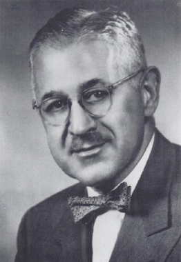
Paul György (April 7, 1893 – March 1, 1976) was a Hungarian-born American biochemist, nutritionist, and pediatrician best known for his discovery of three B vitamins: riboflavin, B6, and biotin. Gyorgy was also well known for his research into the protective factors of human breast milk, particularly for his discoveries of Lactobacillus bifidus growth factor activity in human milk and its anti-staphylococcal properties. He was a recipient of the National Medal of Science in 1975 from President Gerald Ford.

MHC multimers are oligomeric forms of MHC molecules, designed to identify and isolate T-cells with high affinity to specific antigens amid a large group of unrelated T-cells. Multimers generally range in size from dimers to octamers; however, some companies use even higher quantities of MHC per multimer. Multimers may be used to display class 1 MHC, class 2 MHC, or nonclassical molecules from species such as monkeys, mice, and humans.

Chem-seq is a technique that is used to map genome-wide interactions between small molecules and their protein targets in the chromatin of eukaryotic cell nuclei. The method employs chemical affinity capture coupled with massively parallel DNA sequencing to identify genomic sites where small molecules interact with their target proteins or DNA. It was first described by Lars Anders et al. in the January, 2014 issue of "Nature Biotechnology".

Esmond Emerson Snell (September 22, 1914 – December 9, 2003) was an American biochemist who spent his career researching vitamins and nutritional requirements of bacteria and yeast. He is well known for his study of lactic acid-producing bacteria, developing microbiological assays for a number of key nutrients; the discovery of more than half of known vitamins has been attributed to the use of this work. He discovered several B vitamins, including folic acid, and characterized the biochemistry of vitamin B6 (also known as pyrixodal).

<span class="mw-page-title-main">IBA Lifesciences</span>

IBA Lifesciences is a biotechnology company providing products and custom specific services for life science applications in academia and industry worldwide. IBA focusses on two business segments: cell selection and protein purification.

BLESS, also known as breaks labeling, enrichment on streptavidin and next-generation sequencing, is a method used to detect genome-wide double-strand DNA damage. In contrast to chromatin immunoprecipitation (ChIP)-based methods of identifying DNA double-strand breaks (DSBs) by labeling DNA repair proteins, BLESS utilizes biotinylated DNA linkers to directly label genomic DNA in situ which allows for high-specificity enrichment of samples on streptavidin beads and the subsequent sequencing-based DSB mapping to nucleotide resolution.

References

  1. Helppolainen SH, Nurminen KP, Määttä JA, Halling KK, Slotte JP, Huhtala T, et al. (August 2007). "Rhizavidin from Rhizobium etli: the first natural dimer in the avidin protein family". The Biochemical Journal. 405 (3): 397–405. doi:10.1042/BJ20070076. PMC   2267316 . PMID   17447892.
  2. Green NM (December 1963). "Avidin. 1. The Use of (14-C)Biotin for Kinetic Studies and for Assay". The Biochemical Journal. 89 (3): 585–91. doi:10.1042/bj0890585. PMC   1202466 . PMID   14101979.
  3. Korpela J (1984). "Avidin, a high affinity biotin-binding protein, as a tool and subject of biological research". Medical Biology. 62 (1): 5–26. PMID   6379329.
  4. Green NM (1975). Anfinsen CB, Edsall JT, Richards FM (eds.). "Avidin" . Advances in Protein Chemistry. 29: 85–133. doi:10.1016/S0065-3233(08)60411-8. ISBN   978-0-12-034229-7. PMID   237414.
  5. Bruch RC, White HB (October 1982). "Compositional and structural heterogeneity of avidin glycopeptides". Biochemistry. 21 (21): 5334–41. doi:10.1021/bi00264a033. PMID   6816268.
  6. Hendrickson WA, Pähler A, Smith JL, Satow Y, Merritt EA, Phizackerley RP (April 1989). "Crystal structure of core streptavidin determined from multiwavelength anomalous diffraction of synchrotron radiation". Proceedings of the National Academy of Sciences of the United States of America. 86 (7): 2190–4. Bibcode:1989PNAS...86.2190H. doi: 10.1073/pnas.86.7.2190 . JSTOR   33443. PMC   286877 . PMID   2928324.
  7. Hiller Y, Gershoni JM, Bayer EA, Wilchek M (November 1987). "Biotin binding to avidin. Oligosaccharide side chain not required for ligand association". The Biochemical Journal. 248 (1): 167–71. doi:10.1042/bj2480167. PMC   1148514 . PMID   3435435.
  8. 1 2 Eakin RE, McKinley WA, Williams RJ (September 1940). "Egg-White Injury in Chicks and ITS Relationship to a Deficiency of Vitamin H (Biotin)". Science. 92 (2384): 224–5. Bibcode:1940Sci....92..224E. doi:10.1126/science.92.2384.224. PMID   17743857.
  9. Snell EE, Eakin RE, Williams RJ (1940). "A Quantitative Test for Biotin and Observations Regarding its Occurrence and Properties". Journal of the American Chemical Society. 62: 175–8. doi:10.1021/ja01858a052.
  10. 1 2 György P, Rose CS, Eakin RE, Snell EE, Williams RJ (May 1941). "Egg-White Injury as the Result of Nonabsorption or Inactivation of Biotin". Science. 93 (2420): 477–8. Bibcode:1941Sci....93..477G. doi:10.1126/science.93.2420.477. PMID   17757050.
  11. Kresge N, Simoni RD, Hill RL (2004). "The Discovery of Avidin by Esmond E. Snell". The Journal of Biological Chemistry. 279 (41): e5–e6. doi: 10.1016/S0021-9258(20)77095-X .
  12. Hofmann K, Kiso Y (October 1976). "An approach to the targeted attachment of peptides and proteins to solid supports". Proceedings of the National Academy of Sciences of the United States of America. 73 (10): 3516–8. Bibcode:1976PNAS...73.3516H. doi: 10.1073/pnas.73.10.3516 . JSTOR   66631. PMC   431147 . PMID   185617.
  13. Bayer EA, Skutelsky E, Wynne D, Wilchek M (August 1976). "Preparation of ferritin-avidin conjugates by reductive alkylation for use in electron microscopic cytochemistry". The Journal of Histochemistry and Cytochemistry. 24 (8): 933–9. doi: 10.1177/24.8.182877 . PMID   182877.
  14. Angerer L, Davidson N, Murphy W, Lynch D, Attardi G (September 1976). "An electron microscope study of the relative positions of the 4S and ribosomal RNA genes in HeLa cells mitochondrial DNA". Cell. 9 (1): 81–90. doi:10.1016/0092-8674(76)90054-4. PMID   975242. S2CID   54340885.
  15. Heggeness MH, Ash JF (June 1977). "Use of the avidin-biotin complex for the localization of actin and myosin with fluorescence microscopy". The Journal of Cell Biology. 73 (3): 783–8. doi:10.1083/jcb.73.3.783. PMC   2111432 . PMID   326797.
  16. Bayer EA, Zalis MG, Wilchek M (September 1985). "3-(N-Maleimido-propionyl)biocytin: a versatile thiol-specific biotinylating reagent". Analytical Biochemistry. 149 (2): 529–36. doi: 10.1016/0003-2697(85)90609-8 . PMID   3935007.
  17. Wilchek M, Ben-Hur H, Bayer EA (July 1986). "p-Diazobenzoyl biocytin--a new biotinylating reagent for the labeling of tyrosines and histidines in proteins". Biochemical and Biophysical Research Communications. 138 (2): 872–9. doi:10.1016/S0006-291X(86)80577-0. PMID   3741438.
  18. 1 2 Morag E, Bayer EA, Wilchek M (May 1996). "Reversibility of biotin-binding by selective modification of tyrosine in avidin". The Biochemical Journal. 316 (1): 193–9. doi:10.1042/bj3160193. PMC   1217322 . PMID   8645205.
  19. Kohanski RA, Lane MD (1990). Monovalent avidin affinity columns. Methods in Enzymology. Vol. 184. pp. 194–200. doi:10.1016/0076-6879(90)84274-K. ISBN   978-0-12-182085-5. PMID   2388570.
  20. Howarth M, Chinnapen DJ, Gerrow K, Dorrestein PC, Grandy MR, Kelleher NL, et al. (April 2006). "A monovalent streptavidin with a single femtomolar biotin binding site". Nature Methods. 3 (4): 267–73. doi:10.1038/nmeth861. PMC   2576293 . PMID   16554831.
  21. 1 2 Durance TD, Wong NS (1992). "Kinetics of thermal inactivation of avidin". Food Research International. 25 (2): 89–92. doi:10.1016/0963-9969(92)90148-X.
  22. Pritchard AB, McCormick DB, Wright LD (1966). "Optical rotatory dispersion studies of the heat denaturation of avidin and the avidin-biotin complex". Biochemical and Biophysical Research Communications. 25 (5): 524–8. doi:10.1016/0006-291X(66)90623-1.
  23. Durance TD (1991). "Residual Avid in Activity in Cooked Egg White Assayed with Improved Sensitivity". Journal of Food Science. 56 (3): 707–9. doi:10.1111/j.1365-2621.1991.tb05361.x.
  24. Büller HR, Gallus AS, Pillion G, Prins MH, Raskob GE (January 2012). "Enoxaparin followed by once-weekly idrabiotaparinux versus enoxaparin plus warfarin for patients with acute symptomatic pulmonary embolism: a randomised, double-blind, double-dummy, non-inferiority trial". Lancet. 379 (9811): 123–9. doi:10.1016/S0140-6736(11)61505-5. PMID   22130488. S2CID   205964156.