Chondroitin sulfate proteoglycan

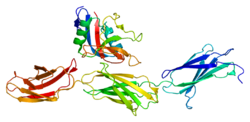
Last updated
Structure of the protein core of aggrecan or chondroitin sulfate proteoglycan 1 Protein ACAN PDB 1tdq.png
Structure of the protein core of aggrecan or chondroitin sulfate proteoglycan 1

Chondroitin sulfate proteoglycans (CSPGs) are proteoglycans consisting of a protein core and a chondroitin sulfate side chain. They are known to be structural components of a variety of human tissues, including cartilage, and also play key roles in neural development and glial scar formation. They are known to be involved in certain cell processes, such as cell adhesion, cell growth, receptor binding, cell migration, and interaction with other extracellular matrix constituents. [1] They are also known to interact with laminin, fibronectin, tenascin, and collagen. [1] CSPGs are generally secreted from cells.

Contents

Importantly, CSPGs are known to inhibit axon regeneration after spinal cord injury. CSPGs contribute to glial scar formation post injury, acting as a barrier against new axons growing into the injury site. [2] CSPGs play a crucial role in explaining why the spinal cord doesn't self-regenerate after an injury.

General structure

Chondroitin sulfate proteoglycans are composed of a core protein and a sugar side chain. The core protein is generally a glycoprotein, and the side chains are glycosaminoglycan (GAG) sugar chains attached through a covalent bond. [1] The GAG side chains are of different lengths depending on the CSPG. Each GAG chain consists of a linear pattern of alternating monosaccharide units: uronic acid and either N-acetylglucosamine or N-acetylgalactosamine. [1]

Types

The following CSPGs have been identified:

Neurocan, brevican, versican, and aggrecan all share similar N-terminal and C-terminal domains. [3]

Neural development

CSPGs play an active role in the neural development of postnatal babies. During development, CSPGs act as guidance cues for developing growth cones. [2] CSPGs guide growth cones through the use of negative signals, as seen by the fact that growing axons avoid CSPG dense areas. [2] Tests done on embryonic roof plates, located on the dorsal midline of developing spinal cords, support this. CSPGs were found near and around the embryonic roof plates that inhibited axon elongation through the spinal cord, and directed the axons in another direction, but were absent in roof plates that attracted axon elongation. [4] These results suggest that CSPGs act in neural development as an inhibitory signal to help guide growing axons.

Spinal cord injury

CSPGs have been implicated in inhibiting axonal regeneration and neurogenesis after central nervous system injury. [5] CSPGs are known to be part of the glial scar that forms post injury, acting as a barrier to prevent axon extension and regrowth. [6] Studies examining CSPG (neurocan, brevican, versican, and phosphacan) levels in rats before spinal cord injury and after spinal cord injury indicate that there is a large up-regulation of these CSPGs after injury is induced. [3] Neurocan, brevican, and versican levels are up-regulated one day post injury, and neurocan and versican remain elevated 4 weeks post injury (brevican remained elevated at 8 weeks post injury, the final time point in the study). [3] Phosphacan showed no up-regulation until 4 weeks post injury. [3] These results, along with previous results showing CSPGs inhibit axon growth, suggest that these four CSPGs work together to inhibit axon growth in spinal cord injury.

Regulation

Inhibition of EGFR inhibits CSPGs

Epidermal growth factor receptor (EGFR) has been suggested to regulate CSPG function. Inhibiting EGFR has been shown to block the activities of certain CSPGs, including neurocan, phosphacan, versican, and aggrecan. [7] When EGFR was inactive, CSPGs had little effect on neurons. [7] As a result, neurogenesis occurred, with significantly longer and many more neurons forming than seen with EGFR active. [7] When EGFR is active, CSPG functioned normally, restricting neurogenesis. [7] Drugs manipulating EGFR may be helpful in preventing the adverse effects CSPGs have during spinal cord injury.

PTP-sigma is a CSPG receptor

PTP-sigma (a transmembrane protein tyrosine phosphatase) is a recently discovered receptor for CSPGs, and is important for proper CSPG function. PTP-sigma binds with very high affinity to CSPGs, specifically neurocan and aggrecan. [8] To simulate more physiological situations, researchers looked at PTP-sigma effects on spinal cord injury sites in mice. Mice with induced spinal cord injury lacking PTP-sigma showed significantly more axon regrowth, with normal amounts of CSPG present. [8] This suggests that without PTP-sigma, CSPGs cannot bind to anything to function properly at the site of a glial scar. [8] Because PTP-sigma is a functional receptor for CSPGs and promotes proper function of CSPGs, drugs manipulating PTP-sigma may help patients with spinal cord injury.

Interferon-gamma

Interferon-gamma (IFN-gamma) is a cytokine that is useful against fighting bacterial infections and helping to suppress tumors. It has also been shown to be beneficial in decreasing CSPG expression after spinal cord injury. Using immunohistochemistry, scientists have shown that CSPGs at the site of spinal cord injury in mice were significantly decreased when treated with IFN-gamma compared to mice without IFN-gamma treatments. [9] Control mice had 80% more levels of CSPGs after spinal cord injury compared to mice treated with IFN-gamma, and scientists suggest that IFN-gamma works by inhibiting mRNA expression. [9]

Rho/ROCK pathway mediates CSPGs

The CSPG inhibition of axon regrowth and neurogenesis post spinal cord injury has been shown to be associated with the rho-associated protein kinase (ROCK) pathway. [6] Studies have shown that when CSPGs inhibit axon growth in the glial scar, the ROCK pathway is activated. [6] However, using C3 transferase and Y27632, two inhibitors of the ROCK signaling pathway, researchers showed that neurogenesis and new neuron length both significantly increased. [6] With C3 transferase, there was a 57% increase in new neuron length, and Y27632 produced a 77% increase in length. [6] Neurogenesis was greatly improved, but not quantifiable. Deactivating the ROCK pathway greatly decreased CSPG inhibition of axon regrowth. These results indicate that the CSPG effect of neurogenesis inhibition is mediated through the ROCK pathway.

In disease

Chondroitin sulfate proteoglycans have been implicated in Alzheimer's disease, stroke, and epilepsy.

Alzheimer's

The two primary markers of Alzheimer's disease are neurofibrillary tangles (NFT) and senile plaques (SP). Studies have shown that CSPGs are present in the frontal cortex and hippocampus NFTs and SPs of postmortem brains of Alzheimer's patients. CSPG-4 and CSPG-6 are both localized on the perimeter of NFTs and SPs, and were also found on dystrophic neurons as well. [10] Given CSPGs inhibitory effects, these results suggest that CSPGs play an important role in Alzheimer's Disease progression, and could be responsible for facilitating the regression of neurons around NFTs and SPs. [10] Medications that target the CSPGs in the NFT and SP may help to alleviate some of the symptoms of Alzheimer's disease.[ clarification needed ][ citation needed ]

Stroke

A stroke is a sudden loss of brain function due to either a blood clot or blood leakage in the brain. Often, a stroke seriously debilitates the patient. However, in those patients that do regain some brain function in affected areas, down-regulations of CSPGs are shown to occur. After stroke, plasticity occurs in some regions of the brain and is associated with some return of brain function. [11] Rats that were able to recover from induced stroke had down-regulations of several CSPGs, including aggrecan, versican, and phosphacan [11] Rats that did not return any brain function did not have significant down-regulation of CSPGs. The reduction of CSPGs in rats that returned some brain function after stroke suggest that more neurological connections could be made with less CSPGs present. Medications that are able to down-regulate CSPGs may help return more brain function to stroke patients.[ clarification needed ][ citation needed ]

Epilepsy

Epilepsy is a neurological disorder characterized by excessive neurological activity in the brain, causing seizures. Researchers have observed that CSPGs are somewhat removed from the brain in epilepsy patients. [11] Research has shown a decrease in phosphacan in both the temporal lobe and the hippocampus in epilepsy cases, suggesting that there CSPGs play a role in the control of axonal regrowth. [11]

Related Research Articles

<span class="mw-page-title-main">Neuron</span> Electrically excitable cell found in the nervous system of animals

Within a nervous system, a neuron, neurone, or nerve cell is an electrically excitable cell that fires electric signals called action potentials across a neural network. Neurons communicate with other cells via synapses - specialized connections that commonly use minute amounts of chemical neurotransmitters to pass the electric signal from the presynaptic neuron to the target cell through the synaptic gap. The neuron is the main component of nervous tissue in all animals except sponges and placozoa. Non-animals like plants and fungi do not have nerve cells.

<span class="mw-page-title-main">Glia</span> Support cells in the nervous system

Glia, also called glial cells(gliocytes) or neuroglia, are non-neuronal cells in the central nervous system (brain and spinal cord) and the peripheral nervous system that do not produce electrical impulses. The neuroglia make up more than one half the volume of neural tissue in our body. They maintain homeostasis, form myelin in the peripheral nervous system, and provide support and protection for neurons. In the central nervous system, glial cells include oligodendrocytes, astrocytes, ependymal cells and microglia, and in the peripheral nervous system they include Schwann cells and satellite cells.

<span class="mw-page-title-main">Astrocyte</span> Type of brain cell

Astrocytes, also known collectively as astroglia, are characteristic star-shaped glial cells in the brain and spinal cord. They perform many functions, including biochemical control of endothelial cells that form the blood–brain barrier, provision of nutrients to the nervous tissue, maintenance of extracellular ion balance, regulation of cerebral blood flow, and a role in the repair and scarring process of the brain and spinal cord following infection and traumatic injuries. The proportion of astrocytes in the brain is not well defined; depending on the counting technique used, studies have found that the astrocyte proportion varies by region and ranges from 20% to around 40% of all glia. Another study reports that astrocytes are the most numerous cell type in the brain. Astrocytes are the major source of cholesterol in the central nervous system.Apolipoprotein E transports cholesterol from astrocytes to neurons and other glial cells, regulating cell signaling in the brain. Astrocytes in humans are more than twenty times larger than in rodent brains, and make contact with more than ten times the number of synapses.

Oligodendrocyte progenitor cells (OPCs), also known as oligodendrocyte precursor cells, NG2-glia, O2A cells, or polydendrocytes, are a subtype of glia in the central nervous system named for their essential role as precursors to oligodendrocytes. They are typically identified in the human by co-expression of PDGFRA and CSPG4.

<span class="mw-page-title-main">Versican</span> Protein-coding gene in the species Homo sapiens

Versican is a large extracellular matrix proteoglycan that is present in a variety of human tissues. It is encoded by the VCAN gene.

Gliosis is a nonspecific reactive change of glial cells in response to damage to the central nervous system (CNS). In most cases, gliosis involves the proliferation or hypertrophy of several different types of glial cells, including astrocytes, microglia, and oligodendrocytes. In its most extreme form, the proliferation associated with gliosis leads to the formation of a glial scar.

<span class="mw-page-title-main">Aggrecan</span>

Aggrecan (ACAN), also known as cartilage-specific proteoglycan core protein (CSPCP) or chondroitin sulfate proteoglycan 1, is a protein that in humans is encoded by the ACAN gene. This gene is a member of the lectican (chondroitin sulfate proteoglycan) family. The encoded protein is an integral part of the extracellular matrix in cartilagenous tissue and it withstands compression in cartilage.

Neuroregeneration involves the regrowth or repair of nervous tissues, cells or cell products. Neuroregenerative mechanisms may include generation of new neurons, glia, axons, myelin, or synapses. Neuroregeneration differs between the peripheral nervous system (PNS) and the central nervous system (CNS) by the functional mechanisms involved, especially in the extent and speed of repair. When an axon is damaged, the distal segment undergoes Wallerian degeneration, losing its myelin sheath. The proximal segment can either die by apoptosis or undergo the chromatolytic reaction, which is an attempt at repair. In the CNS, synaptic stripping occurs as glial foot processes invade the dead synapse.

Chondroitinase treatment is a treatment of proteoglycans, a protein in the fluid among cells where they affect neural activity. Chondroitinase treatment has been shown to allow adults vision to be restored as far as ocular dominance is concerned. Moreover, there is some evidence that Chondroitinase could be used for the treatment of spinal injuries.

<span class="mw-page-title-main">Glial scar</span> Mass formed in response to injury to the nervous system

A glial scar formation (gliosis) is a reactive cellular process involving astrogliosis that occurs after injury to the central nervous system. As with scarring in other organs and tissues, the glial scar is the body's mechanism to protect and begin the healing process in the nervous system.

<span class="mw-page-title-main">ADAMTS4</span> Protein-coding gene in the species Homo sapiens

A disintegrin and metalloproteinase with thrombospondin motifs 4 is an enzyme that in humans is encoded by the ADAMTS4 gene.

<span class="mw-page-title-main">Perineuronal net</span>

Perineuronal nets (PNNs) are specialized extracellular matrix structures responsible for synaptic stabilization in the adult brain. PNNs are found around certain neuron cell bodies and proximal neurites in the central nervous system. PNNs play a critical role in the closure of the childhood critical period, and their digestion can cause restored critical period-like synaptic plasticity in the adult brain. They are largely negatively charged and composed of chondroitin sulfate proteoglycans, molecules that play a key role in development and plasticity during postnatal development and in the adult.

<span class="mw-page-title-main">CSPG4</span> Protein-coding gene in the species Homo sapiens

Chondroitin sulfate proteoglycan 4, also known as melanoma-associated chondroitin sulfate proteoglycan (MCSP) or neuron-glial antigen 2 (NG2), is a chondroitin sulfate proteoglycan that in humans is encoded by the CSPG4 gene.

<span class="mw-page-title-main">PTPRS</span> Protein-coding gene in the species Homo sapiens

Receptor-type tyrosine-protein phosphatase S, also known as R-PTP-S, R-PTP-sigma, or PTPσ, is an enzyme that in humans is encoded by the PTPRS gene.

<span class="mw-page-title-main">XYLT1</span> Protein-coding gene in the species Homo sapiens

Xylosyltransferase 1 is an enzyme that in humans is encoded by the XYLT1 gene.

<span class="mw-page-title-main">Neurocan</span> Protein-coding gene in the species Homo sapiens

Neurocan core protein is a protein that in humans is encoded by the NCAN gene.

Gliogenesis is the generation of non-neuronal glia populations derived from multipotent neural stem cells.

<span class="mw-page-title-main">Olfactory ensheathing cell</span> Type of macroglia that ensheath unmyelinated olfactory neurons

Olfactory ensheathing cells (OECs), also known as olfactory ensheathing glia or olfactory ensheathing glial cells, are a type of macroglia found in the nervous system. They are also known as olfactory Schwann cells, because they ensheath the non-myelinated axons of olfactory neurons in a similar way to which Schwann cells ensheath non-myelinated peripheral neurons. They also share the property of assisting axonal regeneration.

Neural/glial antigen 2, or NG2, is a rat integral membrane proteoglycan found in the plasma membrane of many diverse cell types. Homologous proteins in other species include human CSPG4, also known as melanoma-associated chondroitin sulfate proteoglycan (MCSP), Mouse AN2, and Sea urchin ECM3. This single-pass transmembrane molecule may be plasma membrane-bound or secreted and associated with the extracellular matrix. It is believed to play a role in functions such as cell adhesion, cell-cell and cell-ECM communication, migration and metastasis, proliferation, and axonal growth, guidance and regeneration. NG2-positive cells include oligodendrocyte progenitor cells (OPCs) and other progenitor cell populations, such as chondroblasts, myoblasts, and pericytes, as well as several different tumors including glioblastoma multiforme and melanoma.

Lecticans, also known as hyalectans, are a family of proteoglycans that are components of the extracellular matrix. There are four members of the lectican family: aggrecan, brevican, neurocan, and versican. Lecticans interact with hyaluronic acid and tenascin-R to form a ternary complex.

References

  1. 1 2 3 4 Rhodes, K. E.; Fawcett, J. W. (2004). "Chondroitin sulphate proteoglycans: Preventing plasticity or protecting the CNS?". Journal of Anatomy. 204 (1): 33–48. doi:10.1111/j.1469-7580.2004.00261.x. PMC   1571240 . PMID   14690476.
  2. 1 2 3 Siebert, J. R.; Osterhout, D. J. (2011). "The inhibitory effects of chondroitin sulfate proteoglycans on oligodendrocytes". Journal of Neurochemistry. 119 (1): 176–188. doi: 10.1111/j.1471-4159.2011.07370.x . PMID   21848846. S2CID   205623522.
  3. 1 2 3 4 Jones, L. L.; Margolis, R. U.; Tuszynski, M. H. (2003). "The chondroitin sulfate proteoglycans neurocan, brevican, phosphacan, and versican are differentially regulated following spinal cord injury". Experimental Neurology. 182 (2): 399–411. doi:10.1016/S0014-4886(03)00087-6. PMID   12895450. S2CID   16748373.
  4. Snow, D. M.; Steindler, D. A.; Silver, J. (1990). "Molecular and cellular characterization of the glial roof plate of the spinal cord and optic tectum: A possible role for a proteoglycan in the development of an axon barrier". Developmental Biology. 138 (2): 359–376. doi:10.1016/0012-1606(90)90203-u. PMID   1690673.
  5. Siebert JR, Conta Steencken A, Osterhout DJ (September 2014). "Chondroitin Sulfate Proteoglycans in the Nervous System: Inhibitors to Repair". Biomed Res Int. 2014: 845323. doi: 10.1155/2014/845323 . PMC   4182688 . PMID   25309928.
  6. 1 2 3 4 5 Monnier, P. P.; Sierra, A.; Schwab, J. M.; Henke-Fahle, S.; Mueller, B. K. (2003). "The Rho/ROCK pathway mediates neurite growth-inhibitory activity associated with the chondroitin sulfate proteoglycans of the CNS glial scar". Molecular and Cellular Neurosciences. 22 (3): 319–330. doi:10.1016/s1044-7431(02)00035-0. PMID   12691734. S2CID   35817148.
  7. 1 2 3 4 Koprivica, V.; Cho, K. S.; Park, J. B.; Yiu, G.; Atwal, J.; Gore, B.; Kim, J. A.; Lin, E.; Tessier-Lavigne, M.; Chen, D. F.; He, Z. (2005). "EGFR Activation Mediates Inhibition of Axon Regeneration by Myelin and Chondroitin Sulfate Proteoglycans". Science. 310 (5745): 106–110. Bibcode:2005Sci...310..106K. doi:10.1126/science.1115462. PMID   16210539. S2CID   19188235.
  8. 1 2 3 Shen, Y.; Tenney, A. P.; Busch, S. A.; Horn, K. P.; Cuascut, F. X.; Liu, K.; He, Z.; Silver, J.; Flanagan, J. G. (2009). "PTPσ is a Receptor for Chondroitin Sulfate Proteoglycan, an Inhibitor of Neural Regeneration". Science. 326 (5952): 592–596. Bibcode:2009Sci...326..592S. doi:10.1126/science.1178310. PMC   2811318 . PMID   19833921.
  9. 1 2 Fujiyoshi, T.; Kubo, T.; Chan, C. C. M.; Koda, M.; Okawa, A.; Takahashi, K.; Yamazaki, M. (2010). "Interferon-γ Decreases Chondroitin Sulfate Proteoglycan Expression and Enhances Hindlimb Function after Spinal Cord Injury in Mice". Journal of Neurotrauma. 27 (12): 2283–2294. doi:10.1089/neu.2009.1144. PMID   20925481.
  10. 1 2 Dewitt, D. A.; Silver, J.; Canning, D. R.; Perry, G. (1993). "Chondroitin Sulfate Proteoglycans Are Associated with the Lesions of Alzheimer's Disease". Experimental Neurology. 121 (2): 149–152. doi:10.1006/exnr.1993.1081. PMID   8339766. S2CID   11283695.
  11. 1 2 3 4 Galtrey, C. M.; Fawcett, J. W. (2007). "The role of chondroitin sulfate proteoglycans in regeneration and plasticity in the central nervous system". Brain Research Reviews. 54 (1): 1–18. doi:10.1016/j.brainresrev.2006.09.006. PMID   17222456. S2CID   13913614.