Venous thrombosis

Last updated
Venous thrombosis
Deep vein thrombosis of the right leg.jpg
A deep vein thrombosis in the right leg. There is striking redness and swelling.
Specialty Hematology, pulmonology, cardiology
Frequency1-2 per 1,000 per year [1]

Venous thrombosis is the blockage of a vein caused by a thrombus (blood clot). A common form of venous thrombosis is deep vein thrombosis (DVT), when a blood clot forms in the deep veins. If a thrombus breaks off (embolizes) and flows to the lungs to lodge there, it becomes a pulmonary embolism (PE), a blood clot in the lungs. The conditions of DVT only, DVT with PE, and PE only, are all captured by the term venous thromboembolism (VTE). [2]

Contents

The initial treatment for VTE is typically either low-molecular-weight heparin (LMWH) or unfractionated heparin, or increasingly with direct acting oral anticoagulants (DOAC). Those initially treated with heparins can be switched to other anticoagulants (warfarin, DOACs), although pregnant women and some people with cancer receive ongoing heparin treatment. Superficial venous thrombosis or phlebitis affects the superficial veins of the upper or lower extremity and only require anticoagulation in specific situations, and may be treated with anti-inflammatory pain relief only.

There are other less common forms of venous thrombosis, some of which can also lead to pulmonary embolism. Venous thromboembolism and superficial vein thrombosis account for about 90% of venous thrombosis. Other rarer forms include retinal vein thrombosis, mesenteric vein thrombosis (affecting veins draining blood from the gastrointestinal organs), cerebral venous sinus thrombosis, renal vein thrombosis, and ovarian vein thrombosis. [3]

Classification

Common forms

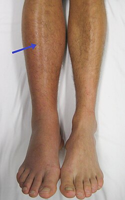
Superficial venous thromboses cause discomfort but generally not serious consequences, as do the deep vein thromboses (DVTs) that form in the deep veins of the legs or in the pelvic veins. Nevertheless, they can progress to the deep veins through the perforator veins or, they can be responsible for a lung embolism mainly if the head of the clot is poorly attached to the vein wall and is situated near the sapheno-femoral junction.[ citation needed ]

When a blood clot breaks loose and travels in the blood, this is called a thromboembolism. The abbreviation DVT/PE refers to a VTE where a deep vein thrombosis (DVT) has moved to the lungs (PE or pulmonary embolism). [4]

Since the veins return blood to the heart, if a piece of a blood clot formed in a vein breaks off it can be transported to the right side of the heart, and from there into the lungs. A piece of thrombus that is transported in this way is an embolus : the process of forming a thrombus that becomes embolic is called a thromboembolism. An embolism that lodges in the lungs is a pulmonary embolism (PE). A pulmonary embolism is a very serious condition that can be fatal depending on the dimensions of the embolus.[ citation needed ]

Rare forms

While venous thrombosis of the legs is the most common form, venous thrombosis may occur in other veins. These may have particular specific risk factors: [5]

Parodoxical embolism

Systemic embolism of venous origin can occur in patients with an atrial or ventricular septal defect, or an arteriovenous connection in the lung, through which an embolus may pass into the arterial system. Such an event is termed a paradoxical embolism. When this affects the blood vessels of the brain it can cause stroke. [6]

Causes

Venous thrombi are caused mainly by a combination of venous stasis and hypercoagulability—but to a lesser extent endothelial damage and activation. [7] The three factors of stasis, hypercoagulability, and alterations in the blood vessel wall represent Virchow's triad, and changes to the vessel wall are the least understood. [8] Various risk factors increase the likelihood of any one individual developing a thrombosis:

Risk factors

Acquired

Inherited

Mixed

The overall absolute risk of venous thrombosis per 100,000 woman years in current use of combined oral contraceptives is approximately 60, compared to 30 in non-users. [23] The risk of thromboembolism varies with different types of birth control pills; Compared with combined oral contraceptives containing levonorgestrel (LNG), and with the same dose of estrogen and duration of use, the rate ratio of deep vein thrombosis for combined oral contraceptives with norethisterone is 0.98, with norgestimate 1.19, with desogestrel (DSG) 1.82, with gestodene 1.86, with drospirenone (DRSP) 1.64, and with cyproterone acetate 1.88. [23] Venous thromboembolism occurs in 100–200 per 100,000 pregnant women every year. [23]

Regarding family history, age has substantial effect modification. For people with two or more affected siblings, the highest incidence rate is found among those ≥70 years of age (390 per 100,000 in men and 370 per 100,000 in women), whereas the highest incidence ratios compared to those without affected siblings occurred at much younger ages (ratio of 4.3 among men 20 to 29 years of age and 5.5 among women 10 to 19 years of age). [24]

Risk of venous thromboembolism (VTE) with hormone therapy and birth control (QResearch/CPRD)
TypeRouteMedications Odds ratio (95% CI Tooltip confidence interval)
Menopausal hormone therapy Oral Estradiol alone
    ≤1 mg/day
    >1 mg/day
1.27 (1.16–1.39)*
1.22 (1.09–1.37)*
1.35 (1.18–1.55)*
Conjugated estrogens alone
    ≤0.625 mg/day
    >0.625 mg/day
1.49 (1.39–1.60)*
1.40 (1.28–1.53)*
1.71 (1.51–1.93)*
Estradiol/medroxyprogesterone acetate 1.44 (1.09–1.89)*
Estradiol/dydrogesterone
    ≤1 mg/day E2
    >1 mg/day E2
1.18 (0.98–1.42)
1.12 (0.90–1.40)
1.34 (0.94–1.90)
Estradiol/norethisterone
    ≤1 mg/day E2
    >1 mg/day E2
1.68 (1.57–1.80)*
1.38 (1.23–1.56)*
1.84 (1.69–2.00)*
Estradiol/norgestrel or estradiol/drospirenone 1.42 (1.00–2.03)
Conjugated estrogens/medroxyprogesterone acetate 2.10 (1.92–2.31)*
Conjugated estrogens/norgestrel
    ≤0.625 mg/day CEEs
    >0.625 mg/day CEEs
1.73 (1.57–1.91)*
1.53 (1.36–1.72)*
2.38 (1.99–2.85)*
Tibolone alone1.02 (0.90–1.15)
Raloxifene alone1.49 (1.24–1.79)*
Transdermal Estradiol alone
   ≤50 μg/day
   >50 μg/day
0.96 (0.88–1.04)
0.94 (0.85–1.03)
1.05 (0.88–1.24)
Estradiol/progestogen 0.88 (0.73–1.01)
Vaginal Estradiol alone0.84 (0.73–0.97)
Conjugated estrogens alone1.04 (0.76–1.43)
Combined birth control Oral Ethinylestradiol/norethisterone 2.56 (2.15–3.06)*
Ethinylestradiol/levonorgestrel 2.38 (2.18–2.59)*
Ethinylestradiol/norgestimate 2.53 (2.17–2.96)*
Ethinylestradiol/desogestrel 4.28 (3.66–5.01)*
Ethinylestradiol/gestodene 3.64 (3.00–4.43)*
Ethinylestradiol/drospirenone 4.12 (3.43–4.96)*
Ethinylestradiol/cyproterone acetate 4.27 (3.57–5.11)*
Notes: (1) Nested case–control studies (2015, 2019) based on data from the QResearch and Clinical Practice Research Datalink (CPRD) databases. (2) Bioidentical progesterone was not included, but is known to be associated with no additional risk relative to estrogen alone. Footnotes: * = Statistically significant (p < 0.01). Sources: See template.
Absolute and relative incidence of venous thromboembolism (VTE) during pregnancy and the postpartum period
Absolute incidence of first VTE per 10,000 person–years during pregnancy and the postpartum period
Swedish data ASwedish data BEnglish dataDanish data
Time periodNRate (95% CI)NRate (95% CI)NRate (95% CI)NRate (95% CI)
Outside pregnancy11054.2 (4.0–4.4)10153.8 (?)14803.2 (3.0–3.3)28953.6 (3.4–3.7)
Antepartum99520.5 (19.2–21.8)69014.2 (13.2–15.3)1569.9 (8.5–11.6)49110.7 (9.7–11.6)
  Trimester 120713.6 (11.8–15.5)17211.3 (9.7–13.1)234.6 (3.1–7.0)614.1 (3.2–5.2)
  Trimester 227517.4 (15.4–19.6)17811.2 (9.7–13.0)305.8 (4.1–8.3)755.7 (4.6–7.2)
  Trimester 351329.2 (26.8–31.9)34019.4 (17.4–21.6)10318.2 (15.0–22.1)35519.7 (17.7–21.9)
Around delivery115154.6 (128.8–185.6)79106.1 (85.1–132.3)34142.8 (102.0–199.8)
Postpartum64942.3 (39.2–45.7)50933.1 (30.4–36.1)13527.4 (23.1–32.4)21817.5 (15.3–20.0)
  Early postpartum58475.4 (69.6–81.8)46059.3 (54.1–65.0)17746.8 (39.1–56.1)19930.4 (26.4–35.0)
  Late postpartum658.5 (7.0–10.9)496.4 (4.9–8.5)187.3 (4.6–11.6)3193.2 (1.9–5.0)
Incidence rate ratios (IRRs) of first VTE during pregnancy and the postpartum period
Swedish data ASwedish data BEnglish dataDanish data
Time periodIRR* (95% CI)IRR* (95% CI)IRR (95% CI)†IRR (95% CI)†
Outside pregnancy
Reference (i.e., 1.00)
Antepartum5.08 (4.66–5.54)3.80 (3.44–4.19)3.10 (2.63–3.66)2.95 (2.68–3.25)
  Trimester 13.42 (2.95–3.98)3.04 (2.58–3.56)1.46 (0.96–2.20)1.12 (0.86–1.45)
  Trimester 24.31 (3.78–4.93)3.01 (2.56–3.53)1.82 (1.27–2.62)1.58 (1.24–1.99)
  Trimester 37.14 (6.43–7.94)5.12 (4.53–5.80)5.69 (4.66–6.95)5.48 (4.89–6.12)
Around delivery37.5 (30.9–44.45)27.97 (22.24–35.17)44.5 (31.68–62.54)
Postpartum10.21 (9.27–11.25)8.72 (7.83–9.70)8.54 (7.16–10.19)4.85 (4.21–5.57)
  Early postpartum19.27 (16.53–20.21)15.62 (14.00–17.45)14.61 (12.10–17.67)8.44 (7.27–9.75)
  Late postpartum2.06 (1.60–2.64)1.69 (1.26–2.25)2.29 (1.44–3.65)0.89 (0.53–1.39)
Notes: Swedish data A = Using any code for VTE regardless of confirmation. Swedish data B = Using only algorithm-confirmed VTE. Early postpartum = First 6 weeks after delivery. Late postpartum = More than 6 weeks after delivery. * = Adjusted for age and calendar year. † = Unadjusted ratio calculated based on the data provided. Source: [25]

Pathophysiology

In contrast to the understanding for how arterial thromboses occur, as with heart attacks, venous thrombosis formation is not well understood. [26] With arterial thrombosis, blood vessel wall damage is required for thrombosis formation, as it initiates coagulation, [26] but the majority of venous thrombi form without any injured epithelium. [7]

Red blood cells and fibrin are the main components of venous thrombi, [7] and the thrombi appear to attach to the blood vessel wall endothelium, normally a non-thrombogenic surface, with fibrin. [26] Platelets in venous thrombi attach to downstream fibrin, while in arterial thrombi, they compose the core. [26] As a whole, platelets constitute less of venous thrombi when compared to arterial ones. [7] The process is thought to be initiated by tissue factor-affected thrombin production, which leads to fibrin deposition. [8]

The valves of veins are a recognized site of VT initiation. Due to the blood flow pattern, the base of the valve sinus is particularly deprived of oxygen (hypoxic). Stasis exacerbates hypoxia, and this state is linked to the activation of white blood cells (leukocytes) and the endothelium. Specifically, the two pathways of hypoxia-inducible factor-1 (HIF-1) and early growth response 1 (EGR-1) are activated by hypoxia, and they contribute to monocyte and endothelial activation. Hypoxia also causes reactive oxygen species (ROS) production that can activate HIF-1, EGR-1, and nuclear factor-κB (NF-κB), which regulates HIF-1 transcription. [8]

HIF-1 and EGR-1 pathways lead to monocyte association with endothelial proteins, such as P-selectin, prompting monocytes to release tissue factor-filled microvesicles, which presumably initiate fibrin deposition (via thrombin) after binding the endothelial surface. [8]

Diagnosis

Prevention

Numerous medications have been shown to reduce the risk of a person having a VTE, however careful decision making is required in order to decide if a person's risk of having a VTE outweighs the risks associated with most thromboprophylaxis treatment approaches (medications to prevent venous thrombosis). It is recommended that people should be assessed at their hospital discharge for persistent high-risk of venous thrombosis and that people who adopt a heart-healthy lifestyle might lower their risk of venous thrombosis. [27] Clinical policy from the American College of Physicians states a lack of support for any performance measures that incentivize physicians to apply universal prophylaxis without regard to the risks. [28]

Surgery

Evidence supports the use of heparin in people following surgery who have a high risk of thrombosis to reduce the risk of DVTs; however, the effect on PEs or overall mortality is not known. [29] In hospitalized non-surgical patients, mortality does not appear to change. [30] [31] [32] It does not appear, however, to decrease the rate of symptomatic DVTs. [30] Using both heparin and compression stockings appears better than either one alone in reducing the rate of DVT. [33]

Non-surgical medical conditions

In hospitalized people who have had a stroke and not had surgery, mechanical measures (compression stockings) resulted in skin damage and no clinical improvement. [30] Data on the effectiveness of compression stockings among hospitalized non-surgical patients without stroke is scarce. [30]

The American College of Physicians (ACP) gave three strong recommendations with moderate quality evidence on VTE prevention in non-surgical patients:

In adults who have had their lower leg casted, braced, or otherwise immobilized for more than a week, LMWH may decrease the risk and severity of deep vein thrombosis, but does not have any effect on the incidence of pulmonary embolism. [34]

Prior VTE

Following the completion of warfarin in those with prior VTE, the use of long-term aspirin has been shown to be beneficial. [35]

Cancer

People who have cancer have a higher risk of VTE and may respond differently to anticoagulant preventative treatments and prevention measures. [36] The American Society of Hematology strongly suggests that people undergoing chemotherapy for cancer who are at low risk of a VTE avoid medications to prevent thrombosis (thromboprophylaxis). [37] For people undergoing chemotherapy for cancer that do not require a hospital stay (those undergoing ambulatory care), there is low certainty evidence to suggest that treatment with direct factor Xa inhibitors may help prevent symptomatic VTEs, however this treatment approach may also lead to an increase in the risk of a major bleed compared to a placebo medication. [38] There is stronger evidence to suggest that LMWH helps prevent symptomatic VTE, however this treatment approach also comes with a higher risk of a major bleed compared to a placebo medication or no treatments to prevent VTE. [38]

For people who are having surgery for cancer, it is recommended that they receive anticoagulation therapy (preferably LMWH) in order to prevent a VTE. [39] LMWH is recommended for at least 7–10 days following cancer surgery, and for one month following surgery for people who have a high risk of VTEs. [40] [39]

Specifically for patients with various types of lymphoma, there is a risk assessment model, ThroLy, to help providers determine how likely a thromboembolic event is to occur. [41]

Treatment

American evidence-based clinical guidelines were published in 2016 for the treatment of VTE. [42] In the UK, guidelines by the National Institute for Health and Care Excellence (NICE) were published in 2012, updated in 2020. [43] These guidelines do not cover rare forms of thrombosis, for which an individualized approach is often needed. [5] Central and branch retinal vein occlusion does not benefit from anticoagulation in the way that other venous thromboses do. [5]

Anticoagulation

If diagnostic testing cannot be performed swiftly, many are commenced on empirical treatment. [43] Traditionally this was heparin, but several of the DOACs are licensed for treatment without initial heparin use. [42]

If heparin is used for initial treatment of VTE, fixed doses with low-molecular-weight heparin (LMWH) may be more effective than adjusted doses of unfractionated heparin (UFH) in reducing blood clots. [44] No differences in mortality, prevention of major bleeding, or preventing VTEs from recurring were observed between LMWH and UFH. [45] No differences have been detected in the route of administration of UFH (subcutaneous or intravenous). [44] LMWH is usually administered by a subcutaneous injection, and a person's blood clotting factors do not have to be monitored as closely as with UFH. [44]

Once the diagnosis is confirmed, a decision needs to be made about the nature of the ongoing treatment and its duration. USA recommendations for those without cancer include anticoagulation (medication that prevents further blood clots from forming) with the DOACs dabigatran, rivaroxaban, apixaban, or edoxaban rather than warfarin or low molecular weight heparin (LMWH). [42]

For those with cancer, LMWH is recommended, [42] although DOACs appear safe in the majority of situations. [43] For long-term treatment in people with cancer, LMWH is probably more effective at reducing VTEs when compared to vitamin K antagonists. [36] People with cancer have a higher risk of experiencing reoccurring VTE episodes ("recurrent VTE"), even while taking preventative anticoagulation medication. These people should be given therapeutic doses of LMWH medication, either by switching from another anticoagulant or by taking a higher dose of LMWH. [46]

In pregnancy, warfarin and DOACs are not considered suitable and LMWH is recommended. [42]

For those with a small pulmonary embolism and few risk factors, no anticoagulation is needed. [42] Anticoagulation is, however, recommended in those who do have risk factors. [42]

Thrombolysis

Thrombolysis is the administration of medication (a recombinant enzyme) that activates plasmin, the body's main enzyme that breaks down blood clots. This carries a risk of bleeding and is therefore reserved for those who have a form of thrombosis that may cause major complications. In pulmonary embolism, this applies in situations where heart function is compromised due to lack of blood flow through the lungs ("massive" or "high risk" pulmonary embolism), leading to low blood pressure. [42] Deep vein thrombosis may require thrombolysis if there is a significant risk of post-thrombotic syndrome. [42] Thrombolysis may be administered by intravenous catheter directly into the clot ("catheter-directed thrombolysis"); this requires a lower dose of the medication and may carry a lower bleeding risk but evidence for its benefit is limited. [42]

Inferior vena cava filters

Inferior vena cava filters (IVCFs) are not recommended in those who are on anticoagulants. [42] IVCFs may be used in clinical situations where a person has a high risk of experiencing a pulmonary embolism, but cannot be on anticoagulants due to a high risk of bleeding, or they have active bleeding. [46] [47] Retrievable IVCFs are recommended if IVCFs must be used, and a plan should be created to remove the filter when it is no longer needed. [46]

Superficial venous thrombosis

While topical treatments for superficial venous thrombosis are widely used, the evidence is strongest for the heparin-like drug fondaparinux (a factor Xa inhibitor), which reduces extension and recurrence of superficial venous thrombosis as well as progression to symptomatic embolism. [48]

Prognosis

After an episode of unprovoked VTE, the risk of further episodes after completing treatment remains elevated, although this risk diminishes over time. Over ten years, 41% of men and 29% of women can expect to experience a further episode. For each episode, the risk of death is 4%. [49]

See also

Related Research Articles

<span class="mw-page-title-main">Anticoagulant</span> Class of drugs

An anticoagulant, commonly known as a blood thinner, is a chemical substance that prevents or reduces coagulation of blood, prolonging the clotting time. Some of them occur naturally in blood-eating animals such as leeches and mosquitoes, where they help keep the bite area unclotted long enough for the animal to obtain some blood.

<span class="mw-page-title-main">Thrombosis</span> Medical condition caused by blood clots

Thrombosis is the formation of a blood clot inside a blood vessel, obstructing the flow of blood through the circulatory system. When a blood vessel is injured, the body uses platelets (thrombocytes) and fibrin to form a blood clot to prevent blood loss. Even when a blood vessel is not injured, blood clots may form in the body under certain conditions. A clot, or a piece of the clot, that breaks free and begins to travel around the body is known as an embolus.

<span class="mw-page-title-main">Thrombus</span> Blood clot

A thrombus, colloquially called a blood clot, is the final product of the blood coagulation step in hemostasis. There are two components to a thrombus: aggregated platelets and red blood cells that form a plug, and a mesh of cross-linked fibrin protein. The substance making up a thrombus is sometimes called cruor. A thrombus is a healthy response to injury intended to stop and prevent further bleeding, but can be harmful in thrombosis, when a clot obstructs blood flow through healthy blood vessels in the circulatory system.

<span class="mw-page-title-main">Pulmonary embolism</span> Blockage of an artery in the lungs

Pulmonary embolism (PE) is a blockage of an artery in the lungs by a substance that has moved from elsewhere in the body through the bloodstream (embolism). Symptoms of a PE may include shortness of breath, chest pain particularly upon breathing in, and coughing up blood. Symptoms of a blood clot in the leg may also be present, such as a red, warm, swollen, and painful leg. Signs of a PE include low blood oxygen levels, rapid breathing, rapid heart rate, and sometimes a mild fever. Severe cases can lead to passing out, abnormally low blood pressure, obstructive shock, and sudden death.

Factor V Leiden is a variant of human factor V, which causes an increase in blood clotting (hypercoagulability). Due to this mutation, protein C, an anticoagulant protein that normally inhibits the pro-clotting activity of factor V, is not able to bind normally to factor V, leading to a hypercoagulable state, i.e., an increased tendency for the patient to form abnormal and potentially harmful blood clots. Factor V Leiden is the most common hereditary hypercoagulability disorder amongst ethnic Europeans. It is named after the Dutch city of Leiden, where it was first identified in 1994 by Rogier Maria Bertina under the direction of Pieter Hendrick Reitsma. Despite the increased risk of venous thromboembolisms, people with one copy of this gene have not been found to have shorter lives than the general population. It is an autosomal dominant genetic disorder with incomplete penetrance.

<span class="mw-page-title-main">Deep vein thrombosis</span> Formation of a blood clot (thrombus) in a deep vein

Deep vein thrombosis (DVT) is a type of venous thrombosis involving the formation of a blood clot in a deep vein, most commonly in the legs or pelvis. A minority of DVTs occur in the arms. Symptoms can include pain, swelling, redness, and enlarged veins in the affected area, but some DVTs have no symptoms.

Low-molecular-weight heparin (LMWH) is a class of anticoagulant medications. They are used in the prevention of blood clots and treatment of venous thromboembolism and in the treatment of myocardial infarction.

<span class="mw-page-title-main">Thromboembolism</span> Obstruction of a blood vessel by a clot

Thromboembolism is a condition in which a blood clot (thrombus) breaks off from its original site and travels through the bloodstream to obstruct a blood vessel, causing tissue ischemia and organ damage. Thromboembolism can affect both the venous and arterial systems, with different clinical manifestations and management strategies.

<span class="mw-page-title-main">Thrombophilia</span> Abnormality of blood coagulation

Thrombophilia is an abnormality of blood coagulation that increases the risk of thrombosis. Such abnormalities can be identified in 50% of people who have an episode of thrombosis that was not provoked by other causes. A significant proportion of the population has a detectable thrombophilic abnormality, but most of these develop thrombosis only in the presence of an additional risk factor.

<span class="mw-page-title-main">Renal vein thrombosis</span> Medical condition

Renal vein thrombosis (RVT) is the formation of a clot in the vein that drains blood from the kidneys, ultimately leading to a reduction in the drainage of one or both kidneys and the possible migration of the clot to other parts of the body. First described by German pathologist Friedrich Daniel von Recklinghausen in 1861, RVT most commonly affects two subpopulations: newly born infants with blood clotting abnormalities or dehydration and adults with nephrotic syndrome.

An embolus, is described as a free-floating mass, located inside blood vessels that can travel from one site in the blood stream to another. An embolus can be made up of solid, liquid, or gas. Once these masses get "stuck" in a different blood vessel, it is then known as an "embolism." An embolism can cause ischemia—damage to an organ from lack of oxygen. A paradoxical embolism is a specific type of embolism in which the embolus travels from the right side of the heart to the left side of the heart and lodges itself in a blood vessel known as an artery. Thus, it is termed "paradoxical" because the embolus lands in an artery, rather than a vein.

<span class="mw-page-title-main">Dalteparin sodium</span> Pharmaceutical drug

Dalteparin is a low molecular weight heparin. It is marketed as Fragmin. Like other low molecular weight heparins, dalteparin is used for prophylaxis or treatment of deep vein thrombosis and pulmonary embolism to reduce the risk of a stroke or heart attack. Dalteparin acts by potentiating the activity of antithrombin III, inhibiting formation of both Factor Xa and thrombin. It is normally administered by self-injection.

<span class="mw-page-title-main">Post-thrombotic syndrome</span> Medical condition

Post-thrombotic syndrome (PTS), also called postphlebitic syndrome and venous stress disorder is a medical condition that may occur as a long-term complication of deep vein thrombosis (DVT).

Hypercoagulability in pregnancy is the propensity of pregnant women to develop thrombosis. Pregnancy itself is a factor of hypercoagulability, as a physiologically adaptive mechanism to prevent post partum bleeding. However, when combined with an additional underlying hypercoagulable states, the risk of thrombosis or embolism may become substantial.

Embolectomy is the emergency interventional or surgical removal of emboli which are blocking blood circulation. It usually involves removal of thrombi, and is then referred to as thromboembolectomy or thrombectomy. Embolectomy is an emergency procedure often as the last resort because permanent occlusion of a significant blood flow to an organ leads to necrosis. Other involved therapeutic options are anticoagulation and thrombolysis.

<span class="mw-page-title-main">Superficial thrombophlebitis</span> Medical condition

Superficial thrombophlebitis is a thrombosis and inflammation of superficial veins which presents as a painful induration (thickening) with erythema, often in a linear or branching configuration; forming a cord-like appearance.

Blood clots are a relatively common occurrence in the general population and are seen in approximately 1-2% of the population by age 60. Typically, blood clots develop in the deep veins of the lower extremities, deep vein thrombosis (DVT) or as a blood clot in the lung, pulmonary embolism. A very small number of people who develop blood clots have a more serious and often life-threatening condition, known as thrombotic storm (TS). TS is characterized by the development of more than one blood clot in a short period of time. These clots often occur in multiple and sometimes unusual locations in the body and are often difficult to treat. TS may be associated with an existing condition or situation that predisposes a person to blood clots, such as injury, infection, or pregnancy. In many cases, a risk assessment will identify interventions that will prevent the formation of blood clots.

Prothrombin G20210A is a genetic condition that increases the risk of blood clots including from deep vein thrombosis, and of pulmonary embolism. One copy of the mutation increases the risk of a blood clot from 1 in 1,000 per year to 2.5 in 1,000. Two copies increases the risk to up to 20 in 1,000 per year. Most people never develop a blood clot in their lifetimes.

<span class="mw-page-title-main">Superficial vein thrombosis</span> Medical condition

Superficial vein thrombosis (SVT) is a blood clot formed in a superficial vein, a vein near the surface of the body. Usually there is thrombophlebitis, which is an inflammatory reaction around a thrombosed vein, presenting as a painful induration with redness. SVT itself has limited significance when compared to a deep vein thrombosis (DVT), which occurs deeper in the body at the deep venous system level. However, SVT can lead to serious complications, and is therefore no longer regarded as a benign condition. If the blood clot is too near the saphenofemoral junction there is a higher risk of pulmonary embolism, a potentially life-threatening complication.

<span class="mw-page-title-main">Thrombosis prevention</span> Medical treatment

Thrombosis prevention or thromboprophylaxis is medical treatment to prevent the development of thrombosis in those considered at risk for developing thrombosis. Some people are at a higher risk for the formation of blood clots than others, such as those with cancer undergoing a surgical procedure. Prevention measures or interventions are usually begun after surgery as the associated immobility will increase a person's risk.

References

  1. Ortel, TL; Neumann, I; Ageno, W; et al. (13 October 2020). "American Society of Hematology 2020 guidelines for management of venous thromboembolism: treatment of deep vein thrombosis and pulmonary embolism". Blood Advances. 4 (19): 4693–4738. doi:10.1182/bloodadvances.2020001830. PMC   7556153 . PMID   33007077.
  2. Heit JA, Spencer FA, White RH (January 2016). "The epidemiology of venous thromboembolism". Journal of Thrombosis and Thrombolysis. 41 (1): 3–14. doi:10.1007/s11239-015-1311-6. PMC   4715842 . PMID   26780736.
  3. 1 2 Abbattista M, Capecchi M, Martinelli I (January 2020). "Treatment of unusual thrombotic manifestations". Blood. 135 (5): 326–334. doi: 10.1182/blood.2019000918 . PMID   31917405.
  4. National Clinical Guideline Centre – Acute and Chronic Conditions (UK) (2010). "Venous Thromboembolism: Reducing the Risk of Venous Thromboembolism (Deep Vein Thrombosis and Pulmonary Embolism) in Patients Admitted to Hospital". PMID   23346611.{{cite journal}}: Cite journal requires |journal= (help)
  5. 1 2 3 Shatzel, Joseph J.; O'Donnell, Matthew; Olson, Sven R.; Kearney, Matthew R.; Daughety, Molly M.; Hum, Justine; Nguyen, Khanh P.; DeLoughery, Thomas G. (January 2019). "Venous thrombosis in unusual sites: A practical review for the hematologist". European Journal of Haematology. 102 (1): 53–62. doi: 10.1111/ejh.13177 . ISSN   0902-4441. PMID   30267448.
  6. Windecker, Stephan; Stortecky, Stefan; Meier, Bernhard (July 2014). "Paradoxical Embolism". Journal of the American College of Cardiology. 64 (4): 403–415. doi: 10.1016/j.jacc.2014.04.063 . PMID   25060377.
  7. 1 2 3 4 5 6 7 8 9 10 11 12 13 14 15 16 Martinelli I, Bucciarelli P, Mannucci PM (2010). "Thrombotic risk factors: basic pathophysiology". Crit Care Med. 38 (suppl 2): S3–9. doi:10.1097/CCM.0b013e3181c9cbd9. PMID   20083911. S2CID   34486553.
  8. 1 2 3 4 5 6 7 Bovill EG, van der Vliet A (2011). "Venous valvular stasis-associated hypoxia and thrombosis: what is the link?". Annu Rev Physiol. 73: 527–45. doi:10.1146/annurev-physiol-012110-142305. PMID   21034220.
  9. 1 2 3 4 5 6 7 8 9 Rosendaal FR, Reitsma PH (2009). "Genetics of venous thrombosis". J. Thromb. Haemost. 7 (suppl 1): 301–4. doi: 10.1111/j.1538-7836.2009.03394.x . PMID   19630821. S2CID   27104496.
  10. Khan NR, Patel PG, Sharpe JP, Lee SL, Sorenson J (2018). "Chemical venous thromboembolism prophylaxis in neurosurgical patients: an updated systematic review and meta-analysis". Journal of Neurosurgery. 129 (4): 906–915. doi: 10.3171/2017.2.JNS162040 . PMID   29192859. S2CID   37464528. Patients requiring cranial and spinal surgery present a unique situation of elevated risk for VTE but also high risk for disastrous outcomes should bleeding complications occur in eloquent areas of the brain or spinal cord. Open Access logo PLoS transparent.svg
  11. Stein PD, Beemath A, Meyers FA, et al. (2006). "Incidence of venous thromboembolism in patients hospitalized with cancer". Am J Med. 119 (1): 60–8. doi:10.1016/j.amjmed.2005.06.058. PMID   16431186.
  12. Jackson E, Curtis KM, Gaffield ME (2011). "Risk of venous thromboembolism during the postpartum period: a systematic review". Obstet Gynecol. 117 (3): 691–703. doi:10.1097/AOG.0b013e31820ce2db. PMID   21343773. S2CID   12561.
  13. Varga EA, Kujovich JL (2012). "Management of inherited thrombophilia: guide for genetics professionals". Clin Genet. 81 (1): 7–17. doi:10.1111/j.1399-0004.2011.01746.x. PMID   21707594. S2CID   9305488.
  14. Turpie AGG (March 2008). "Deep Venous Thrombosis". The Merck's Manuals Online Medical Library. Merck.
  15. Beyer-Westendorf J, Bauersachs R, Hach-Wunderle V, Zotz RB, Rott H. Sex hormones and venous thromboembolism - from contraception to hormone replacement therapy. Vasa. 2018 Jul 16:1-10.2018/07/17
  16. Reitsma PH, Versteeg HH, Middeldorp S (2012). "Mechanistic view of risk factors for venous thromboembolism". Arterioscler Thromb Vasc Biol. 32 (3): 563–8. doi: 10.1161/ATVBAHA.111.242818 . PMID   22345594. S2CID   2624599.
  17. Zöller B, Li X, Sundquist J, et al. (2012). "Risk of pulmonary embolism in patients with autoimmune disorders: a nationwide follow-up study from Sweden". Lancet. 379 (9812): 244–9. doi:10.1016/S0140-6736(11)61306-8. PMID   22119579. S2CID   11612703.
  18. 1 2 3 4 5 6 Lijfering WM, Rosendaal FR, Cannegieter SC (2010). "Risk factors for venous thrombosis – current understanding from an epidemiological point of view". Br J Haematol. 149 (6): 824–33. doi: 10.1111/j.1365-2141.2010.08206.x . PMID   20456358. S2CID   41684138.
  19. Mandalà, M; Falanga, A; Roila, F; ESMO Guidelines Working, Group. (September 2011). "Management of venous thromboembolism (VTE) in cancer patients: ESMO Clinical Practice Guidelines". Annals of Oncology. 22 (Suppl 6): vi85–92. doi: 10.1093/annonc/mdr392 . PMID   21908511.
  20. Tang L, Wu YY, Lip GY, Yin P, Hu Y (January 2016). "Heart failure and risk of venous thromboembolism: a systematic review and meta-analysis". Lancet Haematol. 3 (1): e30–44. doi:10.1016/S2352-3026(15)00228-8. PMID   26765646.
  21. Dentali F, Sironi AP, Ageno W, et al. (2012). "Non-O Blood Type Is the Commonest Genetic Risk Factor for VTE: Results from a Meta-Analysis of the Literature". Semin. Thromb. Hemost. 38 (5): 535–48. doi:10.1055/s-0032-1315758. PMID   22740183. S2CID   5203474.
  22. Jenkins PV, Rawley O, Smith OP, et al. (2012). "Elevated factor VIII levels and risk of venous thrombosis". Br J Haematol. 157 (6): 653–63. doi: 10.1111/j.1365-2141.2012.09134.x . PMID   22530883. S2CID   24467063.
  23. 1 2 3 Eichinger, S.; Evers, J. L. H.; Glasier, A.; La Vecchia, C.; Martinelli, I.; Skouby, S.; Somigliana, E.; Baird, D. T.; Benagiano, G.; Crosignani, P. G.; Gianaroli, L.; Negri, E.; Volpe, A.; Glasier, A.; Crosignani, P. G. (2013). "Venous thromboembolism in women: A specific reproductive health risk". Human Reproduction Update. 19 (5): 471–482. doi: 10.1093/humupd/dmt028 . PMID   23825156.
  24. Eikelboom, J. W.; Weitz, J. I. (2011). "Importance of family history as a risk factor for venous thromboembolism". Circulation. 124 (9): 996–7. doi: 10.1161/CIRCULATIONAHA.111.048868 . PMID   21875920.
  25. Abdul Sultan A, West J, Stephansson O, Grainge MJ, Tata LJ, Fleming KM, Humes D, Ludvigsson JF (November 2015). "Defining venous thromboembolism and measuring its incidence using Swedish health registries: a nationwide pregnancy cohort study". BMJ Open. 5 (11): e008864. doi:10.1136/bmjopen-2015-008864. PMC   4654387 . PMID   26560059.
  26. 1 2 3 4 López JA, Chen J (2009). "Pathophysiology of venous thrombosis". Thromb Res. 123 (Suppl 4): S30–4. doi:10.1016/S0049-3848(09)70140-9. PMID   19303501.
  27. Goldhaber, Samuel Z. (2010). "Risk Factors for Venous Thromboembolism". Journal of the American College of Cardiology. 56 (1): 1–7. doi: 10.1016/j.jacc.2010.01.057 . PMID   20620709.
  28. 1 2 Qaseem A, Chou R, Humphrey LL, Starkey M, Shekelle P (November 2011). "Venous thromboembolism prophylaxis in hospitalized patients: a clinical practice guideline from the American College of Physicians". Ann. Intern. Med. 155 (9): 625–32. CiteSeerX   10.1.1.689.591 . doi:10.7326/0003-4819-155-9-201111010-00011. PMID   22041951. S2CID   7129943.
  29. Roderick, P; Ferris, G; Wilson, K; Halls, H; Jackson, D; Collins, R; Baigent, C (December 2005). "Towards evidence-based guidelines for the prevention of venous thromboembolism: systematic reviews of mechanical methods, oral anticoagulation, dextran and regional anaesthesia as thromboprophylaxis". Health Technology Assessment. 9 (49): iii–iv, ix–x, 1–78. doi: 10.3310/hta9490 . PMID   16336844.
  30. 1 2 3 4 Lederle, FA; Zylla, D; Macdonald, R; Wilt, TJ (2011-11-01). "Venous thromboembolism prophylaxis in hospitalized medical patients and those with stroke: a background review for an american college of physicians clinical practice guideline". Annals of Internal Medicine. 155 (9): 602–15. doi:10.7326/0003-4819-155-9-201111010-00008. PMID   22041949. S2CID   207536371.
  31. Alikhan, R; Bedenis, R; Cohen, AT (7 May 2014). "Heparin for the prevention of venous thromboembolism in acutely ill medical patients (excluding stroke and myocardial infarction)". The Cochrane Database of Systematic Reviews. 2018 (5): CD003747. doi:10.1002/14651858.CD003747.pub4. PMC   6491079 . PMID   24804622.
  32. "[120] Routine VTE prophylaxis: Is there a net health benefit?". Therapeutics Initiative. 16 July 2019.
  33. Zareba, P; Wu, C; Agzarian, J; Rodriguez, D; Kearon, C (Aug 2014). "Meta-analysis of randomized trials comparing combined compression and anticoagulation with either modality alone for prevention of venous thromboembolism after surgery". The British Journal of Surgery. 101 (9): 1053–62. doi: 10.1002/bjs.9527 . PMID   24916118. S2CID   37373926.
  34. Zee, AA; van Lieshout, K; van der Heide, M; Janssen, L; Janzing, HM (Apr 25, 2017). "Low molecular weight heparin for prevention of venous thromboembolism in patients with lower-leg immobilization". The Cochrane Database of Systematic Reviews. 2017 (8): CD006681. doi:10.1002/14651858.CD006681.pub4. PMC   6483324 . PMID   28780771.
  35. Simes, J; Becattini, C; Agnelli, G; Eikelboom, JW; Kirby, AC; Mister, R; Prandoni, P; Brighton, TA; INSPIRE Study Investigators (International Collaboration of Aspirin Trials for Recurrent Venous, Thromboembolism) (23 September 2014). "Aspirin for the prevention of recurrent venous thromboembolism: the INSPIRE collaboration". Circulation. 130 (13): 1062–71. doi: 10.1161/circulationaha.114.008828 . PMID   25156992.
  36. 1 2 Kahale, Lara A.; Hakoum, Maram B.; Tsolakian, Ibrahim G.; Matar, Charbel F.; Terrenato, Irene; Sperati, Francesca; Barba, Maddalena; Yosuico, Victor Ed; Schünemann, Holger (2018). "Anticoagulation for the long-term treatment of venous thromboembolism in people with cancer". The Cochrane Database of Systematic Reviews. 2022 (6): CD006650. doi:10.1002/14651858.CD006650.pub5. ISSN   1469-493X. PMC   6389342 . PMID   29920657.
  37. Lyman, Gary H.; Carrier, Marc; Ay, Cihan; Di Nisio, Marcello; Hicks, Lisa K.; Khorana, Alok A.; Leavitt, Andrew D.; Lee, Agnes Y. Y.; Macbeth, Fergus; Morgan, Rebecca L.; Noble, Simon (2021-02-23). "American Society of Hematology 2021 guidelines for management of venous thromboembolism: prevention and treatment in patients with cancer". Blood Advances. 5 (4): 927–974. doi:10.1182/bloodadvances.2020003442. ISSN   2473-9537. PMC   7903232 . PMID   33570602.
  38. 1 2 Rutjes, Anne Ws; Porreca, Ettore; Candeloro, Matteo; Valeriani, Emanuele; Di Nisio, Marcello (2020-12-18). "Primary prophylaxis for venous thromboembolism in ambulatory cancer patients receiving chemotherapy". The Cochrane Database of Systematic Reviews. 2020 (12): CD008500. doi:10.1002/14651858.CD008500.pub5. ISSN   1469-493X. PMC   8829903 . PMID   33337539.
  39. 1 2 Mandalà, M.; Falanga, A.; Roila, F.; ESMO Guidelines Working Group (2011-09-01). "Management of venous thromboembolism (VTE) in cancer patients: ESMO Clinical Practice Guidelines". Annals of Oncology. 22 (Suppl 6): vi85–92. doi: 10.1093/annonc/mdr392 . ISSN   1569-8041. PMID   21908511.
  40. Christensen, Thomas D.; Vad, Henrik; Pedersen, Søren; Hvas, Anne-Mette; Wotton, Robin; Naidu, Babu; Larsen, Torben B. (2014-02-01). "Venous thromboembolism in patients undergoing operations for lung cancer: a systematic review". The Annals of Thoracic Surgery. 97 (2): 394–400. doi: 10.1016/j.athoracsur.2013.10.074 . ISSN   1552-6259. PMID   24365217.
  41. Antic, Darko; Milic, Natasa; Nikolovski, Srdjan; Todorovic, Milena; Bila, Jelena; Djurdjevic, Predrag; Andjelic, Bosko; Djurasinovic, Vladislava; Sretenovic, Aleksandra; Vukovic, Vojin; Jelicic, Jelena; Hayman, Suzanne; Mihaljevic, Biljana (July 2016). "Development and validation of multivariable predictive model for thromboembolic events in lymphoma patients". American Journal of Hematology. 91 (10): 1014–1019. doi:10.1002/ajh.24466. ISSN   0361-8609.
  42. 1 2 3 4 5 6 7 8 9 10 11 Kearon, C; Akl, EA; Ornelas, J; Blaivas, A; Jimenez, D; Bounameaux, H; Huisman, M; King, CS; Morris, TA; Sood, N; Stevens, SM; Vintch, JR; Wells, P; Woller, SC; Moores, L (February 2016). "Antithrombotic Therapy for VTE Disease: CHEST Guideline and Expert Panel Report". Chest. 149 (2): 315–52. doi:10.1016/j.chest.2015.11.026. PMID   26867832.
  43. 1 2 3 "Venous thromboembolic diseases: diagnosis, management and thrombophilia testing". www.nice.org.uk. National Institute for Health and Care Excellence. 2020. Retrieved 2020-08-31.
  44. 1 2 3 Robertson, Lindsay; Jones, Lauren E. (2017-02-09). "Fixed dose subcutaneous low molecular weight heparins versus adjusted dose unfractionated heparin for the initial treatment of venous thromboembolism". The Cochrane Database of Systematic Reviews. 2017 (2): CD001100. doi:10.1002/14651858.CD001100.pub4. ISSN   1469-493X. PMC   6464611 . PMID   28182249.
  45. Robertson L, Strachan J (February 2017). "Subcutaneous unfractionated heparin for the initial treatment of venous thromboembolism". Cochrane Database Syst Rev. 2 (11): CD006771. doi:10.1002/14651858.CD006771.pub3. PMC   6464347 . PMID   28195640.
  46. 1 2 3 Khorana, Alok A.; Carrier, Marc; Garcia, David A.; Lee, Agnes Y. Y. (2016-01-01). "Guidance for the prevention and treatment of cancer-associated venous thromboembolism". Journal of Thrombosis and Thrombolysis. 41 (1): 81–91. doi:10.1007/s11239-015-1313-4. ISSN   1573-742X. PMC   4715852 . PMID   26780740.
  47. Rajasekhar, Anita (2015-04-01). "Inferior vena cava filters: current best practices". Journal of Thrombosis and Thrombolysis. 39 (3): 315–327. doi:10.1007/s11239-015-1187-5. ISSN   1573-742X. PMID   25680894. S2CID   5868257.
  48. Di Nisio, Marcello; Wichers, Iris M; Middeldorp, Saskia (2018-02-25). Cochrane Vascular Group (ed.). "Treatment for superficial thrombophlebitis of the leg". Cochrane Database of Systematic Reviews. 2018 (2): CD004982. doi:10.1002/14651858.CD004982.pub6. PMC   6491080 . PMID   29478266.
  49. Khan, Faizan; Rahman, Alvi; Carrier, Marc; Kearon, Clive; Weitz, Jeffrey I; Schulman, Sam; Couturaud, Francis; Eichinger, Sabine; Kyrle, Paul A (2019-07-24). "Long term risk of symptomatic recurrent venous thromboembolism after discontinuation of anticoagulant treatment for first unprovoked venous thromboembolism event: systematic review and meta-analysis". BMJ. 366: l4363. doi:10.1136/bmj.l4363. PMC   6651066 . PMID   31340984.