Telangiectasia

Last updated
Telangiectasia
Other namesSpider veins, angioectasias
Dermoscopy nodular basal cell carcinoma.jpg
Telangiectasia as seen in a basal-cell carcinoma
Pronunciation
Specialty Dermatology

Telangiectasias, from Greek: tel- (end) + angi- (blood or lymph vessel) + ectasia (the expansion of a hollow or tubular organ), also known as spider veins, are small dilated blood vessels [1] that can occur near the surface of the skin or mucous membranes, measuring between 0.5 and 1 millimeter in diameter. [2] These dilated blood vessels can develop anywhere on the body, but are commonly seen on the face around the nose, cheeks and chin. Dilated blood vessels can also develop on the legs, although when they occur on the legs, they often have underlying venous reflux or "hidden varicose veins" (see Venous hypertension section below). When found on the legs, they are found specifically on the upper thigh, below the knee joint and around the ankles.

Contents

Many patients with spider veins seek the assistance of physicians who specialize in vein care or peripheral vascular disease. These physicians are called vascular surgeons or phlebologists. More recently, interventional radiologists have started treating venous problems.

Some telangiectasias are due to developmental abnormalities that can closely mimic the behaviour of benign vascular neoplasms. They may be composed of abnormal aggregations of arterioles, capillaries or venules. Because telangiectasias are vascular lesions, they blanch when tested with diascopy.

Telangiectasias, aside from presenting in many other conditions, are one of the features of the acronymically named CREST syndrome, a form of systemic scleroderma. The syndrome recognises the significantly co-presenting symptoms of calcinosis, Raynaud's phenomenon, esophageal dysmotility, sclerodactyly and telangiectasia.

Causes

The causes of telangiectasia can be divided into congenital and acquired factors.

Genetic

Goldman states that "numerous inherited or congenital conditions display cutaneous telangiectasia". [2] These include:

Venous hypertension

In the past, it was believed that leg varicose veins or telangectasia were caused by high venous pressure or "venous hypertension". However it is now understood that venous reflux disease is usually the cause of these problems. [4] [ full citation needed ]

Telangiectasia in the legs is often related to the presence of venous reflux within underlying varicose veins. Flow abnormalities in smaller veins known as reticular veins or feeder veins under the skin can also cause the spider veins to form, thereby making a recurrence of the spider veins in the treated area less likely.

Factors that predispose to the development of varicose and telangiectatic leg veins include

Other acquired causes

Acquired telangiectasia, not related to other venous abnormalities, for example on the face and trunk, can be caused by factors such as

Treatment

Before any treatment of leg telangectasia (spider veins) is considered, it is essential to have duplex ultrasonography, the test that has replaced Doppler ultrasound. The reason for this is that there is a clear association between leg telangectasia (spider veins) and underlying venous reflux. [12] Research has shown that 88–89% of women with telangectasia (spider veins) have refluxing reticular veins close, [13] and 15% have incompetent perforator veins nearby. [14] As such, it is essential to both find and treat underlying venous reflux before considering any treatment at all.

Sclerotherapy is the "gold standard" and is preferred over laser for eliminating telangiectasiae and smaller varicose leg veins. [15] A sclerosant medication is injected into the diseased vein so it hardens and eventually shrinks away. Recent evidence with foam sclerotherapy shows that the foam containing the irritating sclerosant quickly appears in the patient's heart and lungs, and then in some cases travels through a patent foramen ovale to the brain. [16] This has led to concerns about the safety of sclerotherapy for telangectasias and spider veins.

In some cases stroke and transient ischemic attacks have occurred after sclerotherapy. [17] Varicose veins and reticular veins are often treated before treating telangiectasia, although treatment of these larger veins in advance of sclerotherapy for telangiectasia may not guarantee better results. [18] [19] [20] Varicose veins can be treated with foam sclerotherapy, endovenous laser treatment, radiofrequency ablation, or open surgery. The biggest risk, however, seems to occur with sclerotherapy, especially in terms of systemic risk of DVT, pulmonary embolism, and stroke.[ citation needed ] Other issues which arise with the use of sclerotherapy to treat spider veins are staining, shadowing, telangetatic matting, and ulceration. In addition, incompleteness of therapy is common, requiring multiple treatment sessions. [21]

Telangiectasias on the face are often treated with a laser. Laser therapy uses a light beam that is pulsed onto the veins in order to seal them off, causing them to dissolve. These light-based treatments require adequate heating of the veins. These treatments can result in the destruction of sweat glands, and the risk increases with the number of treatments.[ citation needed ]

Related Research Articles

<span class="mw-page-title-main">Varicose veins</span> Medical condition in which superficial veins become large and twisted

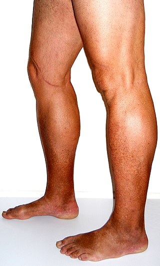
Varicose veins, also known as varicoses, are a medical condition in which superficial veins become enlarged and twisted. These veins typically develop in the legs, just under the skin. Varicose veins usually cause few symptoms. However, some individuals may experience fatigue or pain in the area. Complications can include bleeding or superficial thrombophlebitis. Varices in the scrotum are known as a varicocele, while those around the anus are known as hemorrhoids. Due to the various physical, social, and psychological effects of varicose veins, they can negatively affect one's quality of life.

<span class="mw-page-title-main">Vein</span> Blood vessels that carry blood towards the heart

Veins are blood vessels in the circulatory system of humans and most other animals that carry blood towards the heart. Most veins carry deoxygenated blood from the tissues back to the heart; exceptions are those of the pulmonary and fetal circulations which carry oxygenated blood to the heart. In the systemic circulation, arteries carry oxygenated blood away from the heart, and veins return deoxygenated blood to the heart, in the deep veins.

<span class="mw-page-title-main">Vascular surgery</span> Medical specialty, operative procedures for the treatment of vascular disorders

Vascular surgery is a surgical subspecialty in which vascular diseases involving the arteries, veins, or lymphatic vessels, are managed by medical therapy, minimally-invasive catheter procedures and surgical reconstruction. The specialty evolved from general and cardiovascular surgery where it refined the management of just the vessels, no longer treating the heart or other organs. Modern vascular surgery includes open surgery techniques, endovascular techniques and medical management of vascular diseases - unlike the parent specialities. The vascular surgeon is trained in the diagnosis and management of diseases affecting all parts of the vascular system excluding the coronaries and intracranial vasculature. Vascular surgeons also are called to assist other physicians to carry out surgery near vessels, or to salvage vascular injuries that include hemorrhage control, dissection, occlusion or simply for safe exposure of vascular structures.

Stasis dermatitis refers to the skin changes that occur in the leg as a result of "stasis" or blood pooling from insufficient venous return; the alternative name of varicose eczema comes from a common cause of this being varicose veins.

Endovenous laser treatment (ELT) is a minimally invasive ultrasound-guided technique used for treating varicose veins using laser energy commonly performed by a phlebologist, interventional radiologist or vascular surgeon.

<span class="mw-page-title-main">Sclerotherapy</span> Shrinking of the varicose blood vessels by the injection of medicine

Sclerotherapy is a procedure used to treat blood vessel malformations and also malformations of the lymphatic system. A medication is injected into the vessels, which makes them shrink. It is used for children and young adults with vascular or lymphatic malformations. In adults, sclerotherapy is often used to treat spider veins, smaller varicose veins, hemorrhoids, and hydroceles.

Polidocanol is a local anaesthetic and antipruritic component of ointments and bath additives. It relieves itching caused by eczema and dry skin. It has also been used to treat varicose veins, hemangiomas, and vascular malformations. It is formed by the ethoxylation of dodecanol.

The small saphenous vein is a relatively large superficial vein of the posterior leg.

<span class="mw-page-title-main">Venous ulcer</span> Medical condition

Venous ulcer is defined by the American Venous Forum as "a full-thickness defect of skin, most frequently in the ankle region, that fails to heal spontaneously and is sustained by chronic venous disease, based on venous duplex ultrasound testing." Venous ulcers are wounds that are thought to occur due to improper functioning of venous valves, usually of the legs. They are an important cause of chronic wounds, affecting 1% of the population. Venous ulcers develop mostly along the medial distal leg, and can be painful with negative effects on quality of life.

<span class="mw-page-title-main">Vascular disease</span> Medical condition

Vascular disease is a class of diseases of the vessels of the circulatory system in the body, including blood vessels – the arteries and veins, and the lymphatic vessels. Vascular disease is a subgroup of cardiovascular disease. Disorders in this vast network of blood and lymph vessels can cause a range of health problems that can sometimes become severe, and fatal. Coronary heart disease for example, is the leading cause of death for men and women in the United States.

<span class="mw-page-title-main">Compression stockings</span> Compression garment

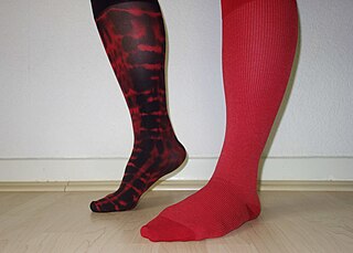
Compression stockings are a specialized hosiery designed to help prevent the occurrence of, and guard against further progression of, venous disorders such as edema, phlebitis and thrombosis. Compression stockings are elastic compression garments worn around the leg, compressing the limb. This reduces the diameter of distended veins and increases venous blood flow velocity and valve effectiveness. Compression therapy helps decrease venous pressure, prevents venous stasis and impairments of venous walls, and relieves heavy and aching legs.

<span class="mw-page-title-main">Venous lake</span> Type of papule usually on the lip, face or ears and among the elderly

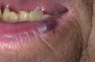
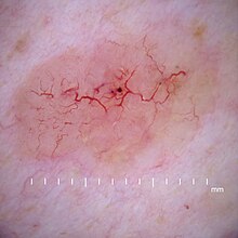
A venous lake is a generally solitary, soft, compressible, dark blue to violaceous, 0.2- to 1-cm papule commonly found on sun-exposed surfaces of the vermilion border of the lip, face and ears. Lesions generally occur among the elderly.

<span class="mw-page-title-main">Chronic venous insufficiency</span> Medical condition

Chronic venous insufficiency (CVI) is a medical condition in which blood pools in the veins, straining the walls of the vein. The most common cause of CVI is superficial venous reflux which is a treatable condition. As functional venous valves are required to provide for efficient blood return from the lower extremities, this condition typically affects the legs. If the impaired vein function causes significant symptoms, such as swelling and ulcer formation, it is referred to as chronic venous disease. It is sometimes called chronic peripheral venous insufficiency and should not be confused with post-thrombotic syndrome in which the deep veins have been damaged by previous deep vein thrombosis.

<span class="mw-page-title-main">Ovarian vein syndrome</span> Medical condition

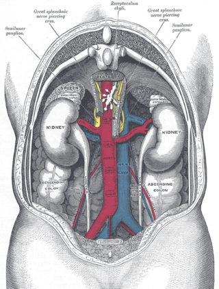
Ovarian vein syndrome is a rare condition in which a dilated ovarian vein compresses the ureter. This causes chronic or colicky abdominal pain, back pain and/or pelvic pain. The pain can worsen on lying down or between ovulation and menstruation. There can also be an increased tendency towards urinary tract infection or pyelonephritis. The right ovarian vein is most commonly involved, although the disease can be left-sided or affect both sides. It is currently classified as a form of pelvic congestion syndrome.

<span class="mw-page-title-main">Klippel–Trénaunay syndrome</span> Medical condition

Klippel–Trénaunay syndrome, formerly Klippel–Trénaunay–Weber syndrome and sometimes angioosteohypertrophy syndrome and hemangiectatic hypertrophy, is a rare congenital medical condition in which blood vessels and/or lymph vessels fail to form properly. The three main features are nevus flammeus, venous and lymphatic malformations, and soft-tissue hypertrophy of the affected limb. It is similar to, though distinctly separate from, the less common Parkes Weber syndrome.

A vascular anomaly is any of a range of lesions from a simple birthmark to a large tumor that may be disfiguring. They are caused by a disorder of the vascular system. A vascular anomaly is a localized defect in blood or lymph vessels. These defects are characterized by an increased number of vessels, and vessels that are both enlarged and sinuous. Some vascular anomalies are congenital, others appear within weeks to years after birth, and others are acquired by trauma or during pregnancy. Inherited vascular anomalies are also described and often present with a number of lesions that increase with age. Vascular anomalies can also be a part of a syndrome.

<span class="mw-page-title-main">Pelvic congestion syndrome</span> Medical condition

Pelvic congestion syndrome, also known as pelvic vein incompetence, is a long-term condition believed to be due to enlarged veins in the lower abdomen. The condition may cause chronic pain, such as a constant dull ache, which can be worsened by standing or sex. Pain in the legs or lower back may also occur.

<span class="mw-page-title-main">Mitchel P. Goldman</span> American dermatologic surgeon (born 1955)

Mitchel P. Goldman, is an American dermatologic surgeon, cosmetic surgeon, dermatologist, and phlebologist, and the founder and director of Cosmetic Laser Dermatology. He is also a past president of the American Society for Dermatologic Surgery, the American College of Phlebology, the San Diego County Dermatology Society, and the Sonoran Dermatology Society.

<span class="mw-page-title-main">Perforator vein</span>

Perforator veins are so called because they perforate the deep fascia of muscles, to connect the superficial veins to the deep veins where they drain.

CLaCS is a treatment for leg vein lesions by combining transdermal laser effect and injection sclerotherapy, all under skin cooling. The laser causes a selective photothermolysis damaging the vein wall. The vein's lumen gets smaller. On a second procedure, sclerosing agent is injected where the vein is still open. This combination can be used treat veins that could be treated by phleboectomy or foam sclerotherapy - more invasive options. To improve results, CLaCS can be guided by Augmented Reality.

References

  1. "telangiectasia" "at Dorland's Medical Dictionary
  2. 1 2 Goldman, Mitchel P (1995). Sclerotherapy treatment of varicose and telangiectatic leg veins (2nd ed.). St. Louis: Mosby. ISBN   0-8151-4011-8.[ page needed ]
  3. Irrthum, Alexandre; Devriendt, Koenraad; Chitayat, David; Matthijs, Gert; Glade, Conrad; Steijlen, Peter M.; Fryns, Jean-Pierre; Van Steensel, Maurice A. M.; Vikkula, Miikka (2003). "Mutations in the Transcription Factor Gene SOX18 Underlie Recessive and Dominant Forms of Hypotrichosis-Lymphedema-Telangiectasia". The American Journal of Human Genetics. 72 (6): 1470–8. doi:10.1086/375614. PMC   1180307 . PMID   12740761.
  4. Whiteley (2011). "Understanding Venous Reflux – the cause of varicose veins and venous leg ulcers".
  5. Ruckley, C.V.; Evans, C.J.; Allan, P.L.; Lee, A.J.; Fowkes, F.G.R. (2008). "Telangiectasia in the Edinburgh Vein Study: Epidemiology and Association with Trunk Varices and Symptoms". European Journal of Vascular and Endovascular Surgery. 36 (6): 719–24. doi: 10.1016/j.ejvs.2008.08.012 . PMID   18848475.
  6. Ismail, Lars; Normahani, Pasha; Standfield, Nigel J.; Jaffer, Usman (2016). "A systematic review and meta-analysis of the risk for development of varicose veins in women with a history of pregnancy". Journal of Vascular Surgery. Venous and Lymphatic Disorders. Elsevier BV. 4 (4): 518–524.e1. doi: 10.1016/j.jvsv.2016.06.003 . ISSN   2213-333X. PMID   27639009.
  7. Smyth, Rebecca MD; Aflaifel, Nasreen; Bamigboye, Anthony A; Pregnancy, Cochrane; Group, Childbirth (2021-06-02). "Interventions for varicose veins and leg oedema in pregnancy". The Cochrane Database of Systematic Reviews. 2015 (10): CD001066. doi:10.1002/14651858.CD001066.pub3. PMC   7050615 . PMID   26477632.
  8. "Varicose veins - Symptoms and causes". Mayo Clinic. Retrieved 2020-08-11.
  9. Lindsley, Kristina; Matsumura, Sueko; Hatef, Elham; Akpek, Esen K (2012). "Interventions for chronic blepharitis". Cochrane Database of Systematic Reviews. 5 (5): CD005556. doi:10.1002/14651858.CD005556.pub2. PMC   4270370 . PMID   22592706.
  10. 1 2 3 Kennedy, Cornelis; Bastiaens, Maarten T.; Willemze, Rein; Bouwes Bavinck, Jan N.; Bajdik, Chris D.; Westendorp, Rudi G.J. (April 2003). "Effect of Smoking and Sun on the Aging Skin". Journal of Investigative Dermatology. 120 (4): 548–554. doi: 10.1046/j.1523-1747.2003.12092.x . PMID   12648216.
  11. Johnson, B. A.; Nunley, J. R. (2000). "Treatment of seborrheic dermatitis". American Family Physician. 61 (9): 2703–10, 2713–4. PMID   10821151.
  12. Ruckley, C. V.; Allan, P. L.; Evans, C. J.; Lee, A. J.; Fowkes, F. G. R. (2011). "Telangiectasia and venous reflux in the Edinburgh Vein Study". Phlebology. 27 (6): 297–302. doi:10.1258/phleb.2011.011007. PMID   22106449. S2CID   29067831.
  13. Weiss, Robert A.; Weiss, Margaret A. (1993). "Doppler Ultrasound Findings in Reticular Veins of the Thigh Subdermic Lateral Venous System and Implications for Sclerotherapy". The Journal of Dermatologic Surgery and Oncology. 19 (10): 947–51. doi:10.1111/j.1524-4725.1993.tb00983.x. PMID   8408914.
  14. Somjen, George M.; Ziegenbein, Robert; Johnston, Andrew H.; Royle, John P. (1993). "Anatomical Examination of Leg Telangiectases with Duplex Scanning". The Journal of Dermatologic Surgery and Oncology. 19 (10): 940–5. doi:10.1111/j.1524-4725.1993.tb00982.x. PMID   8408913.
  15. Sadick N, Sorhaindo L (2007). "16. Laser Treatment of Telangiectatic and Reticular Veins". In Bergan, John J. (ed.). The Vein Book. Amsterdam: Elsevier Academic Press. p. 157. ISBN   978-0-12-369515-4.
  16. Ceulen, Roeland P.M.; Sommer, Anja; Vernooy, Kevin (2008). "Microembolism during Foam Sclerotherapy of Varicose Veins". New England Journal of Medicine. 358 (14): 1525–6. doi: 10.1056/NEJMc0707265 . PMID   18385510.
  17. Forlee, Martin V.; Grouden, Maria; Moore, Dermot J.; Shanik, Gregor (2006). "Stroke after varicose vein foam injection sclerotherapy". Journal of Vascular Surgery. 43 (1): 162–4. doi: 10.1016/j.jvs.2005.09.032 . PMID   16414404.
  18. Duffy, David M. (2012). "Sclerotherapy for Telangiectasia – The impact of small changes in vessel size on treatment outcomes" (PDF). Cosmetic Dermatology. 25 (3): 126–33. Archived from the original (PDF) on 2019-12-15.
  19. Treatment of Leg Veins. Procedures in Cosmetic Dermatology Series. Editors Murad Alam, Sirunya Silapunt. Second Edition Saunders Elsevier Inc. 2011[ page needed ]
  20. Schuller-Petrovic, S.; Pavlovic, M. D.; Schuller, S.; Schuller-Lukic, B.; Adamic, M. (2012). "Telangiectasias resistant to sclerotherapy are commonly connected to a perforating vessel". Phlebology. 28 (6): 320–3. doi:10.1258/phleb.2012.012019. PMID   22865418. S2CID   36994668.
  21. Goldman, Mitchel P.; Bennett, Richard G. (1987-08-01). "Treatment of telangiectasia: A review". Journal of the American Academy of Dermatology. 17 (2): 167–182. doi:10.1016/s0190-9622(87)70187-x. ISSN   0190-9622. PMID   3305603.