Renal vein thrombosis

Last updated
Renal vein thrombosis
Other namesBlood clot in the renal vein [1]
Gray1122.png
The anterior surfaces of the kidneys, showing the areas of contact of neighboring viscera.
Specialty Nephrology   OOjs UI icon edit-ltr-progressive.svg

Renal vein thrombosis (RVT) is the formation of a clot in the vein that drains blood from the kidneys, ultimately leading to a reduction in the drainage of one or both kidneys and the possible migration of the clot to other parts of the body. [1] First described by German pathologist Friedrich Daniel von Recklinghausen in 1861, RVT most commonly affects two subpopulations: newly born infants with blood clotting abnormalities or dehydration and adults with nephrotic syndrome. [2]

Contents

Nephrotic syndrome, a kidney disorder, causes excessive loss of protein in the urine, low levels of albumin in the blood, a high level of cholesterol in the blood and swelling, triggering a hypercoagulable state and increasing chances of clot formation. Other less common causes include hypercoagulable state, cancer, kidney transplantation, Behcet syndrome, antiphospholipid antibody syndrome or blunt trauma to the back or abdomen. [3]

Treatment of RVT mainly focuses on preventing further blood clots in the kidneys and maintaining stable kidney function. The use of anticoagulants has become the standard treatment in treating this abnormality. [2] Membranous glomerulonephritis, the most common cause for nephrotic syndrome in adults, peaks in people ages 40–60 years old and it is twice as likely to occur in men than in women. Since nephrotic syndrome is the most common cause of RVT, people over 40 years old and men are most at risk to develop a renal vein thrombosis. [3]

Signs and symptoms

Aside from the occasional flank or lower back pain caused by a sudden clot in the major veins to the kidneys, RVT produces few symptoms. Some patients may not display any symptoms while other patients may experience bloody urine, decrease in urine output, edema and worsening proteinuria. Usually the diagnoses of RVT is first made when a nephrotic syndrome patient experiences a pulmonary embolism or a sudden decrease in kidney function or kidney failure. These symptoms may vary in duration since a blood clot can resolve itself, but precautions should be taken to prevent the migration of the clot to other parts of the body. The most severe complication of RVT is a pulmonary embolism, caused by a clot, also called a thrombus, that originates from the renal vein or any other vein in the body and migrates to the pulmonary artery. A pulmonary embolism is a serious condition because; it can damage the lungs due to pulmonary hypertension and cause low blood oxygen, damaging other organs in the body. This condition can cause death if left untreated; about 30% percent of patients who have a pulmonary embolism will die, usually within one hour. [4] [5]

Infants and young children experiencing dehydration induced RVT, may experience dehydration symptoms (dry mouth, low urine output, loss of skin turgidity) as while as vomiting, nausea and fever, and the usual RVT symptoms like flank pain, blood in the urine, anaemia, edema, enlarged kidneys and kidney failure. [6]

Mechanism

The mechanism behind RVT is no different from other types of blood clots in other parts of the body. Rudolf Virchow, was the first to describe the physiological mechanism behind venous thrombosis (blood clots) using three related factors, known as Virchow's Triad; damage to the blood vessel (endothelial damage), decrease in blood flow (stasis) and increased coagulability of the blood (thrombophilia or hypercoagulability). it is possible for one of these factors alone to cause a blood clot, but in most cases, a combination or all of these factors induce the formation of a blood clot. Decreased urine output or kidney function may be the only observable symptoms caused by a blood clot renal vein. Other less common causes include hypercoagulable state, invasion by renal cell carcinoma, kidney transplantation, Behcet syndrome, antiphospholipid antibody syndrome or blunt trauma to the back or abdomen. [3]

Vein tissue damage

Damage to the endothelial tissue of the vein can be caused by blunt damage, trauma during venography, a renal transplant, tumors, acute rejection, vasculitis or spontaneous micro-trauma to the endothelium due to homocystinuria. [6] Cystathionine beta synthase deficiency, also known as homocystinuria, is an autosomal recessive inherited disorder in which the body is not able to process certain building blocks of proteins correctly due to a mutation to the CBS gene. This mutation causes the amino acid homocystine not to be used properly thus high levels build up in the blood, damaging the endothelial tissue and increasing the likelihood of RVT. [7]

Decreased blood flow

The most common cause of RVT in infants is dehydration. Dehydration may be caused by reduction in both volume and circulatory blood volume due to water depleting abnormalities like diarrhea or vomiting. The decrease in blood volume due to dehydration will cause blood flow to be diverted away from the kidneys to other organs, resulting in slower blood flow to the kidneys, increasing chances of a blood clot occurrence. "RVT is known to occur in the absence of clinically obvious shock e.g. following neonatal distress and placement of central venous catheters." RVT can also be induced by post transplant distortion or physical distortion or compression of the renal vein, which depending on the shape distortion can affect the rate of flow through the vein. [6]

Hypercoagulability

Hypercoagulability is an abnormality of the blood that increases the risk of the formation blood clots. Nephrotic syndrome patients have a higher risk of RVT development due to hypercoagulability caused by proteinuria. The increased loss of proteins in the urine caused by nephrotic syndrome results in lower osmotic pressure. Reduced osmotic pressure will trigger the liver to produce more proteins like fibrinogen and beta-thromboglobulin, which promote blood clotting. Other than nephrotic syndrome, there are many other factors that can promote hypercoagulability. Hypercoagulability can be promoted by increased platelet count, enhanced platelet aggregation, increased protein S count, and a decrease in coagulation inhibiters like antithrombin. [6] Hypercoagulability can be inherited and/or acquired. Hyperhomocysteinemia, a condition known to promote clots, can be caused by a combination of genetic factors and vitamin B6, vitamin B12 and folic acid deficiency. [8] Factor V Leiden and mutations of the prothrombin gene are the two most common genetic causes of hypercoagulability. About 5% of the general population have these heterozygous mutations and in the thrombophilic population, 45–63% have these mutations. [6]

Membranous glomerulonephritis

The incidence of RVT in people with Nephrotic syndrome ranges from 5% to 65%. Nephrotic syndrome is caused by membranous glomerulonephritis, [6] minimal change disease, and focal segmental glomerulosclerosis.[ citation needed ]

Diagnosis

CT showing dilatation and thrombosis of the left renal vein in a patient with nutcracker syndrome NutCracker2.PNG
CT showing dilatation and thrombosis of the left renal vein in a patient with nutcracker syndrome

There are no laboratory tests used to diagnose RVT.[ citation needed ]

Observing the patient's symptoms, medical history and imaging remain the fundamental source for diagnosing RVT. Imaging is used to detect the presence of a blood clot. In an abnormal kidney with RVT, a blood clot is present in the renal vein. In cases where the renal vein is suddenly and/or fully blocked, the kidneys will enlarge, reaching its maximum size within a week. An ultrasound imaging can be used to observe and track the size of the kidneys in RVT patients. Ultrasound is not efficient for use in detecting blood flow in the renal veins and artery. Instead a color doppler ultrasound may be used to detect renal blood flow. It is most commonly used to detect RVT in patients who have undergone renal transplantation. CT angiography is currently the top choice in diagnosing RVT. It is non-invasive, relatively cheap and fast with high accuracy. CT scanning can be used to detect renal enlargement, renal tumors, blood flow and other renal pathologies. An alternative is magnetic resonance angiography or MRA. It is non-invasive, fast and avoids radiation (unlike a CT scan) but it is relatively expensive. MRA produces detailed images of the renal blood flow, vesicle walls, the kidneys and any surrounding tissue. An inferior venocavography with selective venography can be used to rule out the diagnoses of RVT. [6]

Treatment

Surgery to remove the clot is possible, but rarely performed. In the past, surgical removal of the renal vein clot was the primary treatment but it is very invasive and many complications can occur. In the past decades, treatment has shifted its focus from surgical intervention to medical treatments that include intravenous and oral anticoagulants. The use of anticoagulants may improve kidney function in RVT cases by removing the clot in the vein and preventing further clots from occurring. Patients who already have nephrotic syndrome may not need to take anticoagulants. In this case, patients should keep an eye out and maintain reduced level of proteinuria by reducing salt and excess protein, and intaking diuretics and statins. Depending on the severity of RVT, patients may be on anticoagulants from a year up to a lifetime. As long as the albumin levels in the bloodstream are below 2.5g/L, it is recommended that RVT patients continue taking anticoagulants. Main anticoagulants that can be used to treat RVT include warfarin and low molecular weight heparin. Heparin has become very popular, because of its low risk of complications, its availability and because it can easily be administered. Warfarin is known to interact with many other drugs, so careful monitoring is required. [6] If a nephrotic syndrome patient experiences any of the RVT symptoms (flank or back pain, blood in the urine or decreased kidney function), he or she should immediately see a doctor to avoid further complications.[ citation needed ]

The main side effect of anticoagulants is the risk of excessive bleeding. Other side effects include: blood in the urine or feces, severe bruising, prolonged nosebleeds (lasting longer than 10 minutes), bleeding gum, blood in your vomit or coughing up blood, unusual headaches, sudden severe back pain, difficulty breathing or chest pain, in women, heavy or increased bleeding during the period, or any other bleeding from the vagina. Warfarin can cause rashes, diarrhea, nausea (feeling sick) or vomiting, and hair loss. Heparin can cause hair loss (alopecia) thrombocytopenia – a sudden drop in the number of platelets in the blood. [9]

It has been reported in a case study of 27 patients with nephrotic syndrome caused RVT, there was a 40% mortality rate, mostly due to hemorrhagic complications and sepsis. In 75% of the remaining surviving patients, the RVT was resolved and kidney function returned to normal. It has been concluded that age is not a factor on the survival of RVT patients, although older patient (55 and older) are more likely to develop kidney failure. Heparin is crucial in returning normal kidney function; in patients that did not take heparin, long term kidney damage was observed in 100%. In patients that did take heparin, kidney damage was observed in about 33%. By quickly treating, and receiving the correct medications, patients should increase their chances of survival and reduce the risk of the renal vein clot from migrating to another part of the body. [6]

Research

It is known that diabetes causes changes to factors associated with coagulation and clotting, however not much is known of the risk of thromboembolism, or clots, in diabetic patients. There are some studies that show that diabetes increases the risk of thromboembolism; other studies show that diabetes does not increase the risk of thromboembolism. A study conducted in the Umeå University Hospital, in Sweden, observed patients that were hospitalized due to an thromboembolism from 1997 to 1999. The researchers had access to patient information including age, sex, vein thromboembolism diagnosis, diagnostic methods, diabetes type and medical history. This study concluded that there is, in fact, an increased risk of thromboembolism development in diabetic patients, possibly due to factors associated with diabetes or diabetes itself. Diabetic patients are twice as likely to develop a thromboembolism than are non-diabetic patient. The exact mechanism of how diabetes increases the risk of clot formation remains unclear and could possibly be a future direction for study. [10]

From previous studies, it is known that long-distance air travel is associated with high risk of venous thrombosis. Long periods of inactivity in a limited amount of space may be a reason for the increased risk of blood clot formation. In addition, bent knees compresses the vein behind the knee (the popliteal vein) and the low humidity, low oxygen, high cabin pressure and consumption of alcohol concentrate the blood. [11] A recent study, published in the British Journal of Haematology in 2014, determined which groups of people, are most at risk for developing a clot during or after a long flight. The study focused on 8755 frequent flying employees from international companies and organizations. It found that travelers who have recently undergone a surgical procedure or who have a malignant disease such as cancer or who are pregnant are most at risk. Preventative measures before flying may be taken in these at-risk groups as a solution. [12]

Patients who have undergone kidney transplant have a high risk of developing RVT (about 0.4% to 6%). RVT is known to account for a large proportion of transplanted kidney failures due to technical problems (damage to the renal vein), clotting disorders, diabetes, consumption of ciclosporin or an unknown problem. Patients who have undergone a kidney transplant are commonly prescribed ciclosporin, an immunosuppressant drug which is known to reduce renal blood flow, increase platelet aggregation in the blood and cause damage to the endothelial tissue of the veins. In a clinical study conducted by the Nuffield Department of Surgery at the Oxford Transplant Centre, UK, transplant patients were given low doses of aspirin, which has a some anti-platelet activity. There is risk of bleeding in transplant patients when using anticoagulants like warfarin and herapin. Low dosage of aspirin was used as an alternative. The study concluded that a routine low-dose of aspirin in kidney transplant patients who are also taking ciclosporin significantly reduces the risk of RVT development. [13]

See also

Related Research Articles

<span class="mw-page-title-main">Anticoagulant</span> Class of drugs

Anticoagulants, commonly known as blood thinners, are chemical substances that prevent or reduce coagulation of blood, prolonging the clotting time. Some of them occur naturally in blood-eating animals such as leeches and mosquitoes, where they help keep the bite area unclotted long enough for the animal to obtain some blood. As a class of medications, anticoagulants are used in therapy for thrombotic disorders. Oral anticoagulants (OACs) are taken by many people in pill or tablet form, and various intravenous anticoagulant dosage forms are used in hospitals. Some anticoagulants are used in medical equipment, such as sample tubes, blood transfusion bags, heart–lung machines, and dialysis equipment. One of the first anticoagulants, warfarin, was initially approved as a rodenticide.

<span class="mw-page-title-main">Thrombosis</span> Medical condition caused by blood clots

Thrombosis is the formation of a blood clot inside a blood vessel, obstructing the flow of blood through the circulatory system. When a blood vessel is injured, the body uses platelets (thrombocytes) and fibrin to form a blood clot to prevent blood loss. Even when a blood vessel is not injured, blood clots may form in the body under certain conditions. A clot, or a piece of the clot, that breaks free and begins to travel around the body is known as an embolus.

<span class="mw-page-title-main">Pulmonary embolism</span> Blockage of an artery in the lungs

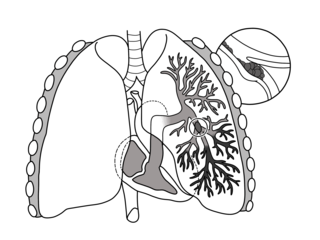
Pulmonary embolism (PE) is a blockage of an artery in the lungs by a substance that has moved from elsewhere in the body through the bloodstream (embolism). Symptoms of a PE may include shortness of breath, chest pain particularly upon breathing in, and coughing up blood. Symptoms of a blood clot in the leg may also be present, such as a red, warm, swollen, and painful leg. Signs of a PE include low blood oxygen levels, rapid breathing, rapid heart rate, and sometimes a mild fever. Severe cases can lead to passing out, abnormally low blood pressure, obstructive shock, and sudden death.

<span class="mw-page-title-main">Venous thrombosis</span> Blood clot (thrombus) that forms within a vein

Venous thrombosis is blockage of a vein caused by a thrombus. A common form of venous thrombosis is deep vein thrombosis (DVT), when a blood clot forms in the deep veins. If a thrombus breaks off (embolizes) and flows to the lungs to lodge there, it becomes a pulmonary embolism (PE), a blood clot in the lungs. The conditions of DVT only, DVT with PE, and PE only, are all captured by the term venous thromboembolism (VTE).

Factor V Leiden is a variant of human factor V, which causes an increase in blood clotting (hypercoagulability). Due to this mutation, protein C, an anticoagulant protein that normally inhibits the pro-clotting activity of factor V, is not able to bind normally to factor V, leading to a hypercoagulable state, i.e., an increased tendency for the patient to form abnormal and potentially harmful blood clots. Factor V Leiden is the most common hereditary hypercoagulability disorder amongst ethnic Europeans. It is named after the Dutch city of Leiden, where it was first identified in 1994 by Rogier Maria Bertina under the direction of Pieter Hendrick Reitsma. Despite the increased risk of venous thromboembolisms, people with one copy of this gene have not been found to have shorter lives than the general population.

<span class="mw-page-title-main">Nephrotic syndrome</span> Collection of symptoms due to kidney damage

Nephrotic syndrome is a collection of symptoms due to kidney damage. This includes protein in the urine, low blood albumin levels, high blood lipids, and significant swelling. Other symptoms may include weight gain, feeling tired, and foamy urine. Complications may include blood clots, infections, and high blood pressure.

<span class="mw-page-title-main">Ischemia</span> Restriction in blood supply to tissues

Ischemia or ischaemia is a restriction in blood supply to any tissue, muscle group, or organ of the body, causing a shortage of oxygen that is needed for cellular metabolism. Ischemia is generally caused by problems with blood vessels, with resultant damage to or dysfunction of tissue i.e. hypoxia and microvascular dysfunction. It also implies local hypoxia in a part of a body resulting from constriction. Ischemia causes not only insufficiency of oxygen, but also reduced availability of nutrients and inadequate removal of metabolic wastes. Ischemia can be partial or total blockage. The inadequate delivery of oxygenated blood to the organs must be resolved either by treating the cause of the inadequate delivery or reducing the oxygen demand of the system that needs it. For example, patients with myocardial ischemia have a decreased blood flow to the heart and are prescribed with medications that reduce chronotrophy and ionotrophy to meet the new level of blood delivery supplied by the stenosed vasculature so that it is adequate.

<span class="mw-page-title-main">Deep vein thrombosis</span> Formation of a blood clot (thrombus) in a deep vein

Deep vein thrombosis (DVT) is a type of venous thrombosis involving the formation of a blood clot in a deep vein, most commonly in the legs or pelvis. A minority of DVTs occur in the arms. Symptoms can include pain, swelling, redness, and enlarged veins in the affected area, but some DVTs have no symptoms. The most common life-threatening concern with DVT is the potential for a clot to embolize, travel as an embolus through the right side of the heart, and become lodged in a pulmonary artery that supplies blood to the lungs. This is called a pulmonary embolism (PE). DVT and PE comprise the cardiovascular disease of venous thromboembolism (VTE). About two-thirds of VTE manifests as DVT only, with one-third manifesting as PE with or without DVT. The most frequent long-term DVT complication is post-thrombotic syndrome, which can cause pain, swelling, a sensation of heaviness, itching, and in severe cases, ulcers. Recurrent VTE occurs in about 30% of those in the ten years following an initial VTE.

Low-molecular-weight heparin (LMWH) is a class of anticoagulant medications. They are used in the prevention of blood clots and treatment of venous thromboembolism and in the treatment of myocardial infarction.

<span class="mw-page-title-main">Budd–Chiari syndrome</span> Medical condition

Budd–Chiari syndrome is a very rare condition, affecting one in a million adults. The condition is caused by occlusion of the hepatic veins that drain the liver. The symptoms are non-specific and vary widely, but it may present with the classical triad of abdominal pain, ascites, and liver enlargement. It is usually seen in younger adults, with the median age at diagnosis between the ages of 35 and 40, and it has a similar incidence in males and females. The syndrome can be fulminant, acute, chronic, or asymptomatic. Subacute presentation is the most common form.

<span class="mw-page-title-main">Thrombophilia</span> Abnormality of blood coagulation

Thrombophilia is an abnormality of blood coagulation that increases the risk of thrombosis. Such abnormalities can be identified in 50% of people who have an episode of thrombosis that was not provoked by other causes. A significant proportion of the population has a detectable thrombophilic abnormality, but most of these develop thrombosis only in the presence of an additional risk factor.

<span class="mw-page-title-main">Dalteparin sodium</span> Pharmaceutical drug

Dalteparin is a low molecular weight heparin. It is marketed as Fragmin. Like other low molecular weight heparins, dalteparin is used for prophylaxis or treatment of deep vein thrombosis and pulmonary embolism to reduce the risk of a stroke or heart attack. Dalteparin acts by potentiating the activity of antithrombin III, inhibiting formation of both Factor Xa and thrombin. It is normally administered by self-injection.

Congenital nephrotic syndrome is a rare kidney disease which manifests in infants during the first 3 months of life, and is characterized by high levels of protein in the urine (proteinuria), low levels of protein in the blood, and swelling. This disease is primarily caused by genetic mutations which result in damage to components of the glomerular filtration barrier and allow for leakage of plasma proteins into the urinary space.

Hypercoagulability in pregnancy is the propensity of pregnant women to develop thrombosis. Pregnancy itself is a factor of hypercoagulability, as a physiologically adaptive mechanism to prevent post partum bleeding. However, when combined with an additional underlying hypercoagulable states, the risk of thrombosis or embolism may become substantial.

<span class="mw-page-title-main">Cerebral venous sinus thrombosis</span> Presence of a blood clot in the dural venous sinuses or cerebral veins

Cerebral venous sinus thrombosis (CVST), cerebral venous and sinus thrombosis or cerebral venous thrombosis (CVT), is the presence of a blood clot in the dural venous sinuses, the cerebral veins, or both. Symptoms may include severe headache, visual symptoms, any of the symptoms of stroke such as weakness of the face and limbs on one side of the body, and seizures, which occur in around 40% of patients.

<span class="mw-page-title-main">Renal biopsy</span> Removal of kidney tissue for medical examination

Renal biopsy is a medical procedure in which a small piece of kidney is removed from the body for examination, usually under a microscope. Microscopic examination of the tissue can provide information needed to diagnose, monitor or treat problems of the kidney.

<span class="mw-page-title-main">Arterial embolism</span> Interruption of blood flow to an organ

Arterial embolism is a sudden interruption of blood flow to an organ or body part due to an embolus adhering to the wall of an artery blocking the flow of blood, the major type of embolus being a blood clot (thromboembolism). Sometimes, pulmonary embolism is classified as arterial embolism as well, in the sense that the clot follows the pulmonary artery carrying deoxygenated blood away from the heart. However, pulmonary embolism is generally classified as a form of venous embolism, because the embolus forms in veins. Arterial embolism is the major cause of infarction.

Blood clots are a relatively common occurrence in the general population and are seen in approximately 1-2% of the population by age 60. Typically, blood clots develop in the deep veins of the lower extremities, deep vein thrombosis (DVT) or as a blood clot in the lung, pulmonary embolism. A very small number of people who develop blood clots have a more serious and often life-threatening condition, known as thrombotic storm (TS). TS is characterized by the development of more than one blood clot in a short period of time. These clots often occur in multiple and sometimes unusual locations in the body and are often difficult to treat. TS may be associated with an existing condition or situation that predisposes a person to blood clots, such as injury, infection, or pregnancy. In many cases, a risk assessment will identify interventions that will prevent the formation of blood clots.

Prothrombin G20210A is a genetic condition that increases the risk of blood clots including from deep vein thrombosis, and of pulmonary embolism. One copy of the mutation increases the risk of a blood clot from 1 in 1,000 per year to 2.5 in 1,000. Two copies increases the risk to up to 20 in 1,000 per year. Most people never develop a blood clot in their lifetimes.

<span class="mw-page-title-main">Thrombosis prevention</span> Medical treatment

Thrombosis prevention or thromboprophylaxis is medical treatment to prevent the development of thrombosis in those considered at risk for developing thrombosis. Some people are at a higher risk for the formation of blood clots than others, such as those with cancer undergoing a surgical procedure. Prevention measures or interventions are usually begun after surgery as the associated immobility will increase a person's risk.

References

  1. 1 2 "Renal vein thrombosis: MedlinePlus Medical Encyclopedia". medlineplus.gov. Retrieved 27 May 2019.
  2. 1 2 Wessels, Hunter; McAninch, Jack W., eds. (2005). Handbook of Urological Emergencies: A Practical Guide. Totowa, N.J.: Humana Press. pp. 171–180. ISBN   978-1-58829-256-8.
  3. 1 2 3 Laskowski, Igor. "Renal Vein Thrombosis". Archived from the original on 31 March 2014. Retrieved 30 March 2014.
  4. "What is a Pulmonary Embolism". National Heart, Lung and Blood Institute. NIH. Retrieved 1 April 2014.
  5. Harrington, John T; Jerome Kassirer (1982). "Renal Vein Thrombosis". Annual Review of Medicine. 33: 255–62. doi:10.1146/annurev.me.33.020182.001351. PMID   7081962.
  6. 1 2 3 4 5 6 7 8 9 Asghar, M.; Ahmed, K.; Shah, S.S.; Siddique, M.K.; Dasgupta, P.; Khan, M.S. (August 2007). "Renal Vein Thrombosis". European Journal of Vascular and Endovascular Surgery. 34 (2): 217–223. doi: 10.1016/j.ejvs.2007.02.017 . PMID   17543556.
  7. "Homocystinuria". Genetics Home Reference. National Institute of Health. Retrieved 30 March 2014.
  8. Deitcher, Seven. "Hypercoagulable States". Disease Management Project. Cleveland Clinic Center for Continuing Education. Retrieved 31 March 2014.
  9. "Anticoagulant medicines - Side effects". NHS Choices. NHS. Retrieved 1 April 2014.
  10. Petrauskiene, V.; Falk, M.; Waernbaum, I.; Norberg, M.; Eriksson, J. W. (19 March 2005). "The risk of venous thromboembolism is markedly elevated in patients with diabetes". Diabetologia. 48 (5): 1017–1021. doi: 10.1007/s00125-005-1715-5 . PMID   15778859.
  11. Kos, Cynthia. "Air Travel and Deep Vein Thrombosis DVT". Stop the Clot Alliance. Retrieved 29 April 2014.
  12. Kuipers, Saskia; Venemans, Annemarie; Middeldorp, Saskia; Büller, Harry R.; Cannegieter, Suzanne C.; Rosendaal, Frits R. (May 2014). "The risk of venous thrombosis after air travel: contribution of clinical risk factors". British Journal of Haematology. 165 (3): 412–413. doi: 10.1111/bjh.12724 . PMID   24428564.
  13. Robertson, Amanda; Vinod Nargund; Derek W.R. Gray; Peter J. Morris (2000). "Low dose aspirin as prophylaxis against renal-vein thrombosis renal-transplant recipients". Nephrology Dialysis Transplantation. 15 (11): 1865–1868. doi: 10.1093/ndt/15.11.1865 . PMID   11071979.