Gray platelet syndrome

Last updated
Gray platelet syndrome
Other namesPlatelet alpha-granule deficiency
Autosomal recessive - en.svg
Gray platelet syndrome is inherited in an autosomal recessive manner.
Specialty Hematology   OOjs UI icon edit-ltr-progressive.svg

Gray platelet syndrome (GPS), or platelet alpha-granule deficiency, [1] is a rare congenital autosomal recessive bleeding disorder caused by a reduction or absence of alpha-granules in blood platelets, and the release of proteins normally contained in these granules into the marrow, causing myelofibrosis. [2] The name derives from the initial observation of gray appearance of platelets with a paucity of granules on blood films from a patient with a lifelong bleeding disorder. [3]

Contents

Signs and symptoms

Signs of GPS typically arise at birth or in childhood, these signs and symptoms include thrombocytopenia, bruising susceptibility, and epistaxis. Typically, the observed low platelet count in individuals is progressive, this can result in fatal hemorrhages later in life. Additionally, females who are affected may experience irregular menstrual cycles and heavy menstrual bleeding. [4] Another common effect of GPS is myelofibrosis, where scar tissue builds up in the bone marrow causing it to be unable to produce a sufficient amount of blood cells. To compensate, other organs such as the spleen start to produce more blood cells which can lead to splenomegaly. [5]

Genetics

GPS is primarily inherited in an autosomal recessive manner. The gene that is mutated in GPS has been mapped to chromosome 3p [6] and identified as NBEAL2 . [7] The location is 3p21.31. [8] This is caused by a homozygous or compound heterozygous mutations in the NBEAL2 gene. [9] NBEAL2 encodes a protein containing a BEACH domain that is predicted to be involved in vesicular trafficking. It is expressed in platelets and megakaryocytes and is required for the development of platelet alpha-granules. [10] NBEAL2 expression is also required for the development of thrombocytes in zebrafish. [11]

In Japan, GPS was found in 24 affected patients in a single family. There was at least 1 instance of male-to-male transmission, consistent with autosomal dominant inheritance. [12]

A study reported of 14 families with GPS, 11 families had clear autosomal recessive inheritance. This was evidenced by consanguinity or multiple affected siblings with unaffected parents. The families had various backgrounds, including Bedouin, Turkish, Mennonite, French, German, Somali, African American, and mixed Northern and Southern European. [13]

GPS is characterized by "thrombocytopenia, and abnormally large agranular platelets in peripheral blood smears." [14] The defect in GPS is the failure of megakaryocytes to package secretory proteins into alpha-granules. Patients with the GPS are affected by mild to moderate bleeding tendencies. Usually these are not major bleeds but there has been some life-threatening cases. Affected women will tend to have heavy, irregular periods. Myelofibrosis is a condition that usually comes with GPS. [15]

Diagnosis

GPS is diagnosed based on clinical findings and requires demonstration of absence or marked reduction of alpha-granules by electron microscopy (EM). [16] High serum vitamin B12 levels are common in patients diagnosed with GPS. Bone marrow sampling, though not required for diagnosis of GPS, might be necessary to evaluate myelofibrosis occurring in GPS and to exclude other disorders. [16]

Management

There is no specific treatment  for GPS, but it can be managed by anticipating and preventing the risks of bleeding. The treatment options include desmopressin. [17] Splenectomy can be used to increase the platelet counts in those whose platelet counts decrease to approximately 30,000/microliter. [18] Prognosis is generally good early in life when thrombocytopenia is mild. [19]

Epidemiology

Currently, only 60 cases have been recorded world wide. While the prevalence of this syndrome is not known, it was found that it affects males and females equally. [20]

See also

Related Research Articles

Thrombocytopenia Medical condition

Thrombocytopenia is a condition characterized by abnormally low levels of platelets, also known as thrombocytes, in the blood. It is the most common coagulation disorder among intensive care patients and is seen in 20% of medical patients and a third of surgical patients.

Autoimmune polyendocrine syndrome Medical condition

Autoimmune polyendocrine syndromes (APSs), also called polyglandular autoimmune syndromes (PGASs) or polyendocrine autoimmune syndromes (PASs), are a heterogeneous group of rare diseases characterized by autoimmune activity against more than one endocrine organ, although non-endocrine organs can be affected. There are three types of APS, and there are a number of other diseases which involve endocrine autoimmunity.

Glanzmanns thrombasthenia Medical condition

Glanzmann's thrombasthenia is an abnormality of the platelets. It is an extremely rare coagulopathy, in which the platelets contain defective or low levels of glycoprotein IIb/IIIa (GpIIb/IIIa), which is a receptor for fibrinogen. As a result, no fibrinogen bridging of platelets to other platelets can occur, and the bleeding time is significantly prolonged.

Gardners syndrome Medical condition

Gardner's syndrome is a subtype of familial adenomatous polyposis (FAP). Gardner syndrome is an autosomal dominant form of polyposis characterized by the presence of multiple polyps in the colon together with tumors outside the colon. The extracolonic tumors may include osteomas of the skull, thyroid cancer, epidermoid cysts, fibromas, as well as the occurrence of desmoid tumors in approximately 15% of affected individuals.

Malouf syndrome is a congenital disorder that causes one or more of the following symptoms: mental retardation, ovarian dysgenesis, congestive cardiomyopathy, broad nasal base, blepharoptosis, and bone abnormalities, and occasionally marfanoid habitus.

Evans syndrome is an autoimmune disease in which an individual's immune system attacks their own red blood cells and platelets, the syndrome can include immune neutropenia. These immune cytopenias may occur simultaneously or sequentially.

Ring chromosome Chromosome whose ends have fused together to form a ring

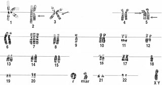
A ring chromosome is an aberrant chromosome whose ends have fused together to form a ring. Ring chromosomes were first discovered by Lilian Vaughan Morgan in 1926. A ring chromosome is denoted by the symbol r in human genetics and R in Drosophila genetics. Ring chromosomes may form in cells following genetic damage by mutagens like radiation, but they may also arise spontaneously during development.

Severe congenital neutropenia (SCN), also often known as Kostmann syndrome or disease, is a group of rare disorders that affect myelopoiesis, causing a congenital form of neutropenia, usually without other physical malformations. SCN manifests in infancy with life-threatening bacterial infections.

Hermansky–Pudlak syndrome Medical condition

Heřmanský–Pudlák syndrome is an extremely rare autosomal recessive disorder which results in oculocutaneous albinism, bleeding problems due to a platelet abnormality, and storage of an abnormal fat-protein compound. It is considered to affect around 1 in 500,000 people worldwide, with a significantly higher occurrence in Puerto Ricans, with a prevalence of 1 in 1800. Many of the clinical research studies on the disease have been conducted in Puerto Rico.

Bernard–Soulier syndrome Medical condition

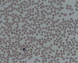
Bernard–Soulier syndrome (BSS), is a rare autosomal recessive bleeding disorder that is caused by a deficiency of the glycoprotein Ib-IX-V complex (GPIb-IX-V), the receptor for von Willebrand factor. The incidence of BSS is estimated to be less than 1 case per million persons, based on cases reported from Europe, North America, and Japan. BSS is a giant platelet disorder, meaning that it is characterized by abnormally large platelets.

Griscelli syndrome Medical condition

Griscelli syndrome is a rare autosomal recessive disorder characterized by albinism (hypopigmentation) with immunodeficiency, that usually causes death by early childhood. Researchers have developed three different classifications of the form of disorder, characterised by different signs and symptoms. Type 1 Griscelli Syndrome is assosciated with severe brain function issues along with distinctive discolouring of the hair and skin. Type 2 Griscelli Syndrome have immune system abnormalities in addition to hypopigmentation of skin and hair. Finally, Type 3 is seen as those only affected by hypopigmentation of the skin and hair. This type is not associated with immune deficiencies or neurological abnormalities.

GATA1

GATA-binding factor 1 or GATA-1 is the founding member of the GATA family of transcription factors. This protein is widely expressed throughout vertebrate species. In humans and mice, it is encoded by the GATA1 and Gata1 genes, respectively. These genes are located on the X chromosome in both species.

Lucey–Driscoll syndrome Medical condition

Lucey–Driscoll syndrome is an autosomal recessive metabolic disorder affecting enzymes involved in bilirubin metabolism. It is one of several disorders classified as a transient familial neonatal unconjugated hyperbilirubinemia.

TNF receptor associated periodic syndrome Medical condition

TNF receptor associated periodic syndrome (TRAPS) is a periodic fever syndrome associated with mutations in a receptor for the molecule tumor necrosis factor (TNF) that is inheritable in an autosomal dominant manner. Individuals with TRAPS have episodic symptoms such as recurrent high fevers, rash, abdominal pain, joint/muscle aches and puffy eyes.

Naegeli–Franceschetti–Jadassohn syndrome Medical condition

Naegeli–Franceschetti–Jadassohn syndrome (NFJS), also known as chromatophore nevus of Naegeli and Naegeli syndrome, is a rare autosomal dominant form of ectodermal dysplasia, characterized by reticular skin pigmentation, diminished function of the sweat glands, the absence of teeth and hyperkeratosis of the palms and soles. One of the most striking features is the absence of fingerprint lines on the fingers.

Pseudo gray platelet syndrome was described by Cockbill, Burmester, and Heptinstall (1988) who reported a 25-year-old woman with a history of mild bruising and bleeding. Another case was described in Japan in 2002.

Platelet storage pool deficiency Medical condition

Platelet storage pool deficiency is a type of coagulopathy characterized by defects in the granules in platelets, particularly a lack of granular non-metabolic adenosine diphosphate. Individuals with adenosine diphosphate deficient storage pool disease present a prolonged bleeding time due to impaired aggregation response to fibrillar collagen.

Paris-Trousseau syndrome (PTS) is an inherited disorder characterized by mild hemorrhagic tendency associated with 11q chromosome deletion. It manifests as a granular defect within an individual's platelets. It is characterized by thrombocytes with defects in α-granule components which affects the cell's surface area and, consequently, its ability to spread when necessary.

Giant platelet disorder Medical condition

Giant platelet disorders, also known as macrothrombocytopenia, are rare disorders featuring abnormally large platelets, thrombocytopenia and a tendency to bleeding. Giant platelets cannot stick adequately to an injured blood vessel walls, resulting in abnormal bleeding when injured. Giant platelet disorder occurs for inherited diseases like Bernard–Soulier syndrome, gray platelet syndrome and May–Hegglin anomaly.

References

  1. Online Mendelian Inheritance in Man (OMIM): 139090
  2. Nurden AT, Nurden P (January 2007). "The gray platelet syndrome: clinical spectrum of the disease". Blood Reviews. 21 (1): 21–36. doi:10.1016/j.blre.2005.12.003. PMID   16442192.
  3. Rao, A. Koneti; Rao, Deepak A. (2020-10-22). "Gray platelet syndrome: immunity goes awry". Blood. 136 (17): 1898–1900. doi:10.1182/blood.2020008196. ISSN   0006-4971. PMC   7582557 . PMID   33091137.
  4. "Gray platelet syndrome | Genetic and Rare Diseases Information Center (GARD) – an NCATS Program". rarediseases.info.nih.gov. Retrieved 2021-04-26.
  5. "Gray platelet syndrome: MedlinePlus Genetics". medlineplus.gov. Retrieved 2021-04-26.
  6. Gunay-Aygun M, Zivony-Elboum Y, Gumruk F, Geiger D, Cetin M, Khayat M, et al. (December 2010). "Gray platelet syndrome: natural history of a large patient cohort and locus assignment to chromosome 3p". Blood. 116 (23): 4990–5001. doi:10.1182/blood-2010-05-286534. PMC   3012593 . PMID   20709904.
  7. Kahr WH, Hinckley J, Li L, Schwertz H, Christensen H, Rowley JW, et al. (July 2011). "Mutations in NBEAL2, encoding a BEACH protein, cause gray platelet syndrome". Nature Genetics. 43 (8): 738–40. doi:10.1038/ng.884. PMC   6050511 . PMID   21765413.
  8. "OMIM Gene Map - Chromosome: 3". www.omim.org. Retrieved 2021-04-26.
  9. "OMIM Entry - # 139090 - GRAY PLATELET SYNDROME; GPS". www.omim.org. Retrieved 2021-04-26.
  10. Gunay-Aygun M, Falik-Zaccai TC, Vilboux T, Zivony-Elboum Y, Gumruk F, Cetin M, et al. (July 2011). "NBEAL2 is mutated in gray platelet syndrome and is required for biogenesis of platelet α-granules". Nature Genetics. 43 (8): 732–4. doi:10.1038/ng.883. PMC   3154019 . PMID   21765412.
  11. Albers CA, Cvejic A, Favier R, Bouwmans EE, Alessi MC, Bertone P, et al. (July 2011). "Exome sequencing identifies NBEAL2 as the causative gene for gray platelet syndrome". Nature Genetics. 43 (8): 735–7. doi:10.1038/ng.885. PMC   3428934 . PMID   21765411.
  12. Mori K, Suzuki S, Sugai K (July 1984). "Electron microscopic and functional studies on platelets in gray platelet syndrome". The Tohoku Journal of Experimental Medicine. 143 (3): 261–87. doi: 10.1620/tjem.143.261 . PMID   6484975.
  13. Gunay-Aygun M, Zivony-Elboum Y, Gumruk F, Geiger D, Cetin M, Khayat M, et al. (December 2010). "Gray platelet syndrome: natural history of a large patient cohort and locus assignment to chromosome 3p". Blood. 116 (23): 4990–5001. doi:10.1182/blood-2010-05-286534. PMC   3012593 . PMID   20709904.
  14. Jantunen E, Hänninen A, Naukkarinen A, Vornanen M, Lahtinen R (July 1994). "Gray platelet syndrome with splenomegaly and signs of extramedullary hematopoiesis: a case report with review of the literature". American Journal of Hematology. 46 (3): 218–24. doi:10.1002/ajh.2830460311. PMID   8192152. S2CID   127060.
  15. "OMIM Entry - # 139090 - GRAY PLATELET SYNDROME; GPS". www.omim.org. Retrieved 2021-04-26.
  16. 1 2 RESERVED, INSERM US14-- ALL RIGHTS. "Orphanet: Gray platelet syndrome". www.orpha.net. Retrieved 2021-04-26.
  17. "Gray platelet syndrome | Genetic and Rare Diseases Information Center (GARD) – an NCATS Program". rarediseases.info.nih.gov. Retrieved 2021-04-27.
  18. "Gray platelet syndrome | Genetic and Rare Diseases Information Center (GARD) – an NCATS Program". rarediseases.info.nih.gov. Retrieved 2021-04-27.
  19. "Gray platelet syndrome | Genetic and Rare Diseases Information Center (GARD) – an NCATS Program". rarediseases.info.nih.gov. Retrieved 2021-04-27.
  20. RESERVED, INSERM US14-- ALL RIGHTS. "Orphanet: Gray platelet syndrome". www.orpha.net. Retrieved 2021-04-27.