Hypoprothrombinemia

Last updated
Hypoprothrombinemia
Other namesCongenital factor II deficiency [1]
Autosomal recessive - en.svg
This condition is inherited in an autosomal recessive manner.
Specialty Hematology   OOjs UI icon edit-ltr-progressive.svg

Hypoprothrombinemia is a rare blood disorder in which a deficiency in immunoreactive prothrombin (Factor II), produced in the liver, results in an impaired blood clotting reaction, leading to an increased physiological risk for spontaneous bleeding. This condition can be observed in the gastrointestinal system, cranial vault, and superficial integumentary system, affecting both the male and female population. Prothrombin is a critical protein that is involved in the process of hemostasis, as well as illustrating procoagulant activities. This condition is characterized as an autosomal recessive inheritance congenital coagulation disorder affecting 1 per 2,000,000 of the population, worldwide, but is also attributed as acquired.

Contents

Signs and symptoms

There are various symptoms that are presented and are typically associated to a specific site that they appear at. Hypoprothrombinemia is characterized by a poor blood clotting function of prothrombin. Some symptoms are presented as severe, while others are mild, meaning that blood clotting is slower than normal. Areas that are usually affected are muscles, joints, and the brain, however, these sites are more uncommon.[ citation needed ]

The most common symptoms include:

  1. Easy bruising [2]
  2. Oral mucosal bleeding - Bleeding of the membrane mucus lining inside of the mouth.
  3. Soft tissue bleeding.
  4. Hemarthrosis - Bleeding in joint spaces.
  5. Epistaxis - Acute hemorrhages from areas of the nasal cavity, nostrils, or nasopharynx.
  6. Women with this deficiency experience menorrhagia: prolonged, abnormal heavy menstrual bleeding. [3] This is typically a symptom of the disorder when severe blood loss occurs.

Other reported symptoms that are related to the condition:

  1. Prolonged periods of bleeding due to surgery, injury, or post birth.
  2. Melena - Associated with acute gastrointestinal bleeding, dark black, tarry feces.
  3. Hematochezia - Lower gastrointestinal bleeding, passage of fresh, bright red blood through the anus secreted in or with stools. If associated with upper gastrointestinal bleeding, suggestive of a more life-threatening issue.

Type I: Severe hemorrhages are indicators of a more severe prothrombin deficiency that account for muscle hematomas, intracranial bleeding, postoperative bleeding, and umbilical cord hemorrhage, which may also occur depending on the severity, respectively.

Type II: Symptoms are usually more capricious, but can include a variety of the symptoms described previously. Less severe cases of the disorder typically do not involve spontaneous bleeding. [2]

Causes

Hypoprothrombinemia can be the result of a genetic defect, may be acquired as the result of another disease process, or may be an adverse effect of medication. For example, 5-10% of patients with systemic lupus erythematosus exhibit acquired hypoprothrombinemia due to the presence of autoantibodies which bind to prothrombin and remove it from the bloodstream (lupus anticoagulant-hypoprothrombinemia syndrome). [4] The most common viral pathogen that is involved is Adenovirus, with a prevalence of 50% in postviral cases. [5]

Inheritance

Autosomal recessive condition in which both parents must carry the recessive gene in order to pass the disease on to offspring. If both parents have the autosomal recessive condition, the chance of mutation in offspring increases to 100%. An individual will be considered a carrier if one mutant copy of the gene is inherited, and will not illustrate any symptoms. The disease affects both men and women equally, and overall, is a very uncommon inherited or acquired disorder. [3]

Non-inheritance and other factors

There are two types of prothrombin deficiencies that occur depending on the mutation: [5]

Type I (true deficiency), includes a missense or nonsense mutation, essentially decreasing prothrombin production. This is associated with bleeding from birth. Here, plasma levels of prothrombin are typically less than 10% of normal levels.[ citation needed ]

Type II, known as dysprothrombinemia, includes a missense mutation at specific Xa factor cleavage sites and serine protease prothrombin regions. Type II deficiency creates a dysfunctional protein with decreased activity and usually normal or low-normal antigen levels. A vitamin K-dependent clotting factor is seldom seen as a contributor to inherited prothrombin deficiencies, but lack of Vitamin K decreases the synthesis of prothrombin in liver cells. [5]

Acquired underlying causes of this condition include severe liver disease, warfarin overdose, platelet disorders, and disseminated intravascular coagulation (DIC).

It may also be a rare adverse effect to ceftriaxone. [6]

Mechanism

Hypoprothrombinemia is found to present itself as either inherited or acquired, and is a decrease in the synthesis of prothrombin. [7] In the process of inheritance, it marks itself as an autosomal recessive disorder, meaning that both parents must be carriers of the defective gene in order for the disorder to be present in a child. Prothrombin is a glycoprotein that occurs in blood plasma and functions as a precursor to the enzyme, thrombin, which acts to convert fibrinogen into fibrin, therefore, fortifying clots. This clotting process is known as coagulation. [8]

The mechanism specific to prothrombin (factor II) includes the proteolytically cleaving, breakdown of proteins into smaller polypeptides or amino acids, of this coagulation factor in order to form thrombin at the beginning of the cascade, leading to stemming of blood loss. A mutation in factor II would essentially lead to hypoprothrombinemia. The mutation is presented on chromosome 11. [9]

Areas where the disease has been shown to present itself at include the liver, since the glycoprotein is stored in this area.

Acquired cases are results from an isolated factor II deficiency. Specific cases include:

  1. Vitamin K deficiency: In the liver, vitamin K plays an important role in the synthesis of coagulation factor II. Body's capacity in the storage of vitamin K is typically very low. Vitamin K-dependent coagulation factors have a very short half-life, sometimes leading to a deficiency when a depletion of vitamin K occurs. The liver synthesizes inactive precursor proteins in the absence of vitamin K (liver disease). [8] Vitamin K deficiency leads to impaired clotting of the blood and in some cases, causes internal bleeding without an associated injury. [10]
  2. Disseminated intravascular coagulation (DIC): Involving abnormal, excessive generation of thrombin and fibrin within the blood. Relative to hypoprothrombinemia, due to increased platelet aggregation and coagulation factor consumption involved in the process. [11]
  3. Anticoagulants: warfarin overdose: Used as a treatment for prevention of blood clots, however, like most drugs, side effects have been shown to increase risk of excessive bleeding by functioning in the disruption of hepatic synthesis of coagulation factors II, VII, IX, and X. Vitamin K is an antagonist to warfarin drug, reversing its activity, causing it to be less effective in the process of blood clotting. [12] Warfarin intake has been shown to interfere with Vitamin-K metabolism. [10]

Diagnosis

Diagnosis of inherited hypoprothrombinemia, relies heavily on a patient's medical history, family history of bleeding issues, and lab exams performed by a hematologist. A physical examination by a general physician should also be performed in order to determine whether the condition is congenital or acquired, as well as ruling out other possible conditions with similar symptoms. For acquired forms, information must be taken regarding current diseases and medications taken by the patient, if applicable.[ citation needed ]

Lab tests that are performed to determine diagnosis:

  1. Factor assays: To observe the performance of specific factors (II) to identify missing/poorly performing factors. These lab tests are typically performed first in order to determine the status of the factor.
  2. Prothrombin blood test: Determines if a patient has deficient or low levels of Factor II.[ citation needed ]
  3. Vitamin K1 test: Performed to evaluate bleeding of unknown causes, nosebleeds, and identified bruising. To accomplish this, a band is wrapped around the patient's arm, 4 inches above the superficial vein site in the elbow pit. The vein is penetrated with the needle and amount of blood required for testing is obtained. Decreased vitamin K levels are suggestive of hypoprothrombinemia. However, this exam is rarely used as a prothrombin blood test is performed beforehand. [13]

Treatment

Treatment is almost always aimed to control hemorrhages, treating underlying causes, and taking preventative steps before performing invasive surgeries.

Hypoprothrombinemia can be treated with periodic infusions of purified prothrombin complexes. [14] These are typically used as treatment methods for severe bleeding cases in order to boost clotting ability and increasing levels of vitamin K-dependent coagulation factors.

  1. A known treatment for hypoprothrombinemia is menadoxime. [15]
  2. Menatetrenone was also listed as an antihemorrhagic vitamin.
  3. 4-Amino-2-methyl-1-naphthol (Vitamin K5) is another treatment for hypoprothrombinemia.
    1. Vitamin K forms are administered orally or intravenously.
  4. Other concentrates include Proplex T, Konyne 80, and Bebulin VH.

Fresh frozen plasma infusion (FFP) is a method used for continuous bleeding episodes, every 3–5 weeks for mention. [2]

  1. Used to treat various conditions related to low blood clotting factors.
  2. Administered by intravenous injection and typically at a 15-20 ml/kg/dose.
  3. Can be used to treat acute bleeding.

Invasive options, such as surgery or clotting factor infusions, are required if previous methods do not suffice. Surgery is to be avoided, as it causes significant bleeding in patients with hypoprothrombinemia.

Prognosis

Prognosis for patients varies and is dependent on severity of the condition and how early the treatment is managed.

  1. With proper treatment and care, most people go on to live a normal and healthy life.
  2. With more severe cases, a hematologist will need to be seen throughout the patient's life in order to deal with bleeding and continued risks.

Related Research Articles

<span class="mw-page-title-main">Coagulation</span> Process of formation of blood clots

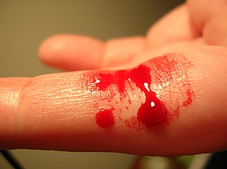
Coagulation, also known as clotting, is the process by which blood changes from a liquid to a gel, forming a blood clot. It potentially results in hemostasis, the cessation of blood loss from a damaged vessel, followed by repair. The mechanism of coagulation involves activation, adhesion and aggregation of platelets, as well as deposition and maturation of fibrin.

<span class="mw-page-title-main">Warfarin</span> Medication

Warfarin is an anticoagulant used as a medication under several brand names including Coumadin. While the drug is described as a "blood thinner", it does not reduce viscosity but rather inhibits coagulation. Accordingly, it is commonly used to prevent blood clots in the circulatory system such as deep vein thrombosis and pulmonary embolism, and to protect against stroke in people who have atrial fibrillation, valvular heart disease, or artificial heart valves. Less commonly, it is used following ST-segment elevation myocardial infarction and orthopedic surgery. It is usually taken by mouth, but may also be administered intravenously.

<span class="mw-page-title-main">Thrombin</span> Enzyme involved in blood coagulation in humans

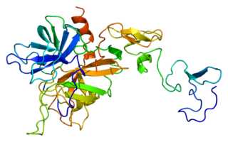
Thrombin is a serine protease, an enzyme that, in humans, is encoded by the F2 gene. Prothrombin is proteolytically cleaved to form thrombin in the clotting process. Thrombin in turn acts as a serine protease that converts soluble fibrinogen into insoluble strands of fibrin, as well as catalyzing many other coagulation-related reactions.

<span class="mw-page-title-main">Prothrombin time</span> Assay for evaluating the extrinsic pathway & common pathway of coagulation

The prothrombin time (PT) – along with its derived measures of prothrombin ratio (PR) and international normalized ratio (INR) – is an assay for evaluating the extrinsic pathway and common pathway of coagulation. This blood test is also called protime INR and PT/INR. They are used to determine the clotting tendency of blood, in such things as the measure of warfarin dosage, liver damage, and vitamin K status. PT measures the following coagulation factors: I (fibrinogen), II (prothrombin), V (proaccelerin), VII (proconvertin), and X.

<span class="mw-page-title-main">Bleeding diathesis</span> Medical condition

In medicine (hematology), bleeding diathesis is an unusual susceptibility to bleed (hemorrhage) mostly due to hypocoagulability, in turn caused by a coagulopathy. Therefore, this may result in the reduction of platelets being produced and leads to excessive bleeding. Several types of coagulopathy are distinguished, ranging from mild to lethal. Coagulopathy can be caused by thinning of the skin, such that the skin is weakened and is bruised easily and frequently without any trauma or injury to the body. Also, coagulopathy can be contributed by impaired wound healing or impaired clot formation.

<span class="mw-page-title-main">Protein S deficiency</span> Medical condition

Protein S deficiency is a disorder associated with increased risk of venous thrombosis. Protein S, a vitamin K-dependent physiological anticoagulant, acts as a nonenzymatic cofactor to activate protein C in the degradation of factor Va and factor VIIIa.

<span class="mw-page-title-main">Protein C</span> Mammalian protein found in Homo sapiens

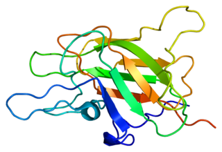
Protein C, also known as autoprothrombin IIA and blood coagulation factor XIX, is a zymogen, that is, an inactive enzyme. The activated form plays an important role in regulating anticoagulation, inflammation, and cell death and maintaining the permeability of blood vessel walls in humans and other animals. Activated protein C (APC) performs these operations primarily by proteolytically inactivating proteins Factor Va and Factor VIIIa. APC is classified as a serine protease since it contains a residue of serine in its active site. In humans, protein C is encoded by the PROC gene, which is found on chromosome 2.

<span class="mw-page-title-main">Thrombophilia</span> Abnormality of blood coagulation

Thrombophilia is an abnormality of blood coagulation that increases the risk of thrombosis. Such abnormalities can be identified in 50% of people who have an episode of thrombosis that was not provoked by other causes. A significant proportion of the population has a detectable thrombophilic abnormality, but most of these develop thrombosis only in the presence of an additional risk factor.

<span class="mw-page-title-main">Factor X</span> Mammalian protein found in Homo sapiens

Factor X, also known by the eponym Stuart–Prower factor, is an enzyme of the coagulation cascade. It is a serine endopeptidase. Factor X is synthesized in the liver and requires vitamin K for its synthesis.

<span class="mw-page-title-main">Factor V</span> Mammalian protein found in humans

Factor V is a protein of the coagulation system, rarely referred to as proaccelerin or labile factor. In contrast to most other coagulation factors, it is not enzymatically active but functions as a cofactor. Deficiency leads to predisposition for hemorrhage, while some mutations predispose for thrombosis.

<span class="mw-page-title-main">Fresh frozen plasma</span> Liquid portion of whole blood

Fresh frozen plasma (FFP) is a blood product made from the liquid portion of whole blood. It is used to treat conditions in which there are low blood clotting factors or low levels of other blood proteins. It may also be used as the replacement fluid in plasma exchange. Using ABO compatible plasma, while not required, may be recommended. Use as a volume expander is not recommended. It is given by slow injection into a vein.

<span class="mw-page-title-main">Renal vein thrombosis</span> Medical condition

Renal vein thrombosis (RVT) is the formation of a clot in the vein that drains blood from the kidneys, ultimately leading to a reduction in the drainage of one or both kidneys and the possible migration of the clot to other parts of the body. First described by German pathologist Friedrich Daniel von Recklinghausen in 1861, RVT most commonly affects two subpopulations: newly born infants with blood clotting abnormalities or dehydration and adults with nephrotic syndrome.

<span class="mw-page-title-main">Warfarin necrosis</span> Medical condition

Warfarin-induced skin necrosis is a condition in which skin and subcutaneous tissue necrosis occurs due to acquired protein C deficiency following treatment with anti-vitamin K anticoagulants.

<span class="mw-page-title-main">Protein C deficiency</span> Medical condition

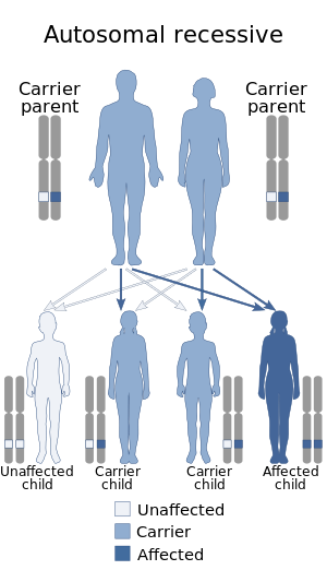
Protein C deficiency is a rare genetic trait that predisposes to thrombotic disease. It was first described in 1981. The disease belongs to a group of genetic disorders known as thrombophilias. Protein C deficiency is associated with an increased incidence of venous thromboembolism, whereas no association with arterial thrombotic disease has been found.

<span class="mw-page-title-main">Factor X deficiency</span> Medical condition

Factor X deficiency is a bleeding disorder characterized by a lack in the production of factor X (FX), an enzyme protein that causes blood to clot in the coagulation cascade. Produced in the liver FX when activated cleaves prothrombin to generate thrombin in the intrinsic pathway of coagulation. This process is vitamin K dependent and enhanced by activated factor V.

<span class="mw-page-title-main">Factor VII deficiency</span> Medical condition

Factor VII deficiency is a bleeding disorder characterized by a lack in the production of Factor VII (FVII) (proconvertin), a protein that causes blood to clot in the coagulation cascade. After a trauma factor VII initiates the process of coagulation in conjunction with tissue factor in the extrinsic pathway.

<span class="mw-page-title-main">Phenprocoumon</span> Drug

Phenprocoumon is a long-acting blood thinner drug to be taken by mouth, and a derivative of coumarin. It acts as a vitamin K antagonist and inhibits blood clotting (coagulation) by blocking synthesis of coagulation factors II, VII, IX and X. It is used for the prophylaxis and treatment of thromboembolic disorders such as heart attacks and pulmonary (lung) embolism. The most common adverse effect is bleeding. The drug interacts with a large number of other medications, including aspirin and St John's Wort. It is the standard coumarin used in Germany, Austria, and other European countries.

Prothrombin complex concentrate (PCC), also known as factor IX complex, sold under the brand name Kcentra among others, is a combination medication made up of blood clotting factors II, IX, and X. Some versions also contain factor VII. It is used to treat and prevent bleeding in hemophilia B if pure factor IX is not available. It may also be used for reversal of warfarin therapy. It is given by slow injection into a vein.

Purpura fulminans is an acute, often fatal, thrombotic disorder which manifests as blood spots, bruising and discolouration of the skin resulting from coagulation in small blood vessels within the skin and rapidly leads to skin necrosis and disseminated intravascular coagulation.

<span class="mw-page-title-main">Vitamin K reaction</span> Medical condition

Vitamin K reactions are adverse side effects that may occur after injection with vitamin K. The liver utilizes vitamin K to produce coagulation factors that help the body form blood clots which prevent excessive bleeding. Vitamin K injections are administered to newborns as a preventative measure to reduce the risk of hemorrhagic disease of the newborn (HDN).

References

  1. RESERVED, INSERM US14-- ALL RIGHTS. "Orphanet: Congenital factor II deficiency". www.orpha.net. Retrieved 5 June 2019.
  2. 1 2 3 "Hypoprothrombinemia Clinical Presentation: History, Physical, Causes". emedicine.medscape.com. Retrieved 2017-12-12.
  3. 1 2 "Factor II Deficiency". DoveMed. Retrieved 2017-12-12.
  4. Bajaj S, Rapaport S, Fierer D, Herbst K, Schwartz D (1983). "A mechanism for the hypoprothrombinemia of the acquired hypoprothrombinemia-lupus anticoagulant syndrome". Blood. 61 (4): 684–92. doi: 10.1182/blood.V61.4.684.684 . PMID   6403077.
  5. 1 2 3 "Hypoprothrombinemia Clinical Presentation: History, Physical, Causes". emedicine.medscape.com. Retrieved 2017-12-12.
  6. [ http://www.gene.com/download/pdf/rocephin_prescribing.pdf Archived 2013-08-16 at the Wayback Machine Rocephin proscribing information]. Accessed October 24, 2013.
  7. "hypoprothrombinemia | pathology". Encyclopedia Britannica. Retrieved 2017-12-12.
  8. 1 2 "Hypoprothrombinemia: Background, Pathophysiology, Epidemiology". 2017-03-30.{{cite journal}}: Cite journal requires |journal= (help)
  9. "ortholog_gene_2147[group] - Gene - NCBI". www.ncbi.nlm.nih.gov. Retrieved 2017-12-12.
  10. 1 2 "nutritional disease - Vitamin K". Encyclopedia Britannica. Retrieved 2017-12-13.
  11. "Disseminated Intravascular Coagulation (DIC) - Hematology and Oncology - Merck Manuals Professional Edition". Merck Manuals Professional Edition. Retrieved 2017-12-12.
  12. Bajaj, SP; Rapaport, SI; Fierer, DS; Herbst, KD; Schwartz, DB (April 1983). "A mechanism for the hypoprothrombinemia of the acquired hypoprothrombinemia-lupus anticoagulant syndrome". Blood. 61 (4): 684–92. doi: 10.1182/blood.V61.4.684.684 . PMID   6403077.
  13. "Vitamin K Deficiency". labtestsonline.org. Retrieved 2017-12-12.
  14. Lechler E (1999). "Use of prothrombin complex concentrates for prophylaxis and treatment of bleeding episodes in patients with hereditary deficiency of prothrombin, factor VII, factor X, protein C protein S, or protein Z.". Thromb Res. 95 (4 Suppl 1): S39–50. doi:10.1016/S0049-3848(99)00083-3. PMID   10499908.
  15. J. Elks, C. R. Ganellin · DoDs page 765.