Placental insufficiency

Last updated
Placental insufficiency
Other namesUtero-placental insufficiency
Specialty Neonatology, obstetrics, maternal–fetal medicine   OOjs UI icon edit-ltr-progressive.svg

Placental insufficiency or utero-placental insufficiency is the failure of the placenta to deliver sufficient nutrients to the fetus during pregnancy, and is often a result of insufficient blood flow to the placenta. The term is also sometimes used to designate late decelerations of fetal heart rate as measured by cardiotocography or an NST, even if there is no other evidence of reduced blood flow to the placenta, normal uterine blood flow rate being 600mL/min.

Contents

Causes

Histopathology of placenta with increased syncytial knotting of chorionic villi, with two knots pointed out. Histopathology of placenta with increased syncytial knotting of chorionic villi, annotated.jpg
Histopathology of placenta with increased syncytial knotting of chorionic villi, with two knots pointed out.

The following characteristics of placentas have been said to be associated with placental insufficiency, however all of them occur in normal healthy placentas and full term healthy births, so none of them can be used to accurately diagnose placental insufficiency:[ citation needed ]

Placental insufficiency should not be confused with complete placental abruption, in which the placenta separates off the uterine wall, which immediately results in no blood flow to the placenta, which leads to immediate fetal demise. In the case of a marginal, incomplete placental abruption of less than 50%, usually weeks of hospitalization precedes delivery and outcomes are not necessarily affected by the partial abruption. [2]

Pathophysiology

Maternal effects

Several aspects of maternal adaptation to pregnancy are affected by dysfunction of placenta. Maternal arteries fail to transform into low-resistance vessels (expected by 22–24 weeks of gestation). [3] [4] This increases vascular resistance in the fetoplacental vascular bed, eventually leading to reduction in metabolically active mass of placenta in a type of vicious cycle.[ citation needed ]

Fetal effects

Placental insufficiency can affect the fetus, causing fetal distress. Placental insufficiency may cause oligohydramnios, preeclampsia, miscarriage or stillbirth. Placental insufficiency is most frequent cause of asymmetric IUGR. [5]

Fetal metabolic changes

Metabolic changes occurring in uteroplacental insufficiency: [6]

SubstrateChange
Glucose Decreases in proportion to degree of fetal hypoglycemia
Amino acids
Fatty acids
Oxygen and Carbon dioxide

Fetal hormonal changes

Decrease in overall thyroid function is correlated with fetal hypoxemia. [7] [8] Serum glucagon, adrenaline, noradrenaline levels increase, eventually causing peripheral glycogenolysis and mobilization of fetal hepatic glycogen stores. [9] [10] [11] [12]

Fetal hematologic changes

Fetal hypoxemia triggers erythropoietin release. This stimulates RBC production from medullary and extramedullary sites and eventually results in polycythemia. [13] [14] [15] [16] Oxygen carrying capacity of blood is thus increased. Prolonged tissue hypoxemia may cause early release of erythrocytes from maturation sites and thus count of nucleated RBCs in blood increases. [17] [18] [19] [20] These factors, increase in blood viscosity, decrease in cell membrane fluidity and platelet aggregation are important precursors in accelerating placental vascular occlusion.[ citation needed ]

Fetal immunological changes

There is decrease in immunoglobulin, absolute B-cell counts [21] and total WBC count. [22] T-helper and cytotoxic T-cells are suppressed [23] in proportion of degree of acidemia. These conditions lead to higher infection susceptibility of infant after delivery.[ citation needed ]

Fetal cardiovascular changes

There is decrease in magnitude of umbilical venous volume flow. [24] In response to this, the proportion of umbilical venous blood diverted to fetal heart increases. [25] This eventually leads to elevation of pulmonary vascular resistance and increased right ventricular afterload. [26] [27] [28] This fetal cerebral redistribution of blood flow is an early response to placental insufficiency. Blood flow is selectively redirected to the myocardium, adrenal glands, and in particular to the brain in a brain-sparing effect. [29]

In late stage, the redistribution becomes ineffective, there is decrease in cardiac output, ineffective preload handling and elevation of central venous pressure. [30] [31] [32] [33] This deterioration in circulation may ultimately lead to tricuspid insufficiency and death of the fetus. [34] [35] Peripheral circulatory disturbances also accompany these central circulatory changes.

Fetal behavioral changes

Chronic hypoxemia leads to delay in all aspects of CNS maturation. [36] [37] [38] [39] With worsening fetal hypoxemia, there is decline in fetal activity. [40] With further hypoxemia, fetal breathing ceases. Gross body movements and tone decrease further. [41] [42] Fetal heart rate decreases due to spontaneous deceleration due to direct depression of cardiac contractility. This leads to intrauterine fetal death.[ citation needed ]

Risk of later metabolic disease

According to the theory of thrifty phenotype, placental insufficiency triggers epigenetic responses in the fetus that are otherwise activated in times of chronic food shortage. If the offspring actually develops in an environment rich in food it may be more prone to metabolic disorders, such as obesity and type II diabetes. [43]

Diagnosis

The following tests have been promoted as supposedly diagnosing placental insufficiency, but all have been unsuccessful at predicting stillbirth due to placental insufficiency: [44] [45]

See also

Related Research Articles

<span class="mw-page-title-main">Placenta</span> Organ that connects the fetus to the uterine wall

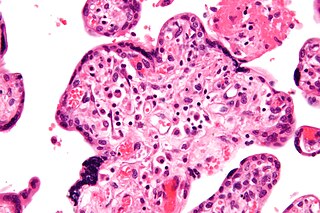
The placenta is a temporary embryonic and later fetal organ that begins developing from the blastocyst shortly after implantation. It plays critical roles in facilitating nutrient, gas and waste exchange between the physically separate maternal and fetal circulations, and is an important endocrine organ, producing hormones that regulate both maternal and fetal physiology during pregnancy. The placenta connects to the fetus via the umbilical cord, and on the opposite aspect to the maternal uterus in a species-dependent manner. In humans, a thin layer of maternal decidual (endometrial) tissue comes away with the placenta when it is expelled from the uterus following birth. Placentas are a defining characteristic of placental mammals, but are also found in marsupials and some non-mammals with varying levels of development.

<span class="mw-page-title-main">Intrauterine growth restriction</span> Medical condition

Intrauterine growth restriction (IUGR), or fetal growth restriction, refers to poor growth of a fetus while in the womb during pregnancy. IUGR is defined by clinical features of malnutrition and evidence of reduced growth regardless of an infant's birth weight percentile. The causes of IUGR are broad and may involve maternal, fetal, or placental complications.

<span class="mw-page-title-main">Placenta praevia</span> Medical condition

Placenta praevia is when the placenta attaches inside the uterus but in a position near or over the cervical opening. Symptoms include vaginal bleeding in the second half of pregnancy. The bleeding is bright red and tends not to be associated with pain. Complications may include placenta accreta, dangerously low blood pressure, or bleeding after delivery. Complications for the baby may include fetal growth restriction.

Oligohydramnios is a medical condition in pregnancy characterized by a deficiency of amniotic fluid, the fluid that surrounds the fetus in the abdomen, in the amniotic sac. It is typically diagnosed by ultrasound when the amniotic fluid index (AFI) measures less than 5 cm or when the single deepest pocket (SDP) of amniotic fluid measures less than 2 cm. Amniotic fluid is necessary to allow for normal fetal movement, lung development, and cushioning from uterine compression. Low amniotic fluid can be attributed to a maternal, fetal, placental or idiopathic cause and can result in poor fetal outcomes including death. The prognosis of the fetus is dependent on the etiology, gestational age at diagnosis, and the severity of the oligohydramnios.

<span class="mw-page-title-main">Twin-to-twin transfusion syndrome</span> Medical condition

Twin-to-twin transfusion syndrome (TTTS), also known as feto-fetal transfusion syndrome (FFTS), twin oligohydramnios-polyhydramnios sequence (TOPS) and stuck twin syndrome, is a complication of monochorionic multiple pregnancies in which there is disproportionate blood supply between the fetuses. This leads to unequal levels of amniotic fluid between each fetus and usually leads to death of the undersupplied twin and, without treatment, usually death or a range of birth defects or disabilities for a surviving twin, such as underdeveloped, damaged or missing limbs, digits or organs, especially cerebral palsy.

<span class="mw-page-title-main">Placental abruption</span> Medical condition

Placental abruption is when the placenta separates early from the uterus, in other words separates before childbirth. It occurs most commonly around 25 weeks of pregnancy. Symptoms may include vaginal bleeding, lower abdominal pain, and dangerously low blood pressure. Complications for the mother can include disseminated intravascular coagulopathy and kidney failure. Complications for the baby can include fetal distress, low birthweight, preterm delivery, and stillbirth.

<span class="mw-page-title-main">Ductus venosus</span> Vein in the human fetus

In the fetus, the ductus venosus shunts a portion of umbilical vein blood flow directly to the inferior vena cava. Thus, it allows oxygenated blood from the placenta to bypass the liver. Compared to the 50% shunting of umbilical blood through the ductus venosus found in animal experiments, the degree of shunting in the human fetus under physiological conditions is considerably less, 30% at 20 weeks, which decreases to 18% at 32 weeks, suggesting a higher priority of the fetal liver than previously realized. In conjunction with the other fetal shunts, the foramen ovale and ductus arteriosus, it plays a critical role in preferentially shunting oxygenated blood to the fetal brain. It is a part of fetal circulation.

<span class="mw-page-title-main">Inferior vena cava syndrome</span> Medical condition

Inferior vena cava syndrome (IVCS) is a very rare constellation of symptoms resulting from either an obstruction, or stenosis of the inferior vena cava. It can be caused by physical invasion or compression by a pathological process or by thrombosis within the vein itself. It can also occur during pregnancy. Pregnancy leads to high venous pressure in the lower limbs, decreased blood return to the heart, decreased cardiac output due to obstruction of the inferior vena cava, sudden rise in venous pressure which can lead to placental separation, and a decrease in kidney function. All of these issues can arise from lying in the supine position during late pregnancy which can cause compression of the inferior vena cava by the uterus. Symptoms of late pregnancy inferior vena cava syndrome consist of intense pain in the right hand side, muscle twitching, hypotension, and fluid retention.

<span class="mw-page-title-main">Abdominal pregnancy</span> Medical condition

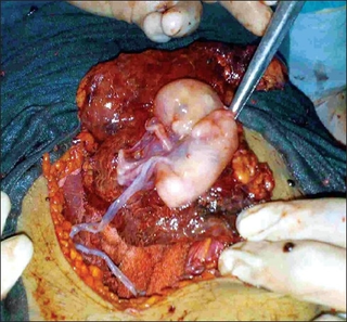
An abdominal pregnancy is a rare type of ectopic pregnancy where the embryo or fetus is growing and developing outside the womb in the abdomen, but not in the Fallopian tube, ovary or broad ligament.

<span class="mw-page-title-main">Intrauterine hypoxia</span> Medical condition when the fetus is deprived of sufficient oxygen

Intrauterine hypoxia occurs when the fetus is deprived of an adequate supply of oxygen. It may be due to a variety of reasons such as prolapse or occlusion of the umbilical cord, placental infarction, maternal diabetes and maternal smoking. Intrauterine growth restriction may cause or be the result of hypoxia. Intrauterine hypoxia can cause cellular damage that occurs within the central nervous system. This results in an increased mortality rate, including an increased risk of sudden infant death syndrome (SIDS). Oxygen deprivation in the fetus and neonate have been implicated as either a primary or as a contributing risk factor in numerous neurological and neuropsychiatric disorders such as epilepsy, attention deficit hyperactivity disorder, eating disorders and cerebral palsy.

<span class="mw-page-title-main">Placenta accreta spectrum</span> Medical condition

Placenta accreta occurs when all or part of the placenta attaches abnormally to the myometrium. Three grades of abnormal placental attachment are defined according to the depth of attachment and invasion into the muscular layers of the uterus:

  1. Accreta – chorionic villi attached to the myometrium, rather than being restricted within the decidua basalis.
  2. Increta – chorionic villi invaded into the myometrium.
  3. Percreta – chorionic villi invaded through the perimetrium.

Postterm pregnancy is when a woman has not yet delivered her baby after 42 weeks of gestation, two weeks beyond the typical 40-week duration of pregnancy. Postmature births carry risks for both the mother and the baby, including fetal malnutrition, meconium aspiration syndrome, and stillbirths. After the 42nd week of gestation, the placenta, which supplies the baby with nutrients and oxygen from the mother, starts aging and will eventually fail. Postterm pregnancy is a reason to induce labor.

<span class="mw-page-title-main">Nuchal scan</span> Routine ultrasound done between 11 and 14 weeks pregnancy

A nuchal scan or nuchal translucency (NT) scan/procedure is a sonographic prenatal screening scan (ultrasound) to detect chromosomal abnormalities in a fetus, though altered extracellular matrix composition and limited lymphatic drainage can also be detected.

<span class="mw-page-title-main">Vasa praevia</span> Condition in which fetal blood vessels cross or run near the internal opening of the uterus.

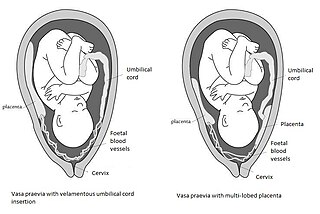
Vasa praevia is a condition in which fetal blood vessels cross or run near the internal opening of the uterus. These vessels are at risk of rupture when the supporting membranes rupture, as they are unsupported by the umbilical cord or placental tissue.

<span class="mw-page-title-main">Percutaneous umbilical cord blood sampling</span>

Percutaneous umbilical cord blood sampling (PUBS), also called cordocentesis, fetal blood sampling, or umbilical vein sampling is a diagnostic genetic test that examines blood from the fetal umbilical cord to detect fetal abnormalities. Fetal and maternal blood supply are typically connected in utero with one vein and two arteries to the fetus. The umbilical vein is responsible for delivering oxygen rich blood to the fetus from the mother; the umbilical arteries are responsible for removing oxygen poor blood from the fetus. This allows for the fetus’ tissues to properly perfuse. PUBS provides a means of rapid chromosome analysis and is useful when information cannot be obtained through amniocentesis, chorionic villus sampling, or ultrasound ; this test carries a significant risk of complication and is typically reserved for pregnancies determined to be at high risk for genetic defect. It has been used with mothers with immune thrombocytopenic purpura.

<span class="mw-page-title-main">Velamentous cord insertion</span> Velamentous placenta

Velamentous cord insertion is a complication of pregnancy where the umbilical cord is inserted in the fetal membranes. It is a major cause of antepartum hemorrhage that leads to loss of fetal blood and associated with high perinatal mortality. In normal pregnancies, the umbilical cord inserts into the middle of the placental mass and is completely encased by the amniotic sac. The vessels are hence normally protected by Wharton's jelly, which prevents rupture during pregnancy and labor. In velamentous cord insertion, the vessels of the umbilical cord are improperly inserted in the chorioamniotic membrane, and hence the vessels traverse between the amnion and the chorion towards the placenta. Without Wharton's jelly protecting the vessels, the exposed vessels are susceptible to compression and rupture.

<span class="mw-page-title-main">Monochorionic twins</span> Identical twins that share the same placenta

Monochorionic twins are monozygotic (identical) twins that share the same placenta. If the placenta is shared by more than two twins, these are monochorionic multiples. Monochorionic twins occur in 0.3% of all pregnancies. Seventy-five percent of monozygotic twin pregnancies are monochorionic; the remaining 25% are dichorionic diamniotic. If the placenta divides, this takes place before the third day after fertilization.

<span class="mw-page-title-main">Circumvallate placenta</span> Medical condition

Circumvallate placenta is a rare condition affecting about 1-2% of pregnancies, in which the amnion and chorion fetal membranes essentially "double back" on the fetal side around the edges of the placenta. After delivery, a circumvallate placenta has a thick ring of membranes on its fetal surface. Circumvallate placenta is a placental morphological abnormality associated with increased fetal morbidity and mortality due to the restricted availability of nutrients and oxygen to the developing fetus.

Amnioinfusion is a method in which isotonic fluid is instilled into the uterine cavity.

The anomaly scan, also sometimes called the anatomy scan, 20-week ultrasound, or level 2 ultrasound, evaluates anatomic structures of the fetus, placenta, and maternal pelvic organs. This scan is an important and common component of routine prenatal care. The function of the ultrasound is to measure the fetus so that growth abnormalities can be recognized quickly later in pregnancy, to assess for congenital malformations and multiple pregnancies, and to plan method of delivery.

References

  1. Brant, William E. (2001). The core curriculum, ultrasound. Philadelphia: Lippincott Williams & Wilkins. p. 265. ISBN   9780683307337.
  2. McCormack, RA; Doherty, DA; Magann, EF; Hutchinson, M; Newnham, JP (October 2008). "Antepartum bleeding of unknown origin in the second half of pregnancy and pregnancy outcomes". BJOG: An International Journal of Obstetrics and Gynaecology. 115 (11): 1451–7. doi:10.1111/j.1471-0528.2008.01856.x. PMID   18715242.
  3. Brosens, I; Dixon, HG; Robertson, WB (September 1977). "Fetal growth retardation and the arteries of the placental bed". British Journal of Obstetrics and Gynaecology. 84 (9): 656–63. doi:10.1111/j.1471-0528.1977.tb12676.x. PMID   911717. S2CID   33620598.
  4. Meekins, JW; Pijnenborg, R; Hanssens, M; McFadyen, IR; van Asshe, A (August 1994). "A study of placental bed spiral arteries and trophoblast invasion in normal and severe pre-eclamptic pregnancies". British Journal of Obstetrics and Gynaecology. 101 (8): 669–74. doi:10.1111/j.1471-0528.1994.tb13182.x. PMID   7947500. S2CID   22676459.
  5. Medical Physiology, 2e. Elsevier Health Sciences. 2012-01-13. ISBN   978-1455711819.
  6. Gabbe, Steven G., ed. (2012-01-01). Obstetrics : normal and problem pregnancies (6th ed.). Philadelphia: Elsevier/Saunders. ISBN   978-1-4377-1935-2.
  7. Thorpe-Beeston, JG; Nicolaides, KH; McGregor, AM (Fall 1992). "Fetal thyroid function". Thyroid . 2 (3): 207–17. doi:10.1089/thy.1992.2.207. PMID   1422233.
  8. Thorpe-Beeston, JG; Nicolaides, KH; Snijders, RJ; Felton, CV; Vyas, S; Campbell, S (November 1991). "Relations between the fetal circulation and pituitary-thyroid function". British Journal of Obstetrics and Gynaecology. 98 (11): 1163–7. doi:10.1111/j.1471-0528.1991.tb15371.x. PMID   1760429. S2CID   2838294.
  9. Hubinont, C; Nicolini, U; Fisk, NM; Tannirandorn, Y; Rodeck, CH (April 1991). "Endocrine pancreatic function in growth-retarded fetuses". Obstetrics & Gynecology . 77 (4): 541–4. PMID   2002976.
  10. Weiner, CP; Robillard, JE (December 1988). "Atrial natriuretic factor, digoxin-like immunoreactive substance, norepinephrine, epinephrine, and plasma renin activity in human fetuses and their alteration by fetal disease". American Journal of Obstetrics and Gynecology . 159 (6): 1353–60. doi:10.1016/0002-9378(88)90555-8. PMID   2974684.
  11. Greenough, A; Nicolaides, KH; Lagercrantz, H (June 1990). "Human fetal sympathoadrenal responsiveness". Early Human Development . 23 (1): 9–13. doi:10.1016/0378-3782(90)90124-2. PMID   2120028.
  12. Ville, Y; Proudler, A; Kuhn, P; Nicolaides, KH (October 1994). "Aldosterone concentration in normal, growth-retarded, anemic, and hydropic fetuses". Obstetrics and Gynecology. 84 (4): 511–4. PMID   8090385.
  13. Weiner, CP; Williamson, RA (February 1989). "Evaluation of severe growth retardation using cordocentesis--hematologic and metabolic alterations by etiology". Obstetrics and Gynecology. 73 (2): 225–9. PMID   2536145.
  14. Thilaganathan, B; Athanasiou, S; Ozmen, S; Creighton, S; Watson, NR; Nicolaides, KH (May 1994). "Umbilical cord blood erythroblast count as an index of intrauterine hypoxia". Archives of Disease in Childhood: Fetal and Neonatal Edition. 70 (3): F192–4. doi:10.1136/fn.70.3.f192. PMC   1061039 . PMID   8198413.
  15. Franz, AR; Pohlandt, F (March 2001). "Red blood cell transfusions in very and extremely low birthweight infants under restrictive transfusion guidelines: is exogenous erythropoietin necessary?". Archives of Disease in Childhood: Fetal and Neonatal Edition. 84 (2): F96–F100. doi:10.1136/fn.84.2.f96. PMC   1721217 . PMID   11207224.
  16. Snijders, RJ; Abbas, A; Melby, O; Ireland, RM; Nicolaides, KH (February 1993). "Fetal plasma erythropoietin concentration in severe growth retardation". American Journal of Obstetrics and Gynecology. 168 (2): 615–9. doi:10.1016/0002-9378(93)90505-d. PMID   8438939.
  17. Thilaganathan, B.; Nicolaides, K. H. (1 January 1992). "Erythroblastosis in birth asphyxia". Ultrasound in Obstetrics and Gynecology. 2 (1): 15–17. doi:10.1046/j.1469-0705.1992.02010015.x. PMID   12797000. S2CID   11457412.
  18. Bernstein, PS; Minior, VK; Divon, MY (November 1997). "Neonatal nucleated red blood cell counts in small-for-gestational age fetuses with abnormal umbilical artery Doppler studies". American Journal of Obstetrics and Gynecology. 177 (5): 1079–84. doi:10.1016/s0002-9378(97)70018-8. PMID   9396897.
  19. Baschat, AA; Gembruch, U; Reiss, I; Gortner, L; Harman, CR; Weiner, CP (July 1999). "Neonatal nucleated red blood cell counts in growth-restricted fetuses: relationship to arterial and venous Doppler studies". American Journal of Obstetrics and Gynecology. 181 (1): 190–5. doi:10.1016/s0002-9378(99)70458-8. PMID   10411818.
  20. Baschat, AA; Gembruch, U; Reiss, I; Gortner, L; Harman, CR (2003). "Neonatal nucleated red blood cell count and postpartum complications in growth restricted fetuses". Journal of Perinatal Medicine. 31 (4): 323–9. doi:10.1515/JPM.2003.046. PMID   12951889. S2CID   608538.
  21. Singh, M; Manerikar, S; Malaviya, AN; Premawathi; Gopalan, R; Kumar, R (July 1978). "Immune status of low birth weight babies". Indian Pediatrics. 15 (7): 563–7. PMID   569131.
  22. Davies, N; Snijders, R; Nicolaides, KH (1991). "Intra-uterine starvation and fetal leucocyte count". Fetal Diagnosis and Therapy. 6 (3–4): 107–12. doi:10.1159/000263633. PMID   1789915.
  23. Thilaganathan, B; Plachouras, N; Makrydimas, G; Nicolaides, KH (November 1993). "Fetal immunodeficiency: a consequence of placental insufficiency". British Journal of Obstetrics and Gynaecology. 100 (11): 1000–4. doi:10.1111/j.1471-0528.1993.tb15141.x. PMID   8251449. S2CID   23159297.
  24. Rigano, S; Bozzo, M; Ferrazzi, E; Bellotti, M; Battaglia, FC; Galan, HL (October 2001). "Early and persistent reduction in umbilical vein blood flow in the growth-restricted fetus: a longitudinal study". American Journal of Obstetrics and Gynecology. 185 (4): 834–8. doi:10.1067/mob.2001.117356. PMID   11641661.
  25. Bellotti, M; Pennati, G; De Gasperi, C; Bozzo, M; Battaglia, FC; Ferrazzi, E (May 2004). "Simultaneous measurements of umbilical venous, fetal hepatic, and ductus venosus blood flow in growth-restricted human fetuses". American Journal of Obstetrics and Gynecology. 190 (5): 1347–58. doi:10.1016/j.ajog.2003.11.018. PMID   15167841.
  26. Rizzo, G; Capponi, A; Chaoui, R; Taddei, F; Arduini, D; Romanini, C (August 1996). "Blood flow velocity waveforms from peripheral pulmonary arteries in normally grown and growth-retarded fetuses". Ultrasound in Obstetrics & Gynecology. 8 (2): 87–92. doi:10.1046/j.1469-0705.1996.08020087.x. PMID   8883309. S2CID   8545352.
  27. Griffin, D; Bilardo, K; Masini, L; Diaz-Recasens, J; Pearce, JM; Willson, K; Campbell, S (October 1984). "Doppler blood flow waveforms in the descending thoracic aorta of the human fetus". British Journal of Obstetrics and Gynaecology. 91 (10): 997–1006. doi:10.1111/j.1471-0528.1984.tb03678.x. PMID   6386040. S2CID   22642248.
  28. Akalin-Sel, T; Nicolaides, KH; Peacock, J; Campbell, S (September 1994). "Doppler dynamics and their complex interrelation with fetal oxygen pressure, carbon dioxide pressure, and pH in growth-retarded fetuses". Obstetrics and Gynecology. 84 (3): 439–44. PMID   8058245.
  29. Reece, E. Albert (2006). Clinical obstetrics : the fetus and mother (3rd ed.). Malden, MA: Blackwell Pub. p. 107. ISBN   978-1-4051-3216-9.
  30. Mäkikallio, K; Jouppila, P; Räsänen, J (February 2002). "Retrograde net blood flow in the aortic isthmus in relation to human fetal arterial and venous circulations". Ultrasound in Obstetrics & Gynecology. 19 (2): 147–52. doi:10.1046/j.0960-7692.2001.00626.x. PMID   11876806.
  31. Rizzo, G; Arduini, D (October 1991). "Fetal cardiac function in intrauterine growth retardation". American Journal of Obstetrics and Gynecology. 165 (4 Pt 1): 876–82. doi:10.1016/0002-9378(91)90431-p. PMID   1951546.
  32. Rizzo, G; Capponi, A; Rinaldo, D; Arduini, D; Romanini, C (April 1995). "Ventricular ejection force in growth-retarded fetuses". Ultrasound in Obstetrics & Gynecology. 5 (4): 247–55. doi: 10.1046/j.1469-0705.1995.05040247.x . PMID   7600206.
  33. Gudmundsson, S; Tulzer, G; Huhta, JC; Marsal, K (April 1996). "Venous Doppler in the fetus with absent end-diastolic flow in the umbilical artery". Ultrasound in Obstetrics & Gynecology. 7 (4): 262–7. doi: 10.1046/j.1469-0705.1996.07040262.x . PMID   8726878.
  34. Hecher, K; Campbell, S; Doyle, P; Harrington, K; Nicolaides, K (Jan 1, 1995). "Assessment of fetal compromise by Doppler ultrasound investigation of the fetal circulation. Arterial, intracardiac, and venous blood flow velocity studies". Circulation. 91 (1): 129–38. doi:10.1161/01.cir.91.1.129. PMID   7805194.
  35. Rizzo, G; Capponi, A; Pietropolli, A; Bufalino, LM; Arduini, D; Romanini, C (Mar 1, 1994). "Fetal cardiac and extracardiac flows preceding intrauterine death". Ultrasound in Obstetrics & Gynecology. 4 (2): 139–42. doi:10.1046/j.1469-0705.1994.04020139.x. PMID   12797208. S2CID   22358224.
  36. Arduini, D; Rizzo, G; Romanini, C; Mancuso, S (1988). "Computerized analysis of behavioural states in asymmetrical growth retarded fetuses". Journal of Perinatal Medicine. 16 (4): 357–63. doi:10.1515/jpme.1988.16.4.357. PMID   3221294. S2CID   21529722.
  37. Arduini, D; Rizzo, G; Caforio, L; Boccolini, MR; Romanini, C; Mancuso, S (June 1989). "Behavioural state transitions in healthy and growth retarded fetuses". Early Human Development. 19 (3): 155–65. doi:10.1016/0378-3782(89)90076-5. PMID   2776681.
  38. Nijhuis, IJ; ten Hof, J; Nijhuis, JG; Mulder, EJ; Narayan, H; Taylor, DJ; Visser, GH (May 1999). "Temporal organization of fetal behavior from 24-weeks gestation onwards in normal and complicated pregnancies". Developmental Psychobiology. 34 (4): 257–68. doi:10.1002/(sici)1098-2302(199905)34:2<257::aid-dev2>3.0.co;2-v. PMID   10331150.
  39. Vindla, S; James, D; Sahota, D (March 1999). "Computerised analysis of unstimulated and stimulated behaviour in fetuses with intrauterine growth restriction". European Journal of Obstetrics, Gynecology, and Reproductive Biology. 83 (1): 37–45. doi:10.1016/s0301-2115(98)00238-3. PMID   10221608.
  40. Ribbert, LS; Nicolaides, KH; Visser, GH (July 1993). "Prediction of fetal acidaemia in intrauterine growth retardation: comparison of quantified fetal activity with biophysical profile score". British Journal of Obstetrics and Gynaecology. 100 (7): 653–6. doi:10.1111/j.1471-0528.1993.tb14233.x. PMID   8369249. S2CID   9439096.
  41. Vintzileos, AM; Fleming, AD; Scorza, WE; Wolf, EJ; Balducci, J; Campbell, WA; Rodis, JF (September 1991). "Relationship between fetal biophysical activities and umbilical cord blood gas values". American Journal of Obstetrics and Gynecology. 165 (3): 707–13. doi:10.1016/0002-9378(91)90314-h. PMID   1822963.
  42. Manning, FA; Snijders, R; Harman, CR; Nicolaides, K; Menticoglou, S; Morrison, I (October 1993). "Fetal biophysical profile score. VI. Correlation with antepartum umbilical venous fetal pH". American Journal of Obstetrics and Gynecology. 169 (4): 755–63. doi:10.1016/0002-9378(93)90002-Z. PMID   8238129.
  43. Barker, D. J. P., ed. (1992). Fetal and infant origins of adult disease. London: British Medical Journal. ISBN   978-0-7279-0743-1.
  44. Haws, Rachel A; Yakoob, Mohammad; Soomro, Tanya; Menezes, Esme V; Darmstadt, Gary L; Bhutta, Zulfiqar A (1 January 2009). "Reducing stillbirths: screening and monitoring during pregnancy and labour". BMC Pregnancy and Childbirth. 9 (Suppl 1): S5. doi: 10.1186/1471-2393-9-S1-S5 . PMC   2679411 . PMID   19426468.
  45. Smith, GC; Fretts, RC (Nov 17, 2007). "Stillbirth". Lancet. 370 (9600): 1715–25. doi:10.1016/S0140-6736(07)61723-1. PMID   18022035. S2CID   208788871.