Nuchal cord

Last updated
Nuchal cord
Tab IX; Foetus in utero with umbilical cord wrapped Wellcome L0064782 (cropped).jpg
Baby in the uterus with umbilical cord wrapped around its neck and arm
Pronunciation
  • /ˈnʲu.kəl/
Specialty Obstetrics, pediatrics
Symptoms Duskiness of face, facial petechia, bleeding in the whites of the eye [1]
Complications Meconium, respiratory distress, anemia, stillbirth [1]
Diagnostic method Suspect based on in the babies heart rate during labor, ultrasound [1]
Differential diagnosis Birth asphyxia [1]
TreatmentUnwrapping the cord during delivery or if this is not possible clamping and cutting the cord [2]
Prognosis Usually good [1]
Frequency25% of deliveries [2]

A nuchal cord is when the umbilical cord becomes wrapped around the fetus's neck. [1] Symptoms present in the baby shortly after birth from a prior nuchal cord may include duskiness of face, facial petechia, and bleeding in the whites of the eye. [1] Complications can include meconium, respiratory distress, anemia, and stillbirth. [1] Multiple wraps are associated with greater risk. [3]

Contents

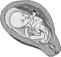
The diagnosis may be suspected if there is a decrease in the baby's heart rate during delivery. [1] Nuchal cords are typically checked for by running the finger over the baby's neck once the head has delivered. [4] Ultrasound may pick up the condition before labor. [1]

If detected during delivery, management includes trying to unwrap the cord or if this is not possible clamping and cutting the cord. [2] Delivery can typically take place as normal and outcomes are generally good. [5] [1] Rarely long term brain damage or cerebral palsy may occur. [1] [6] Nuchal cords occur in about a quarter of deliveries. [2] The condition has been described at least as early as 300 BC by Hippocrates. [1]

Signs and symptoms

Symptoms of a prior nuchal cord shortly after birth in the baby may include duskiness of face, facial petechia, and bleeding in the whites of the eye. [1] Complications can include meconium, respiratory disease, anemia, and still birth. [1]

Diagnosis

Doppler ultrasound showing a nuchal cord USNuchal.gif
Doppler ultrasound showing a nuchal cord

In 1962, J. Selwyn Crawford MD from the British Research Council defined a nuchal cord as one that is wrapped 360 degrees around the fetal neck. Crawford commented "It is all the more remarkable, therefore, that little work has been done. to analyze its effects during labor and delivery".[ citation needed ] To date, there is no prospective case control double-blind study looking at nuchal cords and observational studies vary in opinion as to the degree of poor outcomes. Also not included in these studies is which umbilical cord form (of the 8 different possible structures) was considered a nuchal cord.[ citation needed ]

Ultrasound diagnosis of a cord around the neck was first described in 1982. [7] “Coils occur in about 25% of cases and ordinarily do no harm, but occasionally they may be so tight that constriction of the umbilical vessels and consequent hypoxia result.”[ citation needed ] Williams Obstetrics 16th Edition, has only one single sentence in the entire textbook regarding cords around the neck. [8] By contrast, the First Edition of the Encyclopædia Britannica from 1770 had 20 pages of information about Umbilical Cord Pathology with drawings of Umbilical Cord Entanglement. The Royal College of Obstetricians and Gynaecologists has these images on its brochure. There are currently three recent texts on ultrasonography which demonstrate the ability of ultrasound to identify umbilical cord issues with reliability as of 2009.[ citation needed ]

A study published in 2004 was done to establish the sensitivity of ultrasound in the diagnosis of a nuchal cord. Each of 289 women, induced the same day, underwent a transabdominal ultrasound scan with an Aloka 1700 ultrasound machine with a 3.5 MHz abdominal probe, using gray-scale and color Doppler imaging immediately prior to induction of labor. Presence of the cord was sought in the transverse and sagittal plane of the neck. A nuchal cord was diagnosed if the cord was visualized lying around at least 3 of the 4 sides of the neck. A cord was actually present at delivery in 52 of the 289 women. Only 18 of the 52 cords or 35% of the nuchal cords were detected on ultrasound done immediately before delivery, and 65% of nuchal cords were not detected. Of the 237 cases where there was no cord at delivery, ultrasound had false positive results, i.e. diagnosed a cord in 44 of the 237 cases (19%) in which there was no cord present at all. In this study, ultrasound was only 35% accurate at finding a single loop, and only 60% accurate at detecting a nuchal cord wrapped multiple times around the neck. [9]

In no study was it possible by ultrasound to distinguish between a loose or a tight cord, although at least 3 attempted to do so.[ citation needed ] Peregrine [9] concludes that ultrasound diagnosis of nuchal cords will only be useful if doctors are able to do so reliably and predict which of those fetuses are likely to have a problem., However, perinatologists routinely look for umbilical cord issues in monoamniotic twins. Studies have shown an improvement in outcomes where cord entanglement was prenatally identified in these cases. Ultrasound measurement of the velocity of flow in the cord may be useful in the management of twins and chronically growth-retarded fetuses. Of course this depends on the training of the sonographer. To date there are no ultrasound courses which teach the identification of nuchal cord to physicians or technicians. A recent review by Wilson of the American Academy of Ultrasonography Technicians recommends the documentation of umbilical cord issues. [10]

Classification

Treatment

Management of a presenting nuchal cord should be tailored to prevent umbilical cord compression whenever possible. Techniques to preserve an intact nuchal cord depend on how tightly the cord is wrapped around the infant's neck. If the cord is loose, it can easily be slipped over the infant's head. The infant can be delivered normally and placed on maternal abdomen as desired. If the cord is too tight to go over the infant's head, the provider may be able to slip it over the infant's shoulders and deliver the body through the cord. The cord can then be unwrapped from around the baby after birth. Finally, if the cord is too tight to slip back over the shoulders, one may use the somersault maneuver to allow the body to be delivered. [12] The birth attendant may also choose to clamp and cut the umbilical cord to allow for vaginal delivery if other methods of nuchal cord management are not feasible.

Prognosis

Retrospective data of over 182,000 births, with the statistical power to determine even mild associations, suggest that a single or multiple nuchal cords at the time of delivery is not associated with adverse perinatal outcomes, is associated with higher birthweights and fewer caesarean sections in births. [13] [14] [15] Although some studies have found that a tight nuchal cord is associated with short term morbidity, it is unclear whether such outcomes are actually a result of the presence of the nuchal cord itself, or as a result of clamping and cutting the cord [16]

Related Research Articles

<span class="mw-page-title-main">Stillbirth</span> Death of a fetus before or during delivery, resulting in delivery of a dead baby

Stillbirth is typically defined as fetal death at or after 20 or 28 weeks of pregnancy, depending on the source. It results in a baby born without signs of life. A stillbirth can often result in the feeling of guilt or grief in the mother. The term is in contrast to miscarriage, which is an early pregnancy loss, and Sudden Infant Death Syndrome, where the baby dies a short time after being born alive.

<span class="mw-page-title-main">Umbilical cord</span> Conduit between embryo/fetus and the placenta

In placental mammals, the umbilical cord is a conduit between the developing embryo or fetus and the placenta. During prenatal development, the umbilical cord is physiologically and genetically part of the fetus and normally contains two arteries and one vein, buried within Wharton's jelly. The umbilical vein supplies the fetus with oxygenated, nutrient-rich blood from the placenta. Conversely, the fetal heart pumps low-oxygen, nutrient-depleted blood through the umbilical arteries back to the placenta.

<span class="mw-page-title-main">Obstetric ultrasonography</span> Use of medical ultrasonography in pregnancy

Obstetric ultrasonography, or prenatal ultrasound, is the use of medical ultrasonography in pregnancy, in which sound waves are used to create real-time visual images of the developing embryo or fetus in the uterus (womb). The procedure is a standard part of prenatal care in many countries, as it can provide a variety of information about the health of the mother, the timing and progress of the pregnancy, and the health and development of the embryo or fetus.

Fetal distress, also known as non-reassuring fetal status, is a condition during pregnancy or labor in which the fetus shows signs of inadequate oxygenation. Due to its imprecision, the term "fetal distress" has fallen out of use in American obstetrics. The term "non-reassuring fetal status" has largely replaced it. It is characterized by changes in fetal movement, growth, heart rate, and presence of meconium stained fluid.

<span class="mw-page-title-main">Placenta praevia</span> Medical condition

Placenta praevia is when the placenta attaches inside the uterus but in a position near or over the cervical opening. Symptoms include vaginal bleeding in the second half of pregnancy. The bleeding is bright red and tends not to be associated with pain. Complications may include placenta accreta, dangerously low blood pressure, or bleeding after delivery. Complications for the baby may include fetal growth restriction.

<span class="mw-page-title-main">Umbilical cord prolapse</span> Complication of pregnancy where the umbilical cord slips out of the uterus prior to birth

Umbilical cord prolapse is when the umbilical cord comes out of the uterus with or before the presenting part of the baby. The concern with cord prolapse is that pressure on the cord from the baby will compromise blood flow to the baby. It usually occurs during labor but can occur anytime after the rupture of membranes.

<span class="mw-page-title-main">Twin-to-twin transfusion syndrome</span> Medical condition

Twin-to-twin transfusion syndrome (TTTS), also known as feto-fetal transfusion syndrome (FFTS), twin oligohydramnios-polyhydramnios sequence (TOPS) and stuck twin syndrome, is a complication of monochorionic multiple pregnancies in which there is disproportionate blood supply between the fetuses. This leads to unequal levels of amniotic fluid between each fetus and usually leads to death of the undersupplied twin and, without treatment, usually death or a range of birth defects or disabilities for a surviving twin, such as underdeveloped, damaged or missing limbs, digits or organs, especially cerebral palsy.

<span class="mw-page-title-main">Placental abruption</span> Medical condition

Placental abruption is when the placenta separates early from the uterus, in other words separates before childbirth. It occurs most commonly around 25 weeks of pregnancy. Symptoms may include vaginal bleeding, lower abdominal pain, and dangerously low blood pressure. Complications for the mother can include disseminated intravascular coagulopathy and kidney failure. Complications for the baby can include fetal distress, low birthweight, preterm delivery, and stillbirth.

<span class="mw-page-title-main">Prelabor rupture of membranes</span> Medical condition

Prelabor rupture of membranes (PROM), previously known as premature rupture of membranes, is breakage of the amniotic sac before the onset of labor. Women usually experience a painless gush or a steady leakage of fluid from the vagina. Complications in the baby may include premature birth, cord compression, and infection. Complications in the mother may include placental abruption and postpartum endometritis.

<span class="mw-page-title-main">Chorioamnionitis</span> Medical condition

Chorioamnionitis, also known as intra-amniotic infection (IAI), is inflammation of the fetal membranes, usually due to bacterial infection. In 2015, a National Institute of Child Health and Human Development Workshop expert panel recommended use of the term "triple I" to address the heterogeneity of this disorder. The term triple I refers to intrauterine infection or inflammation or both and is defined by strict diagnostic criteria, but this terminology has not been commonly adopted although the criteria are used.

<span class="mw-page-title-main">EXIT procedure</span>

The EXIT procedure, or ex utero intrapartum treatment procedure, is a specialized surgical delivery procedure used to deliver babies who have airway compression. Causes of airway compression in newborn babies result from a number of rare congenital disorders, including bronchopulmonary sequestration, congenital cystic adenomatoid malformation, mouth or neck tumor such as teratoma, and lung or pleural tumor such as pleuropulmonary blastoma. Airway compression discovered at birth is a medical emergency. In many cases, however, the airway compression is discovered during prenatal ultrasound exams, permitting time to plan a safe delivery using the EXIT procedure or other means.

Placental insufficiency or utero-placental insufficiency is the failure of the placenta to deliver sufficient nutrients to the fetus during pregnancy, and is often a result of insufficient blood flow to the placenta. The term is also sometimes used to designate late decelerations of fetal heart rate as measured by cardiotocography or an NST, even if there is no other evidence of reduced blood flow to the placenta, normal uterine blood flow rate being 600mL/min.

<span class="mw-page-title-main">Vaginal delivery</span> Delivery through the vagina

A vaginal delivery is the birth of offspring in mammals through the vagina. It is the most common method of childbirth worldwide. It is considered the preferred method of delivery, with lower morbidity and mortality than caesarean sections (C-sections).

<span class="mw-page-title-main">Velamentous cord insertion</span> Velamentous placenta

Velamentous cord insertion is a complication of pregnancy where the umbilical cord is inserted in the fetal membranes. It is a major cause of antepartum hemorrhage that leads to loss of fetal blood and associated with high perinatal mortality. In normal pregnancies, the umbilical cord inserts into the middle of the placental mass and is completely encased by the amniotic sac. The vessels are hence normally protected by Wharton's jelly, which prevents rupture during pregnancy and labor. In velamentous cord insertion, the vessels of the umbilical cord are improperly inserted in the chorioamniotic membrane, and hence the vessels traverse between the amnion and the chorion towards the placenta. Without Wharton's jelly protecting the vessels, the exposed vessels are susceptible to compression and rupture.

An obstetric labor complication is a difficulty or abnormality that arises during the process of labor or delivery.

<span class="mw-page-title-main">Monoamniotic twins</span> Identical twins sharing the same amniotic sac in the womb

Monoamniotic twins are identical or semi-identical twins that share the same amniotic sac within their mother's uterus. Monoamniotic twins are always monochorionic and are usually termed Monoamniotic-Monochorionic twins. They share the placenta, but have two separate umbilical cords. Monoamniotic twins develop when an embryo does not split until after formation of the amniotic sac, at about 9–13 days after fertilization. Monoamniotic triplets or other monoamniotic multiples are possible, but extremely rare. Other obscure possibilities include multiples sets where monoamniotic twins are part of a larger gestation such as triplets, quadruplets, or more.

<span class="mw-page-title-main">Single umbilical artery</span> Medical condition

Occasionally, there is a single umbilical artery (SUA) present in the umbilical cord, as opposed to the usual two. This is sometimes also called a two-vessel umbilical cord, or two-vessel cord. Approximately, this affects between 1 in 100 and 1 in 500 pregnancies, making it the most common umbilical abnormality. Its cause is not known.

<span class="mw-page-title-main">Emergency childbirth</span>

Emergency childbirth is the precipitous birth of an infant in an unexpected setting. In planned childbirth, mothers choose the location and obstetric team ahead of time. Options range from delivering at home, at a hospital, a medical facility or a birthing center. Sometimes, birth can occur on the way to these facilities, without a healthcare team. The rates of unplanned childbirth are low. If the birth is imminent, emergency measures may be needed. Emergency services can be contacted for help in some countries.

The anomaly scan, also sometimes called the anatomy scan, 20-week ultrasound, or level 2 ultrasound, evaluates anatomic structures of the fetus, placenta, and maternal pelvic organs. This scan is an important and common component of routine prenatal care. The function of the ultrasound is to measure the fetus so that growth abnormalities can be recognized quickly later in pregnancy, to assess for congenital malformations and multiple pregnancies, and to plan method of delivery.

Epignathus is a rare teratoma of the oropharynx. Epignathus is a form of oropharyngeal teratoma that arises from the palate and, in most cases, results in death. The pathology is thought to be due to unorganized and uncontrolled differentiation of somatic cells leading to formation of the teratoma; sometimes it is also referred to as "fetus-in-fetu", which is an extremely rare occurrence of an incomplete but parasitic fetus located in the body of its twin. This tumor is considered benign but life-threatening because of its atypical features and high risk of airway obstruction, which is the cause of death in 80-100% of the cases at the time of delivery.

References

  1. 1 2 3 4 5 6 7 8 9 10 11 12 13 14 15 16 17 18 Peesay M (6 December 2017). "Nuchal cord and its implications". Maternal Health, Neonatology and Perinatology. 3 (1): 28. doi: 10.1186/s40748-017-0068-7 . PMC   5719938 . PMID   29234502.
  2. 1 2 3 4 "Nuchal Cord". Merck Manuals Consumer Version. June 2018. Retrieved 2 October 2018.
  3. Hasegawa J, Matsuoka R, Ichizuka K, Sekizawa A, Okai T (March 2009). "Ultrasound diagnosis and management of umbilical cord abnormalities". Taiwanese Journal of Obstetrics & Gynecology. 48 (1): 23–7. doi: 10.1016/S1028-4559(09)60031-0 . PMID   19346188.
  4. Ferri FF (2014). Ferri's Clinical Advisor 2015 E-Book: 5 Books in 1. Elsevier Health Sciences. p. e23. ISBN   9780323084307.
  5. Adams JG (2012). Emergency Medicine E-Book: Clinical Essentials (Expert Consult -- Online). Elsevier Health Sciences. p. 1064. ISBN   978-1455733941.
  6. MacLennan AH, Thompson SC, Gecz J (December 2015). "Cerebral palsy: causes, pathways, and the role of genetic variants" (PDF). American Journal of Obstetrics and Gynecology. 213 (6): 779–88. doi: 10.1016/j.ajog.2015.05.034 . PMID   26003063.
  7. Jouppila P, Kirkinen P (February 1982). "Ultrasonic diagnosis of nuchal encirclement by the umbilical cord: a case and methodological report". Journal of Clinical Ultrasound. 10 (2): 59–62. doi:10.1002/jcu.1870100205. PMID   6804502. S2CID   5976372.
  8. Williams JW (1980). Williams Obstetrics 16th Edition . Appleton & Lange, US. pp.  421. ISBN   978-0838597316.
  9. 1 2 Peregrine E, O'Brien P, Jauniaux E (February 2005). "Ultrasound detection of nuchal cord prior to labor induction and the risk of Cesarean section". Ultrasound in Obstetrics & Gynecology. 25 (2): 160–4. doi:10.1097/01.ogx.0000172319.27668.34. PMID   15543520.
  10. Wilson B (March–April 2008). "Sonography of the Placenta And Umbilical Cord". Radiologic Technology. 79: 333S–345S. Retrieved December 26, 2017.
  11. Collins JH (February 2002). "Umbilical cord accidents: human studies". Seminars in Perinatology. 26 (1): 79–82. doi:10.1053/sper.2002.29860. PMID   11876571.
  12. Reynolds L (March 1999). "Practice tips. "Somersault" maneuver for a tight umbilical cord". Canadian Family Physician. 45: 613. PMC   2328444 . PMID   10099799.
  13. Mastrobattista JM, Hollier LM, Yeomans ER, Ramin SM, Day MC, Sosa A, Gilstrap LC (February 2005). "Effects of nuchal cord on birthweight and immediate neonatal outcomes". American Journal of Perinatology. 22 (2): 83–5. doi:10.1055/s-2005-837737. PMID   15731986.
  14. Schäffer L, Burkhardt T, Zimmermann R, Kurmanavicius J (July 2005). "Nuchal cords in term and postterm deliveries--do we need to know?". Obstetrics and Gynecology. 106 (1): 23–8. doi:10.1097/01.AOG.0000165322.42051.0f. PMID   15994613. S2CID   33991885.
  15. Sheiner E, Abramowicz JS, Levy A, Silberstein T, Mazor M, Hershkovitz R (May 2006). "Nuchal cord is not associated with adverse perinatal outcome". Archives of Gynecology and Obstetrics. 274 (2): 81–3. doi:10.1007/s00404-005-0110-2. PMID   16374604. S2CID   31359895.
  16. Reed R, Barnes M, Allan J (February 2009). "Nuchal cords: sharing the evidence with parents". British Journal of Midwifery. 17 (2): 106–109. doi:10.12968/bjom.2009.17.2.39379.