Hemolytic disease of the newborn (anti-Rhc)

Last updated
HDN due to anti-Rhc alloimmunization
Specialty Hematology

Hemolytic disease of the newborn (anti-Rhc) can range from a mild to a severe disease. It is the third most common cause of severe HDN. Rh disease is the most common and hemolytic disease of the newborn (anti-Kell) is the second most common cause of severe HDN. It occurs more commonly in women who are Rh D negative.[ citation needed ]

Contents

Presentation

Complications

Transfusion reactions

Once a woman has antibodies, she is at high risk for a transfusion reaction. [10] For this reason, she must carry a medical alert card at all times and inform all doctors of her antibody status.[ citation needed ]

"Acute hemolytic transfusion reactions may be either immune-mediated or nonimmune-mediated. Immune-mediated hemolytic transfusion reactions caused by immunoglobulin M (IgM) anti-A, anti-B, or anti-A,B typically result in severe, potentially fatal complement-mediated intravascular hemolysis. Immune-mediated hemolytic reactions caused by IgG, Rh, Kell, Duffy, or other non-ABO antibodies typically result in extravascular sequestration, shortened survival of transfused red cells, and relatively mild clinical reactions. Acute hemolytic transfusion reactions due to immune hemolysis may occur in patients who have no antibodies detectable by routine laboratory procedures." [11]

Causes

A Rhc negative mother can become sensitised by red blood cell (RBC) Rhc antigens by her first pregnancy with a Rhc positive fetus. The mother can make IgG anti-Rhc antibodies, which are able to pass through the placenta and enter the fetal circulation. If the fetus is Rhc positive alloimmune hemolysis can occur leading to HDN. This is similar as for Rh disease, which is usually caused when a RhD negative mother is sensitised by her first pregnancy with a RhD positive fetus.[ citation needed ]

Sensitization to Rhc antigens can also be caused by blood transfusion.[ citation needed ]

Diagnosis

Testing for HDN involves blood work from both mother and father, and may also include assessment with amniocentesis and Middle Cerebral Artery scans.[ citation needed ]

Anti-C and anti-c can both show a negative DAT but still have a severely affected infant. [12] [13] An indirect coombs must also be run.

In the case of anti-c, the woman should be checked around 28 weeks to see if she has developed anti-E as well.[ citation needed ]

Mother

Blood testing for the mother is called an Indirect Coombs Test (ICT) or an Indirect Agglutination Test (IAT). This test tells whether there are antibodies in the maternal plasma. If positive, the antibody is identified and given a titer. Critical titers are associated with significant risk of fetal anemia and hydrops. [14] Titers of 1:8 or higher is considered critical for Kell. Titers of 1:16 or higher are considered critical for all other antibodies. After critical titer is reached, care is based on MCA scans. If antibodies are low and have a sudden jump later in pregnancy, an MCA scan is warranted. If the titer undergoes a 4 fold increase, it should be considered significant regardless of if the critical value has been reached. Maternal titers are not useful in predicting fetal anemia after the first affected gestation and should not be used for the basis of care. [15] Titers are tested monthly until 24 weeks, after which they are done every 2 weeks. [16]

"In only 2 situations are patients not monitored identically to patients who are Rh sensitized. The first is that of alloimmunization to the c, E, or, C antigens. Some concern exists that hemolysis may occur in these patients with a lower than 1:16 titer. Thus, if the initial titer is 1:4 and stable but increases at 26 weeks' gestation to 1:8, assessment with MCA Doppler velocity at that point is reasonable. However, if the patient presents in the first trimester with a 1:8 titer that remains stable at 1:8 throughout the second trimester, continued serial antibody titers are appropriate. The second situation in which patients should not be treated identically to patients who are Rh D sensitized is that of Kell isoimmunization because several cases of severe fetal hemolysis with anti-Kell antibodies have occurred in the setting of low titers." [14]

In the case of a positive ICT, the woman must carry a medical alert card or bracelet for life because of the risk of a transfusion reaction. [17]

Father

Blood is generally drawn from the father to help determine fetal antigen status. [18] If he is homozygous for the antigen, there is a 100% chance of all offspring in the pairing to be positive for the antigen and at risk for HDN. If he is heterozygous, there is a 50% chance of offspring to be positive for the antigen. [19] This test can help with knowledge for the current baby, as well as aid in the decision about future pregnancies. With RhD, the test is called the RhD genotype. With RhCE, and Kell antigen it is called an antigen phenotype. [20]

Fetus

There are 3 possible ways to test the fetal antigen status. Free Cell DNA, Amniocentesis, and Chorionic Villus Sampling (CVS). Of the three, CVS is no longer used due to risk of worsening the maternal antibody response. Once antigen status has been determined, assessment may be done with MCA scans.[ citation needed ]

MCA scans: Middle cerebral artery - peak systolic velocity is changing the way sensitized pregnancies are managed. [22] This test is done noninvasively with ultrasound. By measuring the peak velocity of blood flow in the middle cerebral artery, a MoM (multiple of the median) score can be calculated. MoM of 1.5 or greater indicates severe anemia and should be treated with IUT. [23] [22]

Prevention

It has been suggested that women of child-bearing age or young girls should not be given a transfusion with Rhc positive blood (or Kell 1 positive blood for similar reasons). This would require a lot of extra work in blood transfusion departments and it is considered not economical to do the blood group screening at the present time.[ citation needed ]

It is theoretically likely that IgG anti-Rhc antibody injections would prevent sensitization to RBC surface Rhc antigens in a similar way that IgG anti-D antibodies (Rho(D) immune globulin) are used to prevent Rh disease, but the methods for IgG anti-Rhc antibodies have not been developed at the present time.[ citation needed ]

Treatment

There are several intervention options available in early, mid and late pregnancies.

Early pregnancy

Mid to late pregnancy

After Birth

Testing

In some cases, the direct coombs will be negative but severe, even fatal HDN can occur. [12] An indirect coombs needs to be run in cases of anti-C, [13] anti-c, [13] and anti-M. Anti-M also recommends antigen testing to rule out the presence of HDN. [26]

Treatment

History

Hemolytic disease of the fetus and newborn (HDN) is a condition where the passage of maternal antibodies results in the hemolysis of fetal/neonatal red cells. The antibodies can be naturally occurring such as anti-A, and anti-B, or immune antibodies developed following a sensitizing event. [38] Isoimmunization occurs when the maternal immune system is sensitized to red blood cell surface antigens. The most common causes of isoimmunization are blood transfusion, and fetal-maternal hemorrhage. [16] The hemolytic process can result in anemia, hyperbilirubinemia, neonatal thrombocytopenia, and neonatal neutropenia. [5] With the use of RhD Immunoprophylaxis, (commonly called Rhogam), the incidence of anti-D has decreased dramatically and other alloantibodies are now a major cause of HDN. [38]

See also

Related Research Articles

<span class="mw-page-title-main">Blood type</span> Classification of blood based on antibodies and antigens on red blood cell surfaces

A blood type is a classification of blood, based on the presence and absence of antibodies and inherited antigenic substances on the surface of red blood cells (RBCs). These antigens may be proteins, carbohydrates, glycoproteins, or glycolipids, depending on the blood group system. Some of these antigens are also present on the surface of other types of cells of various tissues. Several of these red blood cell surface antigens can stem from one allele and collectively form a blood group system.

Rh disease is a type of hemolytic disease of the fetus and newborn (HDFN). HDFN due to anti-D antibodies is the proper and currently used name for this disease as the Rh blood group system actually has more than 50 antigens and not only the D-antigen. The term "Rh Disease" is commonly used to refer to HDFN due to anti-D antibodies, and prior to the discovery of anti-Rho(D) immune globulin, it was the most common type of HDFN. The disease ranges from mild to severe, and occurs in the second or subsequent pregnancies of Rh-D negative women when the biologic father is Rh-D positive.

<span class="mw-page-title-main">Hemolytic disease of the newborn</span> Fetal and neonatal alloimmune blood condition

Hemolytic disease of the newborn, also known as hemolytic disease of the fetus and newborn, HDN, HDFN, or erythroblastosis foetalis, is an alloimmune condition that develops in a fetus at or around birth, when the IgG molecules produced by the mother pass through the placenta. Among these antibodies are some which attack antigens on the red blood cells in the fetal circulation, breaking down and destroying the cells. The fetus can develop reticulocytosis and anemia. The intensity of this fetal disease ranges from mild to very severe, and fetal death from heart failure can occur. When the disease is moderate or severe, many erythroblasts are present in the fetal blood, earning these forms of the disease the name erythroblastosis fetalis.

A Coombs test, also known as antiglobulin test (AGT), is either of two blood tests used in immunohematology. They are the direct and indirect Coombs tests. The direct Coombs test detects antibodies that are stuck to the surface of the red blood cells. Since these antibodies sometimes destroy red blood cells, a person can be anemic and this test can help clarify the condition. The indirect Coombs detects antibodies that are floating freely in the blood. These antibodies could act against certain red blood cells and the test can be done to diagnose reactions to a blood transfusion.

<span class="mw-page-title-main">Neonatal jaundice</span> Medical condition

Neonatal jaundice is a yellowish discoloration of the white part of the eyes and skin in a newborn baby due to high bilirubin levels. Other symptoms may include excess sleepiness or poor feeding. Complications may include seizures, cerebral palsy, or kernicterus.

Rho(D) immune globulin (RhIG) is a medication used to prevent RhD isoimmunization in mothers who are RhD negative and to treat idiopathic thrombocytopenic purpura (ITP) in people who are Rh positive. It is often given both during and following pregnancy. It may also be used when RhD-negative people are given RhD-positive blood. It is given by injection into muscle or a vein. A single dose lasts 12 weeks. It is made from human blood plasma.

In ABO hemolytic disease of the newborn maternal IgG antibodies with specificity for the ABO blood group system pass through the placenta to the fetal circulation where they can cause hemolysis of fetal red blood cells which can lead to fetal anemia and HDN. In contrast to Rh disease, about half of the cases of ABO HDN occur in a firstborn baby and ABO HDN does not become more severe after further pregnancies.

Hemolytic disease of the newborn (anti-Kell1) is the second most common cause of severe hemolytic disease of the newborn (HDN) after Rh disease. Anti-Kell1 is becoming relatively more important as prevention of Rh disease is also becoming more effective.

The Kell antigen system is a human blood group system, that is, a group of antigens on the human red blood cell surface which are important determinants of blood type and are targets for autoimmune or alloimmune diseases which destroy red blood cells. The Kell antigens are K, k, Kpa, Kpb, Jsa and Jsb. The Kell antigens are peptides found within the Kell protein, a 93-kilodalton transmembrane zinc-dependent endopeptidase which is responsible for cleaving endothelin-3.

<span class="mw-page-title-main">Colton antigen system</span> Blood antigen system

The Colton antigen system (Co) is present on the membranes of red blood cells and in the tubules of the kidney and helps determine a person's blood type. The Co antigen is found on a protein called aquaporin-1 which is responsible for water homeostasis and urine concentration.

<span class="mw-page-title-main">Rh blood group system</span> Human blood group system involving 49 blood antigens

The Rh blood group system is a human blood group system. It contains proteins on the surface of red blood cells. After the ABO blood group system, it is the most likely to be involved in transfusion reactions. The Rh blood group system consisted of 49 defined blood group antigens in 2005. As of 2023, there are over 50 antigens among which the five antigens D, C, c, E, and e are the most important. There is no d antigen. Rh(D) status of an individual is normally described with a positive (+) or negative (−) suffix after the ABO type. The terms Rh factor, Rh positive, and Rh negative refer to the Rh(D) antigen only. Antibodies to Rh antigens can be involved in hemolytic transfusion reactions and antibodies to the Rh(D) and Rh antigens confer significant risk of hemolytic disease of the fetus and newborn

Neonatal alloimmune thrombocytopenia is a disease that affects babies in which the platelet count is decreased because the mother's immune system attacks her fetus' or newborn's platelets. A low platelet count increases the risk of bleeding in the fetus and newborn. If the bleeding occurs in the brain, there may be long-term effects.

Hemolytic disease of the newborn (anti-RhE) is caused by the anti-RhE antibody of the Rh blood group system. The anti-RhE antibody can be naturally occurring, or arise following immune sensitization after a blood transfusion or pregnancy.

Acquired hemolytic anemia can be divided into immune and non-immune mediated forms of hemolytic anemia.

Neonates are defined as babies up to 28 days after birth. Most extremely preterm babies require at least one red cell transfusion; this is partly due to the amount of blood removed with blood samples compared to the baby's total blood volume and partly due to anemia of prematurity. Most transfusions are given as small volume top-up transfusions to increase the baby's hemoglobin above a certain pre-defined level, or because the baby is unwell due to the anemia. Possible side-effects of anemia in babies can be poor growth, lethargy and episodes of apnea. Exchange blood transfusion is used to treat a rapidly rising bilirubin that does not respond to treatment with phototherapy or intravenous immunoglobulin. This is usually due to hemolytic disease of the newborn, but may also be due to other causes, e.g., G6PD deficiency.

Rh factor testing, also known as Rhesus factor testing, is the procedure of determining the rhesus D status of an individual.

An Intrauterine transfusion (IUT) is a procedure that provides blood to a fetus, most commonly through the umbilical cord. It is used in cases of severe fetal anemia, such as when fetal red blood cells are being destroyed by maternal antibodies. IUTs are performed by perinatologists at hospitals or specialized centers.

The Junior blood group system is a human blood group defined by the presence or absence of the Jr(a) antigen, a high-frequency antigen that is found on the red blood cells of most individuals. People with the rare Jr(a) negative blood type can develop anti-Jr(a) antibodies, which may cause transfusion reactions and hemolytic disease of the newborn on subsequent exposures. Jr(a) negative blood is most common in people of Japanese heritage.

<span class="mw-page-title-main">Ruth Darrow</span> American pathologist

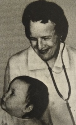
Ruth Renter Darrow (1895–1956) was an American pathologist who was the first to identify the cause of hemolytic disease of the newborn (HDN). In 1938, three years prior to the discovery of antibodies against the Rh antigen, Darrow correctly hypothesized that the disease was caused by destruction of red blood cells due to antibodies in the mother's blood. Darrow's research was inspired by her personal experiences with the disease.

<span class="mw-page-title-main">Blood compatibility testing</span> Testing to identify incompatibilities between blood types

Blood compatibility testing is conducted in a medical laboratory to identify potential incompatibilities between blood group systems in blood transfusion. It is also used to diagnose and prevent some complications of pregnancy that can occur when the baby has a different blood group from the mother. Blood compatibility testing includes blood typing, which detects the antigens on red blood cells that determine a person's blood type; testing for unexpected antibodies against blood group antigens ; and, in the case of blood transfusions, mixing the recipient's plasma with the donor's red blood cells to detect incompatibilities (crossmatching). Routine blood typing involves determining the ABO and RhD type, and involves both identification of ABO antigens on red blood cells and identification of ABO antibodies in the plasma. Other blood group antigens may be tested for in specific clinical situations.

References

  1. 1 2 3 4 5 6 Murray, N. A; Roberts, I. A G (2007). "Haemolytic disease of the newborn". Archives of Disease in Childhood: Fetal and Neonatal Edition. 92 (2): F83–8. doi:10.1136/adc.2005.076794. PMC   2675453 . PMID   17337672.
  2. Shapiro, Steven M (2004). "Definition of the Clinical Spectrum of Kernicterus and Bilirubin-Induced Neurologic Dysfunction (BIND)". Journal of Perinatology. 25 (1): 54–9. doi:10.1038/sj.jp.7211157. PMID   15578034. S2CID   19663259.
  3. Blair, Eve; Watson, Linda (2006). "Epidemiology of cerebral palsy". Seminars in Fetal and Neonatal Medicine. 11 (2): 117–25. doi:10.1016/j.siny.2005.10.010. PMID   16338186.
  4. Lande, Lottie (1948). "Clinical signs and development of survivors of kernicterus due to Rh sensitization". The Journal of Pediatrics. 32 (6): 693–705. doi:10.1016/S0022-3476(48)80225-8. PMID   18866937.
  5. 1 2 3 4 5 Koenig, J. M.; Christensen, R. D. (1989). "Neutropenia and thrombocytopenia in infants with Rh hemolytic disease". The Journal of Pediatrics. 114 (4 Pt 1): 625–31. doi:10.1016/s0022-3476(89)80709-7. PMID   2494315.
  6. 1 2 Lalezari, P; Nussbaum, M; Gelman, S; Spaet, T. H. (1960). "Neonatal neutropenia due to maternal isoimmunization". Blood. 15 (2): 236–43. doi: 10.1182/blood.V15.2.236.236 . PMID   14413526.
  7. 1 2 Rath, M. E. A.; Smits-Wintjens, V. E. H. J.; Oepkes, D.; Walther, F. J.; Lopriore, E. (2013). "Iron status in infants with alloimmune haemolytic disease in the first three months of life". Vox Sanguinis. 105 (4): 328–33. doi:10.1111/vox.12061. PMID   23802744. S2CID   24789324.
  8. Mitchell, S; James, A (1999). "Severe late anemia of hemolytic disease of the newborn". Paediatrics & Child Health. 4 (3): 201–3. doi:10.1093/pch/4.3.201. PMC   2828194 . PMID   20212966.
  9. Al-Alaiyan, S.; Al Omran, A. (1999). "Late hyporegenerative anemia in neonates with rhesus hemolytic disease". Journal of Perinatal Medicine. 27 (2): 112–5. doi:10.1515/JPM.1999.014. PMID   10379500. S2CID   32155893.
  10. Strobel, Erwin (2008). "Hemolytic Transfusion Reactions". Transfusion Medicine and Hemotherapy. 35 (5): 346–353. doi:10.1159/000154811. PMC   3076326 . PMID   21512623.
  11. Transfusion Reactions at eMedicine
  12. 1 2 Heddle, N. M.; Wentworth, P.; Anderson, D. R.; Emmerson, D.; Kelton, J. G.; Blajchman, M. A. (1995). "Three examples of Rh haemolytic disease of the newborn with a negative direct antiglobulin test". Transfusion Medicine. 5 (2): 113–6. doi:10.1111/j.1365-3148.1995.tb00197.x. PMID   7655573. S2CID   21936425.
  13. 1 2 3 4 Hemolytic Disease of Newborn~workup at eMedicine
  14. 1 2 3 4 Erythrocyte Alloimmunization and Pregnancy at eMedicine
  15. 1 2 3 Hemolytic Disease of Newborn~treatment at eMedicine
  16. 1 2 3 4 Cacciatore, A; Rapiti, S; Carrara, S; Cavaliere, A; Ermito, S; Dinatale, A; Imbruglia, L; Recupero, S; La Galia, T; Pappalardo, E. M.; Accardi, M. C. (2009). "Obstetric management in Rh alloimmunizated pregnancy". Journal of Prenatal Medicine. 3 (2): 25–7. PMC   3279102 . PMID   22439037.
  17. Strobel, Erwin (2008). "Hemolytic Transfusion Reactions". Transfusion Medicine and Hemotherapy. 35 (5): 346–353. doi: 10.1159/000154811 . PMC   3076326 . PMID   21512623.
  18. Scheffer, PG; Van Der Schoot, CE; Page-Christiaens, Gcml; De Haas, M (2011). "Noninvasive fetal blood group genotyping of rhesus D, c, E and of K in alloimmunised pregnant women: Evaluation of a 7-year clinical experience". BJOG: An International Journal of Obstetrics & Gynaecology. 118 (11): 1340–8. doi:10.1111/j.1471-0528.2011.03028.x. PMID   21668766. S2CID   32946225.
  19. Transfusion Medicine and Hemostasis: Clinical and Laboratory Aspects ISBN   978-0-12-397788-5 [ page needed ][ full citation needed ]
  20. https://www.aacc.org/publications/cln/articles/2015/march/molecular-typing-for-red-blood-cell-antigens%5B%5D
  21. Finning, Kirstin; Martin, Peter; Summers, Joanna; Daniels, Geoff (2007). "Fetal genotyping for the K (Kell) and Rh C, c, and E blood groups on cell-free fetal DNA in maternal plasma". Transfusion. 47 (11): 2126–33. doi:10.1111/j.1537-2995.2007.01437.x. PMID   17958542. S2CID   8292568.
  22. 1 2 Mari, Giancarlo; Deter, Russell L.; Carpenter, Robert L.; Rahman, Feryal; Zimmerman, Roland; Moise, Kenneth J.; Dorman, Karen F.; Ludomirsky, Avi; Gonzalez, Rogelio; Gomez, Ricardo; Oz, Utku; Detti, Laura; Copel, Joshua A.; Bahado-Singh, Ray; Berry, Stanley; Martinez-Poyer, Juan; Blackwell, Sean C. (2000). "Noninvasive Diagnosis by Doppler Ultrasonography of Fetal Anemia Due to Maternal Red-Cell Alloimmunization". New England Journal of Medicine. 342 (1): 9–14. doi:10.1056/NEJM200001063420102. PMID   10620643.
  23. Mari, G. (2005). "Middle cerebral artery peak systolic velocity for the diagnosis of fetal anemia: The untold story". Ultrasound in Obstetrics and Gynecology. 25 (4): 323–30. doi:10.1002/uog.1882. PMID   15789353. S2CID   12342034.
  24. Voto, L. S.; Mathet, E. R.; Zapaterio, J. L.; Orti, J; Lede, R. L.; Margulies, M (1997). "High-dose gammaglobulin (IVIG) followed by intrauterine transfusions (IUTs): A new alternative for the treatment of severe fetal hemolytic disease". Journal of Perinatal Medicine. 25 (1): 85–8. doi:10.1515/jpme.1997.25.1.85. PMID   9085208. S2CID   22822621.
  25. Novak, Deborah J.; Tyler, Lisa N.; Reddy, Ramakrishna L.; Barsoom, Michael J. (2008). "Plasmapheresis and intravenous immune globulin for the treatment of D alloimmunization in pregnancy". Journal of Clinical Apheresis. 23 (6): 183–5. doi:10.1002/jca.20180. PMID   19003884. S2CID   206013087.
  26. 1 2 Arora, Satyam; Doda, Veena; Maria, Arti; Kotwal, Urvershi; Goyal, Saurabh (2015). "Maternal anti-M induced hemolytic disease of newborn followed by prolonged anemia in newborn twins". Asian Journal of Transfusion Science. 9 (1): 98–101. doi:10.4103/0973-6247.150968. PMC   4339947 . PMID   25722586.
  27. Palfi, Miodrag; Hildén, Jan-Olof; Matthiesen, Leif; Selbing, Anders; Berlin, Gösta (2006). "A case of severe Rh (D) alloimmunization treated by intensive plasma exchange and high-dose intravenous immunoglobulin". Transfusion and Apheresis Science. 35 (2): 131–6. doi:10.1016/j.transci.2006.07.002. PMID   17045529.
  28. Ruma, Michael S.; Moise, Kenneth J.; Kim, Eunhee; Murtha, Amy P.; Prutsman, Wendy J.; Hassan, Sonia S.; Lubarsky, Suzanne L. (2007). "Combined plasmapheresis and intravenous immune globulin for the treatment of severe maternal red cell alloimmunization". American Journal of Obstetrics and Gynecology. 196 (2): 138.e1–6. doi:10.1016/j.ajog.2006.10.890. PMID   17306655.
  29. Deka, Dipika (2016). "Intrauterine Transfusion". Journal of Fetal Medicine. 27 (3): 13–17. doi: 10.1007/s40556-016-0072-4 . PMID   26811110. S2CID   42005756.
  30. 1 2 3 "UpToDate".
  31. https://www.mombaby.org/wp-content/uploads/2016/03/UNC-Isoimmunization-Detection-Prevention.pdf%5B%5D%5B%5D
  32. Rimon, E.; Peltz, R.; Gamzu, R.; Yagel, S.; Feldman, B.; Chayen, B.; Achiron, R.; Lipitz, S. (2006). "Management of Kell isoimmunization — evaluation of a Doppler-guided approach". Ultrasound in Obstetrics and Gynecology. 28 (6): 814–20. doi:10.1002/uog.2837. PMID   16941575.
  33. "Archived copy" (PDF). Archived from the original (PDF) on 2021-03-09. Retrieved 2017-02-15.{{cite web}}: CS1 maint: archived copy as title (link)[ full citation needed ]
  34. 1 2 3 American Academy of Pediatrics Subcommittee on Hyperbilirubinemia. (2004). "Management of hyperbilirubinemia in the newborn infant 35 or more weeks of gestation". Pediatrics. 114 (1): 297–316. doi:10.1542/peds.114.1.297. PMID   15231951.
  35. Onesimo, Roberta; Rizzo, Daniela; Ruggiero, Antonio; Valentini, Piero (2010). "Intravenous Immunoglobulin therapy for anti-E hemolytic disease in the newborn". The Journal of Maternal-Fetal & Neonatal Medicine. 23 (9): 1059–61. doi:10.3109/14767050903544751. PMID   20092394. S2CID   25144401.
  36. Gottstein, R; Cooke, R. W. (2003). "Systematic review of intravenous immunoglobulin in haemolytic disease of the newborn". Archives of Disease in Childhood: Fetal and Neonatal Edition. 88 (1): F6–10. doi:10.1136/fn.88.1.F6. PMC   1755998 . PMID   12496219.
  37. Hemolytic Disease of Newborn~followup at eMedicine
  38. 1 2 Basu, Sabita; Kaur, Ravneet; Kaur, Gagandeep (2011). "Hemolytic disease of the fetus and newborn: Current trends and perspectives". Asian Journal of Transfusion Science. 5 (1): 3–7. doi:10.4103/0973-6247.75963. PMC   3082712 . PMID   21572705.

Further reading