Chorion

Last updated
Chorion
Chicken egg diagram.svg
Diagram showing the chorion of a chicken egg
Gray30.png
Human fetus enclosed in the amnion
Details
Identifiers
Latin chorion
MeSH D002823
TE E5.11.3.1.1.0.3
Anatomical terminology

The chorion is the outermost fetal membrane around the embryo in mammals, birds and reptiles (amniotes). It develops from an outer fold on the surface of the yolk sac, which lies outside the zona pellucida (in mammals), known as the vitelline membrane in other animals. In insects, it is developed by the follicle cells while the egg is in the ovary. [1] Some mollusks also have chorions as part of their eggs. For example, fragile octopus eggs have only a chorion as their envelope. [2]

Contents

Structure

In humans and other mammals (excluding monotremes), the chorion is one of the fetal membranes that exist during pregnancy between the developing fetus and mother. The chorion and the amnion together form the amniotic sac. In humans it is formed by extraembryonic mesoderm and the two layers of trophoblast that surround the embryo and other membranes; the chorionic villi emerge from the chorion, invade the endometrium, and allow the transfer of nutrients from maternal blood to fetal blood.

Layers

The chorion consists of two layers: an outer formed by the trophoblast, and an inner formed by the somatic mesoderm.

The trophoblast is made up of an internal layer of cubical or prismatic cells, the cytotrophoblast or layer of Langhans, and an external multinucleated layer, the syncytiotrophoblast.

Growth

The chorion undergoes rapid proliferation and forms numerous processes, the chorionic villi , which invade and destroy the uterine decidua, while simultaneously absorbing nutritive materials from it for the growth of the embryo.

The chorionic villi are at first small and non-vascular, and consist of the trophoblast only, but they increase in size and ramify, whereas the mesoderm, carrying branches of the umbilical vessels, grows into them, and they are vascularized.

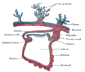
Blood is carried to the villi by the paired umbilical arteries, which branch into chorionic arteries and enter the chorionic villi as cotyledon arteries. After circulating through the capillaries of the villi, the blood is returned to the embryo by the umbilical vein. Until about the end of the second month of pregnancy, the villi cover the entire chorion, and are almost uniform in size; but, after this, they develop unequally.

Parts

Placenta with attached fetal membranes (ruptured at the margin at the left in the image), which consists of the chorion (outer layer) and amnion (inner layer). Placenta with fetal membranes.jpg
Placenta with attached fetal membranes (ruptured at the margin at the left in the image), which consists of the chorion (outer layer) and amnion (inner layer).

The part of the chorion that is in contact with the decidua capsularis undergoes atrophy, so that by the fourth month scarcely a trace of the villi is left. This part of the chorion becomes smooth, [3] and is named the chorion laeve (from the Latin word levis, meaning smooth). As it takes no share in the formation of the placenta, this is also named the non-placental part of the chorion. As the chorion grows, the chorion laeve comes in contact with the decidua parietalis and these layers fuse.

The villi at the embryonic pole, which is in contact with the decidua basalis, increase greatly in size and complexity, and hence this part is named the chorion frondosum. [3]

Thus the placenta develops from the chorion frondosum and the decidua basalis.

Monochorionic twins

Monochorionic twins are twins that share the same placenta. This occurs in 0.3% of all pregnancies, [4] and in 75% of monozygotic (identical) twins, when the split takes place on or after the third day after fertilization. [5] The remaining 25% of monozygous twins become dichorionic diamniotic. [5] The condition may affect any type of multiple birth, resulting in monochorionic multiples.

Infections

Recent studies indicate that the chorion may be susceptible to pathogenic infections. [6] Recent findings indicate that Ureaplasma parvum bacteria can infect the chorion tissue, thereby impacting pregnancy outcome. [7] In addition, footprints of JC polyomavirus and Merkel cell polyomavirus have been detected in chorionic villi from females affected by spontaneous abortion as well as pregnant women. [8] [9] Another virus, BK polyomavirus has been detected in the same tissues, but with lesser extent. [10]

Other animals

Amniotic embryo. a=embryo, b=yolk, c=allantois, d=amnion, e=chorion Amniote embryo.jpg
Amniotic embryo. a=embryo, b=yolk, c=allantois, d=amnion, e=chorion

In reptiles, birds, and monotremes, the chorion is one of the four extraembryonic membranes that make up the amniotic egg that provide for the nutrients and protection needed for the embryo's survival. It is located inside the albumen, which is the white of the egg. It encloses the embryo and the rest of the embryonic system. The chorion is also present in insects. During growth and development of the embryo, there is an increased need for oxygen. To compensate for this, the chorion and the allantois fuse together to form the chorioallantoic membrane. Together these form a double membrane, which functions to remove carbon dioxide and to replenish oxygen through the porous shell. At the time of hatching, the fetus becomes detached from the chorion as it emerges from the shell.

Additional images

See also

Related Research Articles

<span class="mw-page-title-main">Amnion</span> Innermost membranous sac that surrounds and protects the developing embryo

The amnion is a membrane that closely covers the human and various other embryos when first formed. It fills with amniotic fluid, which causes the amnion to expand and become the amniotic sac that provides a protective environment for the developing embryo. The amnion, along with the chorion, the yolk sac and the allantois protect the embryo. In birds, reptiles and monotremes, the protective sac is enclosed in a shell. In marsupials and placental mammals, it is enclosed in a uterus.

<span class="mw-page-title-main">Blastocyst</span> Structure formed around day 5 of mammalian embryonic development

The blastocyst is a structure formed in the early embryonic development of mammals. It possesses an inner cell mass (ICM) also known as the embryoblast which subsequently forms the embryo, and an outer layer of trophoblast cells called the trophectoderm. This layer surrounds the inner cell mass and a fluid-filled cavity known as the blastocoel. In the late blastocyst the trophectoderm is known as the trophoblast. The trophoblast gives rise to the chorion and amnion, the two fetal membranes that surround the embryo. The placenta derives from the embryonic chorion and the underlying uterine tissue of the mother.

<i>Human polyomavirus 2</i> Species of virus

Human polyomavirus 2, commonly referred to as the JC virus or John Cunningham virus, is a type of human polyomavirus. It was identified by electron microscopy in 1965 by ZuRhein and Chou, and by Silverman and Rubinstein, and later isolated in culture and named using the two initials of a patient, John Cunningham, with progressive multifocal leukoencephalopathy (PML). The virus causes PML and other diseases only in cases of immunodeficiency, as in AIDS or during treatment with immunosuppressive drugs.

<i>Ureaplasma urealyticum</i> Species of bacterium

Ureaplasma urealyticum is a bacterium belonging to the genus Ureaplasma and the family Mycoplasmataceae in the order Mycoplasmatales. This family consists of the genera Mycoplasma and Ureaplasma. Its type strain is T960. There are two known biovars of this species; T960 and 27. These strains of bacteria are commonly found as commensals in the urogenital tracts of human beings, but overgrowth can lead to infections that cause the patient discomfort. Unlike most bacteria, Ureaplasma urealyticum lacks a cell wall making it unique in physiology and medical treatment.

<span class="mw-page-title-main">Trophoblast</span> Early embryonic structure that gives rise to the placenta

The trophoblast is the outer layer of cells of the blastocyst. Trophoblasts are present four days after fertilization in humans. They provide nutrients to the embryo and develop into a large part of the placenta. They form during the first stage of pregnancy and are the first cells to differentiate from the fertilized egg to become extraembryonic structures that do not directly contribute to the embryo. After blastulation, the trophoblast is contiguous with the ectoderm of the embryo and is referred to as the trophectoderm. After the first differentiation, the cells in the human embryo lose their totipotency because they can no longer form a trophoblast. They become pluripotent stem cells.

<span class="mw-page-title-main">Gestational sac</span> Cavity of fluid surrounding an embryo

The gestational sac is the large cavity of fluid surrounding the embryo. During early embryogenesis it consists of the extraembryonic coelom, also called the chorionic cavity. The gestational sac is normally contained within the uterus. It is the only available structure that can be used to determine if an intrauterine pregnancy exists until the embryo can be identified.

A peripheral blood mononuclear cell (PBMC) is any peripheral blood cell having a round nucleus. These cells consist of lymphocytes and monocytes, whereas erythrocytes and platelets have no nuclei, and granulocytes have multi-lobed nuclei. In humans, lymphocytes make up the majority of the PBMC population, followed by monocytes, and only a small percentage of dendritic cells.

<span class="mw-page-title-main">Decidua</span> Part of uterus modified in pregnancy

The decidua is the modified mucosal lining of the uterus that forms every month, in preparation for pregnancy. It is shed off each month when there is no fertilised egg to support. The decidua is under the influence of progesterone. Endometrial cells become highly characteristic. The decidua forms the maternal part of the placenta and remains for the duration of the pregnancy. After birth the decidua is shed together with the placenta.

<span class="mw-page-title-main">BK virus</span> Member of the polyomavirus family

The BK virus, also known as Human polyomavirus 1, is a member of the polyomavirus family. Past infection with the BK virus is widespread, but significant consequences of infection are uncommon, with the exception of the immunocompromised and the immunosuppressed. BK virus is an abbreviation of the name of the first patient, from whom the virus was isolated in 1971.

<span class="mw-page-title-main">Placentation</span> Formation and structure of the placenta

Placentation refers to the formation, type and structure, or arrangement of the placenta. The function of placentation is to transfer nutrients, respiratory gases, and water from maternal tissue to a growing embryo, and in some instances to remove waste from the embryo. Placentation is best known in live-bearing mammals (theria), but also occurs in some fish, reptiles, amphibians, a diversity of invertebrates, and flowering plants. In vertebrates, placentas have evolved more than 100 times independently, with the majority of these instances occurring in squamate reptiles.

<span class="mw-page-title-main">Cytotrophoblast</span>

"Cytotrophoblast" is the name given to both the inner layer of the trophoblast or the cells that live there. It is interior to the syncytiotrophoblast and external to the wall of the blastocyst in a developing embryo.

<span class="mw-page-title-main">Chorionic villi</span> Villi that sprout from the chorion

Chorionic villi are villi that sprout from the chorion to provide maximal contact area with maternal blood.

<span class="mw-page-title-main">Implantation (embryology)</span> First stage of pregnancy

Implantation, also known as nidation is the stage in the embryonic development of mammals in which the blastocyst hatches, attaches, adheres, and invades into the wall of the female's uterus. Implantation is the first stage of gestation, and, when successful, the female is considered to be pregnant. An implanted embryo is detected by the presence of increased levels of human chorionic gonadotropin (hCG) in a pregnancy test. The implanted embryo will receive oxygen and nutrients in order to grow.

<span class="mw-page-title-main">Bilaminar embryonic disc</span>

The bilaminar embryonic disc, bilaminar blastoderm or embryonic disc is the distinct two-layered structure of cells formed in an embryo. In the development of the human embryo this takes place by day eight. It is formed when the inner cell mass, also known as the embryoblast, forms a bilaminar disc of two layers, an upper layer called the epiblast and a lower layer called the hypoblast, which will eventually form into fetus. These two layers of cells are stretched between two fluid-filled cavities at either end: the primitive yolk sac and the amniotic sac.

<span class="mw-page-title-main">Human embryonic development</span> Development and formation of the human embryo

Human embryonic development, or human embryogenesis, is the development and formation of the human embryo. It is characterised by the processes of cell division and cellular differentiation of the embryo that occurs during the early stages of development. In biological terms, the development of the human body entails growth from a one-celled zygote to an adult human being. Fertilization occurs when the sperm cell successfully enters and fuses with an egg cell (ovum). The genetic material of the sperm and egg then combine to form the single cell zygote and the germinal stage of development commences. Embryonic development in the human, covers the first eight weeks of development; at the beginning of the ninth week the embryo is termed a fetus. The eight weeks has 23 stages.

In developmental biology, choriogenesis is the formation of the chorion, an outer membrane of the placenta that eventually forms chorionic villi that allow the transfer of blood and nutrients from mother to fetus.

Merkel cell polyomavirus was first described in January 2008 in Pittsburgh, Pennsylvania. It was the first example of a human viral pathogen discovered using unbiased metagenomic next-generation sequencing with a technique called digital transcriptome subtraction. MCV is one of seven currently known human oncoviruses. It is suspected to cause the majority of cases of Merkel cell carcinoma, a rare but aggressive form of skin cancer. Approximately 80% of Merkel cell carcinoma (MCC) tumors have been found to be infected with MCV. MCV appears to be a common—if not universal—infection of older children and adults. It is found in respiratory secretions, suggesting that it might be transmitted via a respiratory route. However, it has also been found elsewhere, such as in shedded healthy skin and gastrointestinal tract tissues, thus its precise mode of transmission remains unknown. In addition, recent studies suggest that this virus may latently infect the human sera and peripheral blood mononuclear cells.

<span class="mw-page-title-main">Fetal membranes</span> Amnion and chorion which surround and protect a developing fetus

The fetal membranes are the four extraembryonic membranes, associated with the developing embryo, and fetus in humans and other mammals. They are the amnion, chorion, allantois, and yolk sac. The amnion and the chorion are the chorioamniotic membranes that make up the amniotic sac which surrounds and protects the embryo. The fetal membranes are four of six accessory organs developed by the conceptus that are not part of the embryo itself, the other two are the placenta, and the umbilical cord.

<span class="mw-page-title-main">Products of conception</span>

Products of conception, abbreviated POC, is a medical term used for the tissue derived from the union of an egg and a sperm. It encompasses anembryonic gestation which does not have a viable embryo.

References

PD-icon.svgThis article incorporates text in the public domain from page 60 of the 20th edition of Gray's Anatomy (1918)

  1. Chapman, R.F. (1998) "The insects: structure and function", Section The egg and embryology. Previewed in Google Books on 26 Sep 2009.
  2. “The Octopoda are characterized by eggs that have only a chorion as an envelope”https://www.sciencedirect.com/topics/agricultural-and-biological-sciences/octopoda
  3. 1 2 Genbačev, O; Vićovac, L; Larocque, N (July 2015). "The role of chorionic cytotrophoblasts in the smooth chorion fusion with parietal decidua". Placenta. 36 (7): 716–22. doi:10.1016/j.placenta.2015.05.002. PMC   4476638 . PMID   26003500.
  4. Cordero L, Franco A, Joy SD, O'shaughnessy RW (December 2005). "Monochorionic diamniotic infants without twin-to-twin transfusion syndrome". Journal of Perinatology. 25 (12): 753–8. doi: 10.1038/sj.jp.7211405 . PMID   16281049.
  5. 1 2 Shulman, Lee S.; van Vugt, John M. G. (2006). Prenatal medicine . Washington, DC: Taylor & Francis. p.  447. ISBN   0-8247-2844-0.
  6. Contini C, Rotondo JC, Magagnoli F, Maritati M, Seraceni S, Graziano A (2019). "Investigation on silent bacterial infections in specimens from pregnant women affected by spontaneous miscarriage". J Cell Physiol. 34 (3): 433–440. doi: 10.1002/jcp.26952 . hdl: 11392/2393176 . PMID   30078192.
  7. Contini C, Rotondo JC, Magagnoli F, Maritati M, Seraceni S, Graziano A, Poggi A, Capucci R, Vesce F, Tognon M, Martini F (2018). "Investigation on silent bacterial infections in specimens from pregnant women affected by spontaneous miscarriage". J Cell Physiol. 234 (1): 100–9107. doi: 10.1002/jcp.26952 . hdl: 11392/2393176 . PMID   30078192.
  8. Tagliapietra A, Rotondo JC, Bononi I, Mazzoni E, Magagnoli F, Maritati M (2019). "Footprints of BK and JC polyomaviruses in specimens from females affected by spontaneous abortion". Hum Reprod. 34 (3): 433–440. doi:10.1002/jcp.27490. hdl: 11392/2397717 . PMID   30590693. S2CID   53106591.
  9. Tagliapietra A, Rotondo JC, Bononi I, Mazzoni E, Magagnoli F, Maritati M (2020). "Droplet-digital PCR assay to detect Merkel cell polyomavirus sequences in chorionic villi from spontaneous abortion affected females". J Cell Physiol. 235 (3): 1888–1894. doi: 10.1002/jcp.29213 . hdl: 11392/2409453 . PMID   31549405.
  10. Tagliapietra A, Rotondo JC, Bononi I, Mazzoni E, Magagnoli F, Maritati M (2019). "Footprints of BK and JC polyomaviruses in specimens from females affected by spontaneous abortion". Hum Reprod. 34 (3): 433–440. doi:10.1002/jcp.27490. hdl: 11392/2397717 . PMID   30590693. S2CID   53106591.