In human anatomy, the subclavian arteries are paired major arteries of the upper thorax, below the clavicle. They receive blood from the aortic arch. The left subclavian artery supplies blood to the left arm and the right subclavian artery supplies blood to the right arm, with some branches supplying the head and thorax. On the left side of the body, the subclavian comes directly off the aortic arch, while on the right side it arises from the relatively short brachiocephalic artery when it bifurcates into the subclavian and the right common carotid artery.

In human anatomy, the thoracic duct is the larger of the two lymph ducts of the lymphatic system. The thoracic duct usually begins from the upper aspect of the cisterna chyli, passing out of the abdomen through the aortic hiatus into first the posterior mediastinum and then the superior mediastinum, extending as high up as the root of the neck before descending to drain into the systemic (blood) circulation at the venous angle.

In neuroanatomy, dura mater is a thick membrane made of dense irregular connective tissue that surrounds the brain and spinal cord. It is the outermost of the three layers of membrane called the meninges that protect the central nervous system. The other two meningeal layers are the arachnoid mater and the pia mater. It envelops the arachnoid mater, which is responsible for keeping in the cerebrospinal fluid. It is derived primarily from the neural crest cell population, with postnatal contributions of the paraxial mesoderm.

The azygos vein is a vein running up the right side of the thoracic vertebral column draining itself towards the superior vena cava. It connects the systems of superior vena cava and inferior vena cava and can provide an alternative path for blood to the right atrium when either of the venae cavae is blocked.

The yolk sac is a membranous sac attached to an embryo, formed by cells of the hypoblast layer of the bilaminar embryonic disc. This is alternatively called the umbilical vesicle by the Terminologia Embryologica (TE), though yolk sac is far more widely used. In humans, the yolk sac is important in early embryonic blood supply, and much of it is incorporated into the primordial gut during the fourth week of embryonic development.

A genital tubercle or phallic tubercle is a body of tissue present in the development of the reproductive system. It forms in the ventral, caudal region of mammalian embryos of both sexes, and eventually develops into a primordial phallus. In the human fetus, the genital tubercle develops around week 4 of gestation, and by week 9 becomes recognizably either a clitoris or penis. This should not be confused with the sinus tubercle which is a proliferation of endoderm induced by paramesonephric ducts. Even after the phallus is developed, the term genital tubercle remains, but only as the terminal end of it, which develops into either the glans penis or the glans clitoridis.

The subclavian vein is a paired large vein, one on either side of the body, that is responsible for draining blood from the upper extremities, allowing this blood to return to the heart. The left subclavian vein plays a key role in the absorption of lipids, by allowing products that have been carried by lymph in the thoracic duct to enter the bloodstream. The diameter of the subclavian veins is approximately 1–2 cm, depending on the individual.

The coronary sinus is the largest vein of the heart. It drains over half of the deoxygenated blood from the heart muscle into the right atrium. It begins on the backside of the heart, in between the left atrium, and left ventricle; it begins at the junction of the great cardiac vein, and oblique vein of the left atrium. It receives multiple tributaries. It passes across the backside of the heart along a groove between left atrium and left ventricle, then drains into the right atrium at the orifice of the coronary sinus.

The superior ophthalmic vein is a vein of the orbit that drains venous blood from structures of the upper orbit. It is fromed by the union of the angular vein, and supraorbital vein. It passes backwards within the orbit alongside the ophthalmic artery, then exits the orbit through the superior orbital fissure to drain into the cavernous sinus.

The hemiazygos vein is a vein running superiorly in the lower thoracic region, just to the left side of the vertebral column.
The inferior ophthalmic vein is a vein of the orbit that - together with the superior ophthalmic vein - represents the principal drainage system of the orbit. It begins from a venous network in the front of the orbit, then passes backwards through the lower orbit. It drains several structures of the orbit. It may end by splitting into two branches, one draining into the pterygoid venous plexus and the other ultimately into the cavernous sinus.

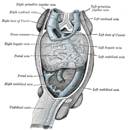
The sinus venosus is a large quadrangular cavity which precedes the atrium on the venous side of the chordate heart.

The angular vein is a vein of the face. It is the upper part of the facial vein, above its junction with the superior labial vein. It is formed by the junction of the supratrochlear vein and supraorbital vein, and joins with the superior labial vein. It drains the medial canthus, and parts of the nose and the upper lip. It can be a route of spread of infection from the danger triangle of the face to the cavernous sinus.

The vitelline veins are veins that drain blood from the yolk sac and the gut tube during gestation.

The right lymphatic duct is an important lymphatic vessel that drains the right upper quadrant of the body. It forms various combinations with the right subclavian vein and right internal jugular vein.

The superior intercostal veins are two veins that drain the 2nd, 3rd, and 4th intercostal spaces, one vein for each side of the body.

The ascending lumbar vein is a vein that runs up through the lumbar region on the side of the vertebral column.

The vaginal venous plexus is a group of veins draining blood from the vagina. It lies around the sides of the vagina. Its blood is eventually into the internal iliac veins.
The uterine vein is a vein of the uterus. It is found in the cardinal ligament. It drains into the internal iliac vein. It follows a similar course to the uterine artery. It helps to drain blood from the uterus, and removes waste from blood in the placenta during pregnancy.

The liver is a major organ only found in vertebrates which performs many essential biological functions such as detoxification of the organism, and the synthesis of proteins and biochemicals necessary for digestion and growth. In humans, it is located in the right upper quadrant of the abdomen, below the diaphragm. Its other roles in metabolism include the regulation of glycogen storage, decomposition of red blood cells, and the production of hormones.