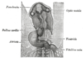
The liver and the veins in connection with it, of a human embryo, twenty-four or twenty-five days old, as seen from the ventral surface. (Vitelline veins visible at center bottom.)
The portions of the veins above the upper ring become interrupted by the developing liver and broken up by it into a plexus of small capillary-like vessels termed sinusoids.
Derivatives
Illustration of early development of veins and portal venous system. VV Vitelline veins, UV Umbilical veins, CV Cardinal veins, SV Sinus venosus
The branches conveying the blood to the plexus are named the venae advehentes, and become the branches of the portal vein. The vessels draining the plexus into the sinus venosus are termed the venae revehentes, and form the future hepatic veins.[3] Ultimately the left vena revehens no longer communicates directly with the sinus venosus, but opens into the right vena revehens. The persistent part of the upper venous ring, above the opening of the superior mesenteric vein, forms the trunk of the portal vein.
This page is based on this Wikipedia article Text is available under the CC BY-SA 4.0 license; additional terms may apply. Images, videos and audio are available under their respective licenses.