Foramen secundum

Last updated

Foramen secundum
Foramen ovale.png
Blood, shown in the red arrow, travels through the foramen ovale and the foramen secundum. HH: right ventricle, VH: left ventricle, HF: right atrium, VF: left atrium, LV: pulmonary vein
Details
Days33
Identifiers
Latin foramen secundum
TE secundum_by_E5.11.1.5.2.1.2 E5.11.1.5.2.1.2
Anatomical terminology

The foramen secundum or ostium secundum is a foramen in the septum primum, a precursor to the interatrial septum of the human heart.

Contents

It is not the same as the foramen ovale, which is an opening in the septum secundum.

Development

The foramen secundum (from Latin  'second opening') is formed from small perforations that develop in the septum primum. The septum primum is a septum that grows down between the single primitive atrium of the developing heart to separate it into left and right atria. [1]

Closure

Once a baby is born, blood should flow through the lungs, which now function to provide oxygen to the blood. The foramen secundum and foramen ovale act as a shunt where blood bypasses the lungs and does not become oxygenated. To provide proper blood flow as a newborn, the foramen secundum and foramen ovale must close at birth. Since the lungs now require a significant amount of blood flow, the vessels going to and from the lungs undergo dilation. While the pulmonary artery and pulmonary veins are dilating, the umbilical artery and umbilical vein are severed at the cutting of the umbilical cord, or the funiculus umbilicalis. This combination results in a reversal of pressure differences between the atria, and the septum primum is permanently forced against the septum secundum. This holds true even during atrial diastole, when the pressure is significantly less than atrial systole. [1]

Function

The septum primum is on the left side of the heart in the left atrium while the septum secundum is much thicker and is located on the right side, in the right atrium. During development, blood shunts from the floor of the right atrium through the foramen ovale in the septum secundum then up through the foramen secundum in the septum primum. [2] The foramen secundum is positioned so that blood exits in the ceiling of the left atrium and then out through the left ventricle and the aorta. The position of the foramen secundum and the size of the septum primum are crucial to ensuring that blood not flow backwards from the left atrium to the right atrium. The septum primum, being much thinner, is easily pressed against the septum secundum if blood attempts to flow in the reverse direction, effectively sealing off both the foramen secundum and the foramen ovale. [1]

Clinical significance

An ostium secundum that persists at large size can be a source of atrial septal defects. [3] Foramen secundum atrial septal defects are the most common atrial septal defects. This defect can arise as a result of defects of the septum primum and the septum secundum. For the septum primum, the problem can arise as a result of excess resorption of the septum during the process of apoptosis in order to form the foramen secundum. For the septum secundum, its inadequate growth can cause atrial septal defect since it is supposed to grow and eventually overlap the foramen secundum so as to form the oval foramen. As the right atrial pressure is normally lower than the mean left atrial pressure, a persisting ostium secundum causes usually a left-to-right shunt (meaning that blood flows from the left to the right atrium, wherefore it is an azyanotic heart defect). [4]

Treatment

Newborns with small foramen secundum atrial septal defects have been shown to spontaneously correct by the third or fourth year of life. [5] Therefore, medical supervision is generally accepted as a preventive measure for those diagnosed in infancy, rather than surgical intervention or use of other medical devices. [6]

If surgery is required, it is performed using minimally invasive techniques via robotic surgery that often requires only a few days of hospital stay. [7] Surgical intervention should result in full closure of the foramen secundum, and mortality rates are similar to those for general anesthesia. [8] The repair can be made by suturing the atrial septum or, if the foramen secundum is large in size, a patch can be made from the patient's pericardium to fully separate each atrium. The synthetic material Dacron may also be used to create a repair patch. [9]

Inserting a catheter has proven to be a safe and successful method for closing the foramen secundum in children. [10] This method avoids the symptoms that accompany most ostium secundum atrial septal defects. The catheter is inserted into the femoral vein in the leg and moved into place in the atrial septum. Transesophageal echocardiography is accepted as the method to monitor this procedure which, when performed correctly, has shorter recovery times than surgical intervention. [11] Complications of catheter insertion often include nausea and vomiting, blood vessel obstruction, pain, and hemorrhage. The most common problem with this preventive measure is the incomplete closing of the foramen secundum. [12]

There is at present no drug therapy for foramen secundum atrial septal defects, although infective endocarditis is a postoperative concern. To prevent this condition, a prophylactic is used for six months after the operation. [13]

Prognosis

Many patients with a foramen secundum that persists into adulthood will remain asymptomatic throughout their lives. A mortality rate of less than one tenth of one percent is expected if the operation is performed correctly. Some argue that if the operation is performed before eight years of age, few cardiac abnormalities such as cardiac dysrhythmia are expected later in life. [14] Others argue that the operation can take place as late as age 24, to limit cardiac complications in middle age or later. Some sources have argued that mitral regurgitation and mitral valve prolapse are common after age 40, if the ostium secundum is not repaired by age 24. [15] Operative closure of atrial septal defects after age 40, and the ability to diminish symptoms at all remains controversial. Some data does suggest that even after that age, symptoms can be alleviated via surgical intervention, including prevention of Arrhythmogenic right ventricular dysplasia and other associated cardiac abnormalities. [16]

History

The discovery of the formation of the foramen secundum and septum secundum was published by P. N. B. Odgers at the University of Oxford in the Journal of Anatomy in 1935. [17]

Related Research Articles

dextro-Transposition of the great arteries Medical condition

dextro-Transposition of the great arteries is a potentially life-threatening birth defect in the large arteries of the heart. The primary arteries are transposed.

<span class="mw-page-title-main">Atrial septal defect</span> Human heart defect present at birth

Atrial septal defect (ASD) is a congenital heart defect in which blood flows between the atria of the heart. Some flow is a normal condition both pre-birth and immediately post-birth via the foramen ovale; however, when this does not naturally close after birth it is referred to as a patent (open) foramen ovale (PFO). It is common in patients with a congenital atrial septal aneurysm (ASA).

<span class="mw-page-title-main">Ostium primum atrial septal defect</span> Medical condition

The ostium primum atrial septal defect is a defect in the atrial septum at the level of the tricuspid and mitral valves. This is sometimes known as an endocardial cushion defect because it often involves the endocardial cushion, which is the portion of the heart where the atrial septum meets the ventricular septum and the mitral valve meets the tricuspid valve.

<span class="mw-page-title-main">Congenital heart defect</span> Defect in the structure of the heart that is present at birth

A congenital heart defect (CHD), also known as a congenital heart anomaly, congenital cardiovascular malformation, and congenital heart disease, is a defect in the structure of the heart or great vessels that is present at birth. A congenital heart defect is classed as a cardiovascular disease. Signs and symptoms depend on the specific type of defect. Symptoms can vary from none to life-threatening. When present, symptoms are variable and may include rapid breathing, bluish skin (cyanosis), poor weight gain, and feeling tired. CHD does not cause chest pain. Most congenital heart defects are not associated with other diseases. A complication of CHD is heart failure.

<span class="mw-page-title-main">Ventricular septal defect</span> Medical condition

A ventricular septal defect (VSD) is a defect in the ventricular septum, the wall dividing the left and right ventricles of the heart. The extent of the opening may vary from pin size to complete absence of the ventricular septum, creating one common ventricle. The ventricular septum consists of an inferior muscular and superior membranous portion and is extensively innervated with conducting cardiomyocytes.

<span class="mw-page-title-main">Cardiac catheterization</span> Insertion of a catheter into a chamber or vessel of the heart

Cardiac catheterization is the insertion of a catheter into a chamber or vessel of the heart. This is done both for diagnostic and interventional purposes.

<span class="mw-page-title-main">Atrium (heart)</span> Part of the human heart

The atrium is one of the two upper chambers in the heart that receives blood from the circulatory system. The blood in the atria is pumped into the heart ventricles through the atrioventricular mitral and tricuspid heart valves.

<span class="mw-page-title-main">Atrioventricular septal defect</span> Medical condition

Atrioventricular septal defect (AVSD) or atrioventricular canal defect (AVCD), also known as "common atrioventricular canal" or "endocardial cushion defect" (ECD), is characterized by a deficiency of the atrioventricular septum of the heart that creates connections between all four of its chambers. It is a very specific combination of 3 defects:

<span class="mw-page-title-main">Foramen ovale (heart)</span> Passageway between the atria of the human heart

In the fetal heart, the foramen ovale, also foramen Botalli or the ostium secundum of Born, allows blood to enter the left atrium from the right atrium. It is one of two fetal cardiac shunts, the other being the ductus arteriosus. Another similar adaptation in the fetus is the ductus venosus. In most individuals, the foramen ovale closes at birth. It later forms the fossa ovalis.

<span class="mw-page-title-main">Interatrial septum</span> Wall of tissue separating atria of human heart

The interatrial septum is the wall of tissue that separates the right and left atria of the heart.

<span class="mw-page-title-main">Balloon septostomy</span> Medical procedure

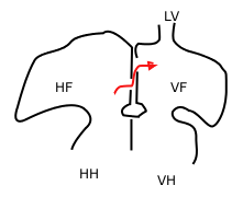
Balloon septostomy is the widening of a foramen ovale, patent foramen ovale (PFO), or atrial septal defect (ASD) via cardiac catheterization using a balloon catheter. This procedure allows a greater amount of oxygenated blood to enter the systemic circulation in some cases of cyanotic congenital heart defect (CHD).

<span class="mw-page-title-main">Primitive atrium</span>

The primitive atrium is a stage in the embryonic development of the human heart. It grows rapidly and partially encircles the bulbus cordis; the groove against which the bulbus cordis lies is the first indication of a division into right and left atria.

<span class="mw-page-title-main">Septum primum</span>

During heart development of a human embryo, the single primitive atrium becomes divided into right and left by a septum, the septum primum. The septum primum grows downward into the single atrium.

<span class="mw-page-title-main">Septum secundum</span>

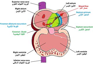
The septum secundum is a muscular flap that is important in heart development. It is semilunar in shape, and grows downward from the upper wall of the atrium immediately to the right of the septum primum and ostium secundum. It is important in the closure of the foramen ovale after birth.

<span class="mw-page-title-main">Primary interatrial foramen</span>

In the developing heart, the atria are initially open to each other, with the opening known as the primary interatrial foramen or ostium primum. The foramen lies beneath the edge of septum primum and the endocardial cushions. It progressively decreases in size as the septum grows downwards, and disappears with the formation of the atrial septum.

The heart is the first functional organ in a vertebrate embryo. There are 5 stages to heart development.

<span class="mw-page-title-main">Lutembacher's syndrome</span> Medical condition

Lutembacher's syndrome is a very rare form of congenital heart disease that affects one of the chambers of the heart as well as a valve. It is commonly known as both congenital atrial septal defect (ASD) and acquired mitral stenosis (MS). Congenital atrial septal defect refers to a hole being in the septum or wall that separates the two atria; this condition is usually seen in fetuses and infants. Mitral stenosis refers to mitral valve leaflets sticking to each other making the opening for blood to pass from the atrium to the ventricles very small. With the valve being so small, blood has difficulty passing from the left atrium into the left ventricle. Septal defects that may occur with Lutembacher's syndrome include: Ostium primum atrial septal defect or ostium secundum which is more prevalent.

<span class="mw-page-title-main">Atrial septostomy</span> Surgical procedure on the heart

Atrial septostomy is a surgical procedure in which a small hole is created between the upper two chambers of the heart, the atria. This procedure is primarily used to palliate dextro-Transposition of the great arteries or d-TGA, a life-threatening cyanotic congenital heart defect seen in infants. It is performed prior to an arterial switch operation. Atrial septostomy has also seen limited use as a surgical treatment for pulmonary hypertension. The first atrial septostomy was developed by Vivien Thomas in a canine model and performed in humans by Alfred Blalock. The Rashkind balloon procedure, a common atrial septostomy technique, was developed in 1966 by American cardiologist William Rashkind at the Children's Hospital of Philadelphia.

<span class="mw-page-title-main">Left atrial appendage occlusion</span> Medical treatment

Left atrial appendage occlusion (LAAO), also referred to as left atrial appendage closure (LAAC), is a procedure used to reduce the risk of blood clots from the left atrial appendage entering the bloodstream and causing a stroke in those with non-valvular atrial fibrillation.

<span class="mw-page-title-main">Heart development</span> Prenatal development of the heart

Heart development, also known as cardiogenesis, refers to the prenatal development of the heart. This begins with the formation of two endocardial tubes which merge to form the tubular heart, also called the primitive heart tube. The heart is the first functional organ in vertebrate embryos.

References

  1. 1 2 3 Schoenwolf, Gary C., and William J. Larsen. Larsen's Human Embryology. 4th ed. Philadelphia: Churchill Livingstone/Elsevier, 2009. 363. Print.
  2. Understanding Patent Foramen Ovale. St, Jude Medical, n.d. Web. 27 November 2012.
  3. ped/1686 at eMedicine
  4. Rigatelli, Gianluca (September 2014). "Should we consider patent foramen ovale and secundum atrial septal defect as different steps of a single anatomo-clinical continuum?". Journal of geriatric cardiology: JGC. 11 (3): 177–179. doi:10.11909/j.issn.1671-5411.2014.03.004. ISSN   1671-5411. PMC   4178506 . PMID   25278963.
  5. Hanslik A, Pospisil U, Salzer-Muhar U, Greber-Platzer S, Male C. Predictors of spontaneous closure of isolated secundum atrial septal defect in children: a longitudinal study. Pediatrics. October 2006; 118(4):1560-5
  6. Rigatelli G, Dell' Avvocata F, Cardaioli P, Giordan M, Vassiliev D, Nghia NT, et al. Five-year Follow-up of Intracardiac Echocardiography-assisted Transcatheter Closure of Complex Ostium Secundum Atrial Septal Defect. Congenital Heart Diseases. 20 October 2011.
  7. Argenziano M, Oz MC, Kohmoto T, et al. Totally endoscopic atrial septal defect repair with robotic assistance. Circulation. 9 September 2003; 108 Suppl 1:II191-4.
  8. Bolz D, Lacina T, Buser P, et al. Long-term outcome after surgical closure of atrial septal defect in childhood with extensive assessment including MRI measurement of the ventricles. Pediatric Cardiology. Sep-Oct 2005; 26(5):614-21
  9. Shah D, Azhar M, Oakley CM, et al. Natural history of secundum atrial septal defect in adults after medical or surgical treatment: a historical prospective study. British Heart Journal. March 1994; 71(3):224-7
  10. Jones TK, Latson LA, Zahn E, et al. Results of the U.S. multicenter pivotal study of the HELEX septal occluder for percutaneous closure of secundum atrial septal defects. Journal of the American College of Cardiologists. 5 June 2007; 49(22):2215-21.
  11. Post MC, Suttorp MJ, Jaarsma W, Plokker HW. Comparison of outcome and complications using different types of devices for percutaneous closure of a secundum atrial septal defect in adults: a single-center experience. Catheter Cardiovascular Intervention. March 2006; 67(3):438-43.
  12. Butera G, Carminati M, Chessa M, et al. Percutaneous versus surgical closure of secundum atrial septal defect: comparison of early results and complications. American Heart Journal. January 2006; 151(1):228-34
  13. Gessner MD, Neish MD, et al. Ostium Secundum Atrial Septal Defects Medication. Medscape Reference. 1 December 2011.
  14. Brochu MC, Baril JF, Dore A, et al. Improvement in exercise capacity in asymptomatic and mildly symptomatic adults after atrial septal defect percutaneous closure. Circulation. 1 October 2002; 106(14):1821-6.
  15. Groundstroem, et al. Late postoperative follow-up of ostium secundum defect. European Heart Journal (1999). 20, 904-909
  16. M. Jemielity, W. Dyszkiewicz, et al. Do patients over 40 years of age benefit from surgical closure of atrial septal defects? 1 September 2000. Heart 2001; 85: 300-303
  17. Anatomical Society of Great Britain and Ireland. J Anat. 1935 July; 69 (Pt 4): 412–422.5