Chorionic hematoma

Last updated
Chorionic hematoma
Other namesChorionic hemorrhage, chorionic bleed
Ultrasound showing a subchorionic hemorrhage [1]

Chorionic hematoma is the pooling of blood (hematoma) between the chorion, a membrane surrounding the embryo, and the uterine wall. [2] It occurs in about 3.1% of all pregnancies, [2] it is the most common sonographic abnormality and the most common cause of first trimester bleeding. [3]

Contents

Cause and diagnosis

Chorion, amnion and gestational (yolk) sac Gray24.png
Chorion, amnion and gestational (yolk) sac

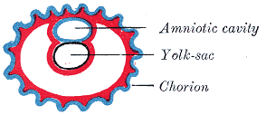
Chorionic hematomas can be caused by the separation of the chorion from the endometrium (inner membrane of the uterus). Hematomas are classified by their location between tissue layers: [4]

Most patients with a small subchorionic hematoma are asymptomatic. [5] Symptoms include vaginal bleeding, abdominal pain, premature labor and threatened miscarriage. [6]

Ultrasonography is the preferred method of diagnosis. [7] A chorionic hematoma appears on ultrasound as a hypoechoic crescent adjacent to the gestational sac. The hematoma is considered small if it is under 20% of the size of the sac and large if it is over 50%. [2]

Prognosis and treatment

The presence of subchorionic bleeding around the gestational sac does not have a significant association with miscarriage overall. [8] [9] However, the case of intrauterine hematoma observed before 9 weeks of gestational age has been associated with an increased risk of miscarriage. [10] In one study women who complied with instructions for bed rest for the duration of bleeding had a lower rate of miscarriage and a higher rate of term pregnancy than non-compliant women. The study had several limitations; results were severely confounded by inherent differences between compliant and non-compliant women. [11]

Related Research Articles

Obstetrics is the field of study concentrated on pregnancy, childbirth and the postpartum period. As a medical specialty, obstetrics is combined with gynecology under the discipline known as obstetrics and gynecology (OB/GYN), which is a surgical field.

<span class="mw-page-title-main">Ectopic pregnancy</span> Female reproductive system health issue

Ectopic pregnancy is a complication of pregnancy in which the embryo attaches outside the uterus. Signs and symptoms classically include abdominal pain and vaginal bleeding, but fewer than 50 percent of affected women have both of these symptoms. The pain may be described as sharp, dull, or crampy. Pain may also spread to the shoulder if bleeding into the abdomen has occurred. Severe bleeding may result in a fast heart rate, fainting, or shock. With very rare exceptions, the fetus is unable to survive.

<span class="mw-page-title-main">Miscarriage</span> Natural death and expulsion of an embryo or fetus before its independent survival

Miscarriage, also known in medical terms as a spontaneous abortion, is the death and expulsion of an embryo or fetus before it is able to survive independently. The term miscarriage is sometimes used to refer to all forms of pregnancy loss and pregnancy with abortive outcome before 20 weeks of gestation.

<span class="mw-page-title-main">Amniocentesis</span> Sampling of amniotic fluid done mainly to detect fetal chromosomal abnormalities

Amniocentesis is a medical procedure used primarily in the prenatal diagnosis of genetic conditions. It has other uses such as in the assessment of infection and fetal lung maturity. Prenatal diagnostic testing, which includes amniocentesis, is necessary to conclusively diagnose the majority of genetic disorders, with amniocentesis being the gold-standard procedure after 15 weeks' gestation.

<span class="mw-page-title-main">Obstetric ultrasonography</span> Use of medical ultrasonography in pregnancy

Obstetric ultrasonography, or prenatal ultrasound, is the use of medical ultrasonography in pregnancy, in which sound waves are used to create real-time visual images of the developing embryo or fetus in the uterus (womb). The procedure is a standard part of prenatal care in many countries, as it can provide a variety of information about the health of the mother, the timing and progress of the pregnancy, and the health and development of the embryo or fetus.

<span class="mw-page-title-main">Placenta praevia</span> Medical condition

Placenta praevia is when the placenta attaches inside the uterus but in a position near or over the cervical opening. Symptoms include vaginal bleeding in the second half of pregnancy. The bleeding is bright red and tends not to be associated with pain. Complications may include placenta accreta, dangerously low blood pressure, or bleeding after delivery. Complications for the baby may include fetal growth restriction.

<span class="mw-page-title-main">Placental abruption</span> Medical condition

Placental abruption is when the placenta separates early from the uterus, in other words separates before childbirth. It occurs most commonly around 25 weeks of pregnancy. Symptoms may include vaginal bleeding, lower abdominal pain, and dangerously low blood pressure. Complications for the mother can include disseminated intravascular coagulopathy and kidney failure. Complications for the baby can include fetal distress, low birthweight, preterm delivery, and stillbirth.

<span class="mw-page-title-main">Vaginal bleeding</span> Medical condition

Vaginal bleeding is any expulsion of blood from the vagina. This bleeding may originate from the uterus, vaginal wall, or cervix. Generally, it is either part of a normal menstrual cycle or is caused by hormonal or other problems of the reproductive system, such as abnormal uterine bleeding.

<span class="mw-page-title-main">Gynecologic ultrasonography</span>

Gynecologic ultrasonography or gynecologic sonography refers to the application of medical ultrasonography to the female pelvic organs as well as the bladder, the adnexa, and the recto-uterine pouch. The procedure may lead to other medically relevant findings in the pelvis.This technique is useful to detect myomas or mullerian malformations.

<span class="mw-page-title-main">Placenta accreta spectrum</span> Medical condition

Placenta accreta occurs when all or part of the placenta attaches abnormally to the myometrium. Three grades of abnormal placental attachment are defined according to the depth of attachment and invasion into the muscular layers of the uterus:

  1. Accreta – chorionic villi attached to the myometrium, rather than being restricted within the decidua basalis.
  2. Increta – chorionic villi invaded into the myometrium.
  3. Percreta – chorionic villi invaded through the perimetrium.
<span class="mw-page-title-main">Nuchal scan</span> Routine ultrasound done between 11 and 14 weeks pregnancy

A nuchal scan or nuchal translucency (NT) scan/procedure is a sonographic prenatal screening scan (ultrasound) to detect chromosomal abnormalities in a fetus, though altered extracellular matrix composition and limited lymphatic drainage can also be detected.

<span class="mw-page-title-main">Velamentous cord insertion</span> Velamentous placenta

Velamentous cord insertion is a complication of pregnancy where the umbilical cord is inserted in the fetal membranes. It is a major cause of antepartum hemorrhage that leads to loss of fetal blood and associated with high perinatal mortality. In normal pregnancies, the umbilical cord inserts into the middle of the placental mass and is completely encased by the amniotic sac. The vessels are hence normally protected by Wharton's jelly, which prevents rupture during pregnancy and labor. In velamentous cord insertion, the vessels of the umbilical cord are improperly inserted in the chorioamniotic membrane, and hence the vessels traverse between the amnion and the chorion towards the placenta. Without Wharton's jelly protecting the vessels, the exposed vessels are susceptible to compression and rupture.

The following outline is provided as an overview of and topical guide to obstetrics:

<span class="mw-page-title-main">Circumvallate placenta</span> Medical condition

Circumvallate placenta is a rare condition affecting about 1-2% of pregnancies, in which the amnion and chorion fetal membranes essentially "double back" on the fetal side around the edges of the placenta. After delivery, a circumvallate placenta has a thick ring of membranes on its fetal surface. Circumvallate placenta is a placental morphological abnormality associated with increased fetal morbidity and mortality due to the restricted availability of nutrients and oxygen to the developing fetus.

<span class="mw-page-title-main">Interstitial pregnancy</span> Medical condition

An interstitial pregnancy is a uterine but ectopic pregnancy; the pregnancy is located outside the uterine cavity in that part of the fallopian tube that penetrates the muscular layer of the uterus. The term cornual pregnancy is sometimes used as a synonym, but remains ambiguous as it is also applied to indicate the presence of a pregnancy located within the cavity in one of the two upper "horns" of a bicornuate uterus. Interstitial pregnancies have a higher mortality than ectopics in general.

Ovarian pregnancy refers to an ectopic pregnancy that is located in the ovary. Typically the egg cell is not released or picked up at ovulation, but fertilized within the ovary where the pregnancy implants. Such a pregnancy usually does not proceed past the first four weeks of pregnancy. An untreated ovarian pregnancy causes potentially fatal intra-abdominal bleeding and thus may become a medical emergency.

Theca lutein cyst is a type of bilateral functional ovarian cyst filled with clear, straw-colored fluid. These cysts result from exaggerated physiological stimulation due to elevated levels of beta-human chorionic gonadotropin (beta-hCG) or hypersensitivity to beta-hCG. On ultrasound and MRI, theca lutein cysts appear in multiples on ovaries that are enlarged.

<span class="mw-page-title-main">Products of conception</span>

Products of conception, abbreviated POC, is a medical term used for the tissue derived from the union of an egg and a sperm. It encompasses anembryonic gestation which does not have a viable embryo.

Early pregnancy bleeding refers to vaginal bleeding before 14 weeks of gestational age. If the bleeding is significant, hemorrhagic shock may occur. Concern for shock is increased in those who have loss of consciousness, chest pain, shortness of breath, or shoulder pain.

The anomaly scan, also sometimes called the anatomy scan, 20-week ultrasound, or level 2 ultrasound, evaluates anatomic structures of the fetus, placenta, and maternal pelvic organs. This scan is an important and common component of routine prenatal care. The function of the ultrasound is to measure the fetus so that growth abnormalities can be recognized quickly later in pregnancy, to assess for congenital malformations and multiple pregnancies, and to plan method of delivery.

References

  1. "UOTW #3 Answer - Ultrasound of the Week". Ultrasound of the Week. Retrieved 27 May 2017.
  2. 1 2 3 Nagy, Sándor MD; Bush, Melissa MD; Stone, Joanne MD; Lapinski, Robert H. PhD; Gardó, Sándor MD, DSci. Clinical Significance of Subchorionic and Retroplacental Hematomas Detected in the First Trimester of Pregnancy . Obstetrics & Gynecology: July 2003 - Volume 102 - Issue 1 - p 94-100
  3. Avneesh Chhabra, MD et al. "Subchorionic Hemorrhage" , Medscape.
  4. Trop, Isabelle and Levine, Deborah. Hemorrhage During Pregnancy: Sonography and MR Imaging . Amer J Roentgenology 2001; 176:607-615.
  5. Trop I, Levine D. Hemorrhage during pregnancy: sonography and MR imaging. AJR Am J Roentgenol. Mar 2001;176(3):607-15.
  6. Hodgson DT, Lotfipour S, Fox JC. Vaginal bleeding before 20 weeks gestation due to placental abruption leading to disseminated intravascular coagulation and fetal loss after appearing to satisfy criteria for routine threatened abortion: a case report and brief review of the literature. J Emerg Med. May 2007;32(4):387-92
  7. Abu-Yousef MM, Bleicher JJ, Williamson RA, Weiner CP. Subchorionic hemorrhage: sonographic diagnosis and clinical significance. AJR Am J Roentgenol. Oct 1987;149(4):737-40.
  8. "Miscarriage risk is not increased with subchorionic hematoma". www.mdedge.com.
  9. N. Stamatopoulos; C. Lu; F. Infante; U. Menakaya; I. Casikar; S. Reid; M. Mongelli; G. Condous (2013). "OP03.03: Does the presence of subchorionic haematoma increase the risk of miscarriage?". Ultrasound in Obstetrics & Gynecology. 42 (s1): 54. doi:10.1002/uog.12736.
  10. Maso, G.; Dʼottavio, G.; De Seta, F.; Sartore, A.; Piccoli, M.; Mandruzzato, G. (2005). "First-Trimester Intrauterine Hematoma and Outcome of Pregnancy". Obstetrics & Gynecology. 105 (2): 339–44. doi:10.1097/01.AOG.0000152000.71369.bd. PMID   15684162. S2CID   12502344.
  11. Ben-Haroush A, Yogev Y, Mashiach R, Meizner I. Pregnancy outcome of threatened abortion with subchorionic hematoma: possible benefit of bed-rest? Journal Isr Med Assoc J. 2003 Jun;5(6):422-4