Interstitial pregnancy

Last updated
Interstitial pregnancy
Other namesCornual pregnancy
The science and art of midwifery (1897) (14740436436).jpg
Specialty Obstetrics

An interstitial pregnancy is a uterine but ectopic pregnancy; the pregnancy is located outside the uterine cavity in that part of the fallopian tube that penetrates the muscular layer of the uterus. [1] The term cornual pregnancy is sometimes used as a synonym, [2] [3] but remains ambiguous as it is also applied to indicate the presence of a pregnancy located within the cavity in one of the two upper "horns" of a bicornuate uterus. [1] Interstitial pregnancies have a higher mortality than ectopics in general.

Contents

Anatomy

The part of the Fallopian tube that is located in the uterine wall and connects the remainder of the tube to the endometrial cavity is called its "interstitial" part, hence the term "interstitial pregnancy"; it has a length of 1–2 cm and a width of 0.7 cm. [1] Its borders are the opening (ostium) of the tube to the endometrial cavity within the uterus and, laterally, the visible narrow segment of the tube. The area is well supplied by the Sampson artery which is connected to both the uterine and the ovarian arteries. Surrounded by uterine muscle (myometrium) it can expand significantly when it hosts a pregnancy.

Interstitial pregnancies can be confused with angular pregnancies; the latter, however, are located within the endometrial cavity in the corner where the tube connects; typically those pregnancies are viable although a high rate of miscarriage has been reported. [1] A pregnancy located next to the interstitial section laterally is an isthmic tubal pregnancy.

The definition of an ectopic pregnancy is a pregnancy outside the uterine cavity, not outside the uterus, as the interstitial pregnancy is still a uterine pregnancy. [4]

Diagnosis

Early diagnosis is important and today facilitated by the use of sonography and the quantitative human chorionic gonadotropin (hCG) assay. As in other cases of ectopic pregnancy, risk factors are: previous tubal pregnancy, IVF therapy, tubal surgery, and a history of sexual infection. [5] Typical symptoms of an interstitial pregnancy are the classic signs of ectopic pregnancy, namely abdominal pain and vaginal bleeding. Hemorrhagic shock is found in almost a quarter of patients.; [2] this explains the relatively high mortality rate.

In pregnant patients, sonography is the primary method to make the diagnosis, even when patients have no symptoms. The paucity of myometrium around the gestational sac is diagnostic, while, in contrast, the angular pregnancy has at least 5 mm of myometrium on all of its sides. [6] Ultrasonic criteria for the diagnosis include an empty uterine cavity, a gestational sac separate from the uterine cavity, and a myometrial thinning of less than 5 mm around the gestational sac; typically the interstitial line sign—an echogenic line from the endometrial cavity to the corner next to the gestational mass—is seen. [3] MRI can be used particularly when it is important to distinguish between an interstitial and angular pregnancy.

On average, the gestational age at presentation is about 7–8 weeks. [1] In a 2007 series, 22% of patients presented with rupture and hemorrhagic shock, while a third of the patients were asymptomatic; the remainder had abdominal pain and/or vaginal bleeding. [2] Cases that are not diagnosed until surgery show an asymmetrical bulge in the upper corner of the uterus. [1]

Treatment

Choice of treatment is largely dictated by the clinical situation. A ruptured interstitial pregnancy is a medical emergency that requires an immediate surgical intervention either by laparoscopy or laparotomy to stop the bleeding and remove the pregnancy. [1]

Surgical methods to remove the pregnancy include cornual evacuation, incision of the cornua with removal of the pregnancy (cornuostomy), resection of the cornual area or a cornual wedge resection, typically combined with an ipsilateral salpingectomy, and hysterectomy. [1] Because of the vascularity of the interstitial region particularly during pregnancy, blood loss during surgery may be substantial. Postoperatively, patients with conservative surgical therapy are at risk for development of a persistent ectopic pregnancy due to the presence of deeply embedded surviving trophoblastic tissue; thus, monitoring of hCG levels is indicated until they become undetectable.

In patients with an asymptomatic interstitial pregnancy methotrexate has been successfully used, however, this approach may fail and result in cornual rupture of the pregnancy. [7] Selective uterine artery embolization has been successfully performed to treat interstitial pregnancies. [8]

Subsequent pregnancies

Patients with an ectopic pregnancy are generally at higher risk for a recurrence, however, there are no specific data for patients with an interstitial pregnancy. When a new pregnancy is diagnosed it is important to monitor the pregnancy by transvaginal sonography to assure that is it properly located, and that the surgically repaired area remains intact. [9] Cesarean delivery is recommended to avoid uterine rupture during labor. [9]

Epidemiology

Interstitial pregnancies account for 2–4% of all tubal pregnancies, or for 1 in 2,500 to 5,000 live births. [10] About one in fifty women with an interstitial pregnancy dies. [11] Patients with an interstitial pregnancies have a seven-times higher mortality than those with ectopics in general. [10] With the growing use of assisted reproductive technologies, the incidence of interstitial pregnancy is rising. [1]

Related Research Articles

<span class="mw-page-title-main">Uterus</span> Female sex organ in mammals

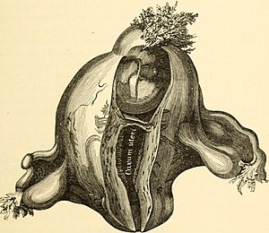
The uterus or womb is the organ in the reproductive system of most female mammals, including humans, that accommodates the embryonic and fetal development of one or more embryos until birth. The uterus is a hormone-responsive sex organ that contains glands in its lining that secrete uterine milk for embryonic nourishment.

<span class="mw-page-title-main">Ectopic pregnancy</span> Female reproductive system health issue

Ectopic pregnancy is a complication of pregnancy in which the embryo attaches outside the uterus. Signs and symptoms classically include abdominal pain and vaginal bleeding, but fewer than 50 percent of affected women have both of these symptoms. The pain may be described as sharp, dull, or crampy. Pain may also spread to the shoulder if bleeding into the abdomen has occurred. Severe bleeding may result in a fast heart rate, fainting, or shock. With very rare exceptions, the fetus is unable to survive.

<span class="mw-page-title-main">Tubal ligation</span> Surgical clipping,removal or blocking of the fallopian tubes

Tubal ligation is a surgical procedure for female sterilization in which the fallopian tubes are permanently blocked, clipped or removed. This prevents the fertilization of eggs by sperm and thus the implantation of a fertilized egg. Tubal ligation is considered a permanent method of sterilization and birth control.

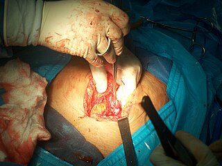
A hysterotomy is an incision made in the uterus. This surgical incision is used in several medical procedures, including during termination of pregnancy in the second trimester and delivering the fetus during caesarean section. It is also used to gain access and perform surgery on a fetus during pregnancy to correct birth defects, and it is an option to achieve resuscitation if cardiac arrest occurs during pregnancy and it is necessary to remove the fetus from the uterus.

<span class="mw-page-title-main">Asherman's syndrome</span> Medical condition

Asherman's syndrome (AS) is an acquired uterine condition that occurs when scar tissue (adhesions) forms inside the uterus and/or the cervix. It is characterized by variable scarring inside the uterine cavity, where in many cases the front and back walls of the uterus stick to one another. AS can be the cause of menstrual disturbances, infertility, and placental abnormalities. Although the first case of intrauterine adhesion was published in 1894 by Heinrich Fritsch, it was only after 54 years that a full description of Asherman syndrome was carried out by Joseph Asherman. A number of other terms have been used to describe the condition and related conditions including: uterine/cervical atresia, traumatic uterine atrophy, sclerotic endometrium, and endometrial sclerosis.

<span class="mw-page-title-main">Adenomyosis</span> Extension of endometrial tissue into the myometrium

Adenomyosis is a medical condition characterized by the growth of cells that proliferate on the inside of the uterus (endometrium) atypically located among the cells of the uterine wall (myometrium), as a result, thickening of the uterus occurs. As well as being misplaced in patients with this condition, endometrial tissue is completely functional. The tissue thickens, sheds and bleeds during every menstrual cycle.

<span class="mw-page-title-main">Vaginal bleeding</span> Medical condition

Vaginal bleeding is any expulsion of blood from the vagina. This bleeding may originate from the uterus, vaginal wall, or cervix. Generally, it is either part of a normal menstrual cycle or is caused by hormonal or other problems of the reproductive system, such as abnormal uterine bleeding.

<span class="mw-page-title-main">Gynecologic ultrasonography</span>

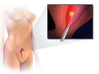
Gynecologic ultrasonography or gynecologic sonography refers to the application of medical ultrasonography to the female pelvic organs as well as the bladder, the adnexa, and the recto-uterine pouch. The procedure may lead to other medically relevant findings in the pelvis.This technique is useful to detect myomas or mullerian malformations.

<span class="mw-page-title-main">Uterine myomectomy</span> Surgical removal of uterine fibroid

Myomectomy, sometimes also called fibroidectomy, refers to the surgical removal of uterine leiomyomas, also known as fibroids. In contrast to a hysterectomy, the uterus remains preserved and the woman retains her reproductive potential. It still may impact hormonal regulation and the menstrual cycle.

Tuboplasty refers to a number of surgical operations that attempt to restore patency and functioning of the fallopian tube(s) so that a pregnancy could be achieved. As tubal infertility is a common cause of infertility, tuboplasties were commonly performed prior to the development of effective in vitro fertilization (IVF) or repair of any type of tube-like structure, including the Eustachian tube in the head and neck.

<span class="mw-page-title-main">Abdominal pregnancy</span> Medical condition

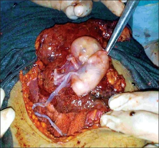
An abdominal pregnancy is a rare type of ectopic pregnancy where the embryo or fetus is growing and developing outside the womb in the abdomen, but not in the Fallopian tube, ovary or broad ligament.

Adenomyoma is a tumor (-oma) including components derived from glands (adeno-) and muscle (-my-). It is a type of complex and mixed tumor, and several variants have been described in the medical literature. Uterine adenomyoma, the localized form of uterine adenomyosis, is a tumor composed of endometrial gland tissue and smooth muscle in the myometrium. Adenomyomas containing endometrial glands are also found outside of the uterus, most commonly on the uterine adnexa but can also develop at distant sites outside of the pelvis. Gallbladder adenomyoma, the localized form of adenomyomatosis, is a polypoid tumor in the gallbladder composed of hyperplastic mucosal epithelium and muscularis propria.

<span class="mw-page-title-main">Endometrial ablation</span>

Endometrial ablation is a surgical procedure that is used to remove (ablate) or destroy the endometrial lining of the uterus. The goal of the procedure is to decrease the amount of blood loss during menstrual periods. Endometrial ablation is most often employed in people with excessive menstrual bleeding, who do not wish to undergo a hysterectomy, following unsuccessful medical therapy.

<span class="mw-page-title-main">Placenta accreta spectrum</span> Medical condition

Placenta accreta occurs when all or part of the placenta attaches abnormally to the myometrium. Three grades of abnormal placental attachment are defined according to the depth of attachment and invasion into the muscular layers of the uterus:

  1. Accreta – chorionic villi attached to the myometrium, rather than being restricted within the decidua basalis.
  2. Increta – chorionic villi invaded into the myometrium.
  3. Percreta – chorionic villi invaded through the perimetrium.
<span class="mw-page-title-main">Unicornuate uterus</span> Medical condition

A unicornuate uterus represents a uterine malformation where the uterus is formed from one only of the paired Müllerian ducts while the other Müllerian duct does not develop or only in a rudimentary fashion. The sometimes called hemi-uterus has a single horn linked to the ipsilateral fallopian tube that faces its ovary.

Tubal reversal, also called tubal sterilization reversal, tubal ligation reversal, or microsurgical tubal reanastomosis, is a surgical procedure that can restore fertility to women after a tubal ligation. By rejoining the separated segments of the fallopian tube, tubal reversal can give women the chance to become pregnant again. In some cases, however, the separated segments cannot actually be reattached to each other. In some cases the remaining segment of tube needs to be re-implanted into the uterus. In other cases, when the end of the tube has been removed, a procedure called a neofimbrioplasty must be performed to recreate a functional end of the tube which can then act like the missing fimbria and retrieve the egg that has been released during ovulation.

Uterine clear-cell carcinoma (CC) is a rare form of endometrial cancer with distinct morphological features on pathology; it is aggressive and has high recurrence rate. Like uterine papillary serous carcinoma CC does not develop from endometrial hyperplasia and is not hormone sensitive, rather it arises from an atrophic endometrium. Such lesions belong to the type II endometrial cancers.

<span class="mw-page-title-main">Cervical pregnancy</span> Medical condition

A cervical pregnancy is an ectopic pregnancy that has implanted in the uterine endocervix. Such a pregnancy typically aborts within the first trimester, however, if it is implanted closer to the uterine cavity – a so-called cervico-isthmic pregnancy – it may continue longer. Placental removal in a cervical pregnancy may result in major hemorrhage.

Ovarian pregnancy refers to an ectopic pregnancy that is located in the ovary. Typically the egg cell is not released or picked up at ovulation, but fertilized within the ovary where the pregnancy implants. Such a pregnancy usually does not proceed past the first four weeks of pregnancy. An untreated ovarian pregnancy causes potentially fatal intra-abdominal bleeding and thus may become a medical emergency.

Pregnancy in a rudimentary horn of the uterus is a very rare type of ectopic pregnancy. This type of pregnancy can be life-threatening, as the rudimentary horn is not meant to sustain a pregnancy and is at risk of rupturing.

References

  1. 1 2 3 4 5 6 7 8 9 Moawad, Nash S.; Mahajan, Sangeeta T.; Moniz, Michelle H.; Taylor, Sarah E.; Hurd, William W. (January 2010). "Current diagnosis and treatment of interstitial pregnancy". American Journal of Obstetrics & Gynecology. 202 (1): 15–29. doi:10.1016/j.ajog.2009.07.054. PMID   20096253. PII  S0002-9378(09)00840-0.
  2. 1 2 3 Soriano, D.; Vicus, D.; Mashiach, R.; Schiff, E.; Seidman, D.; Goldenberg, M. (2008). "Laparoscopic treatment of cornual pregnancy: a series of 20 consecutive cases". Journal of Reproductive Immunology. 90 (3): 839–843. doi: 10.1016/j.fertnstert.2007.07.1288 . PMID   17936282.
  3. 1 2 Ackerman, T. E.; Levi, C. S.; Dashefsky, S. M.; Holt, S. C.; Lindsay, D. J. (1993). "Interstitial line: sonographic finding in interstitial (cornual) ectopic pregnancy". Radiology. 189 (1): 83–87. doi:10.1148/radiology.189.1.8372223. PMID   8372223.
  4. Page, E. W.; Villee, C. A.; Villee, D. B. (1976). Human Reproduction (2nd ed.). Philadelphia: W. B. Saunders. p.  211. ISBN   0-7216-7042-3.
  5. Tulandi, T.; Al-Jaroudi, D. (2004). "Interstitial pregnancy: results generated from the Society of Reproductive Surgeons Registry". Obstetrics & Gynecology. 103 (1): 47–50. doi: 10.1097/01.AOG.0000109218.24211.79 . PMID   14704243. S2CID   25708025.
  6. Doubilet, P.; Benson, C. B. (2003). Atlas of Ultrasound in Obstetrics and Gynecology. Philadelphia: Lippincott Williams & Wilkins. ISBN   0-7817-3633-1.
  7. Jermy, K.; Thomas, J.; Doo, A.; Bourne, T. (2004). "The conservative treatment of interstitial pregnancy". BJOG. 111 (11): 1283–1288. doi: 10.1111/j.1471-0528.2004.00442.x . PMID   15521876.
  8. Deruelle, P.; Closset, E. (2006). "Management of interstitial pregnancy using selective uterine artery embolization". Obstet Gynecol. 107 (2 Pt 1): 427–428. doi:10.1097/01.AOG.0000199425.78203.e4. PMID   16449137.
  9. 1 2 Loret de Mola, J. R.; Austin, C. M.; Judge, N. E.; Assel, B. G.; Peskin, B.; Goldfarb, J. M. (1995). "Cornual heterotopic pregnancy and cornual resection after in vitro fertilization/embryo transfer: a report of two cases". J Reprod Med. 40 (8): 606–610. PMID   7473461.
  10. 1 2 Damario, M. A.; Rock, J. A. (2003). "Ectopic Pregnancy". In Rock, J. A.; Jones, H. W. III (eds.). Te Linde's Operative Gynecology (9th ed.). Philadelphia: Lippincott Williams & Wilkins. pp. 507–536. ISBN   0-7817-2859-2.
  11. Lau, S.; Tulandi, T. (1999). "Conservative medical and surgical management of interstitial ectopic pregnancy". Fertil Steril. 72 (2): 207–215. doi: 10.1016/s0015-0282(99)00242-3 . PMID   10438980.