Gestational pemphigoid

Last updated
Gestational pemphigoid
Other namesPemphigoid gestationis, herpes gestationis [1]
Pemphigoid gestationis - high mag.jpg
Micrograph of gestational pemphigoid showing the characteristic subepidermal blisters and abundant eosinophils. HPS stain.
Specialty Dermatology
Symptoms Blisters, itch, hives [1]
Complications Premature delivery of a small baby, a few who may be born with blisters and urticaria [1]
Usual onsetMiddle of pregnancy or shortly after [1]
DurationAround 6-months [1]
CausesAutoimmune [1]
Risk factors Pregnancy, molar pregnancy, choriocarcinoma, oral contraceptive pill [1]
Diagnostic method Appearance, skin biopsy, immunofluorescence [1]
Differential diagnosis Pruritic urticarial papules and plaques of pregnancy, erythema multiforme, drug reactions, blistering scabies [1]
Treatment Corticosteroid [1]
Frequency1 in 20,000 to 50,000 pregnancies [1]

Gestational pemphigoid (GP) is a rare autoimmune variant of the skin disease bullous pemphigoid, and first appears in pregnancy. [2] It presents with tense blisters, small bumps, hives and intense itching, usually starting around the navel before spreading to limbs in mid-pregnancy or shortly after delivery. [1] The head, face and mouth are not usually affected. [3]

Contents

It may flare after delivery before resolving around three to six months after the pregnancy. [1] It can be triggered by subsequent pregnancies, menstrual periods and oral contraceptive pill. [1] A molar pregnancy and choriocarcinoma can provoke it. [1] In some people, it persists long-term. [1] It is associated with premature delivery of a small baby, a few who may be born with blisters and urticaria, which generally resolves within six weeks. [3] It does not spread from one person to another, and does not run in families. [3]

Diagnosis is by visulaization, biopsy and immunofluorescence. [4] It can resemble pruritic urticarial papules and plaques of pregnancy, erythema multiforme, drug reactions and blistering scabies. [1]

Around 1 in 20,000 to 50,000 pregnancies are affected. [1] It was originally called herpes gestationis because of the blistering appearance, although it is not associated with the herpes virus. [3]

Signs and symptoms

Diagnosis of GP becomes clear when skin lesions progress to tense blisters during the second or third trimester. The face and mucous membranes are usually spared. GP typically starts as a blistering rash in the navel area and then spreads over the entire body. It is sometimes accompanied by raised, hot, painful welts called plaques. After one to two weeks, large, tense blisters typically develop on the red plaques, containing clear or blood-stained fluid. [5] GP creates a histamine response that causes extreme relentless itching (pruritus). GP is characterized by flaring and remission during the gestational and sometimes post partum period. Usually after delivery, lesions will heal within months, but may reoccur during menstruation.[ citation needed ]

Causes

Pathogenically, it is a type II hypersensitivity reaction where circulating complement-fixing IgG antibodies bind to an antigen (a 180-kDa protein, BP-180) in the hemidesmosomes (attach basal cells of epidermis to the basal lamina and hence to dermis) of the dermoepidermal junction[ further explanation needed ], leading to blister formation as loss of hemidesmosomes causes the epidermis to separate from dermis. The immune response is even more highly restricted to the NC16A domain. The primary site of autoimmunity seems not to be the skin, but the placenta, as antibodies bind not only to the basement membrane zone of the epidermis, but also to that of chorionic and amniotic epithelia. Aberrant expression of MHC class II molecules on the chorionic villi suggests an allogenic immune reaction to a placental matrix antigen, thought to be of paternal origin. Recently, both IgA22 and IgE24 antibodies to either BP180 or BP230 have also been detected in pemphigoid gestationis.[ citation needed ]

Risk

Pregnant women with GP should be monitored for conditions that may affect the fetus, including, but not limited to, low or decreasing volume of amniotic fluid, preterm labor, and intrauterine growth retardation. Onset of GP in the first or second trimester and presence of blisters may lead to adverse pregnancy outcomes including decreased gestational age at delivery, preterm birth, and low birth weight children. Such pregnancies should be considered high risk and appropriate obstetric care should be provided. Systemic corticosteroid treatment, in contrast, does not substantially affect pregnancy outcomes, and its use for GP in pregnant women is justified. [6] GP typically reoccurs in subsequent pregnancies. [7] Passive transfer of the mother’s antibodies to the fetus causes some (about 10%) newborns to develop mild skin lesions, but these typically will resolve within weeks of parturition.[ citation needed ]

Diagnosis

Differential diagnosis

GP often is confused with pruritic urticarial papules and plaques of pregnancy (PUPPP), especially if it occurs in a first pregnancy. PUPPP typically begins in stretch mark areas of the abdomen and usually ends within two weeks after delivery. PUPPP is not an autoimmune disease.[ citation needed ]

Diagnosing GP is done by biopsy using direct immunofluorescence, appearance, and blood studies. [8]

Treatment

The most accepted way to treat GP is with the use of corticosteroids, [9] i.e. prednisone; and/or topical steroids, i.e. clobetasol and betamethasone. Suppressing the immune system with corticosteroids helps by decreasing the number of antibodies attacking the skin. Treating GP can be difficult and can take several months. Some cases of GP persist for many years. In the post partum period, if necessary, the full range of immunosuppressive treatment may be administered for cases unresponsive to corticosteroid treatments, such as tetracyclines, nicotinamide, cyclophosphamide, ciclosporin, goserelin, azathioprine, dapsone, rituximumab, or plasmaphoresis, or intravenous immunoglobulin may sometimes be considered when the symptoms are severe.[ citation needed ]

There is no cure for GP. Women who have GP are considered in remission if they are no longer blistering. Remission can last indefinitely, or until a subsequent pregnancy. GP usually occurs in subsequent pregnancies; however, it often seems more manageable because it is anticipated.[ citation needed ]

See also

Related Research Articles

<span class="mw-page-title-main">Hemidesmosome</span>

Hemidesmosomes are very small stud-like structures found in keratinocytes of the epidermis of skin that attach to the extracellular matrix. They are similar in form to desmosomes when visualized by electron microscopy, however, desmosomes attach to adjacent cells. Hemidesmosomes are also comparable to focal adhesions, as they both attach cells to the extracellular matrix. Instead of desmogleins and desmocollins in the extracellular space, hemidesmosomes utilize integrins. Hemidesmosomes are found in epithelial cells connecting the basal epithelial cells to the lamina lucida, which is part of the basal lamina. Hemidesmosomes are also involved in signaling pathways, such as keratinocyte migration or carcinoma cell intrusion.

<span class="mw-page-title-main">Pemphigus</span> Blistering autoimmune diseases

Pemphigus is a rare group of blistering autoimmune diseases that affect the skin and mucous membranes. The name is derived from the Greek root pemphix, meaning "blister".

<span class="mw-page-title-main">Bullous pemphigoid</span> Autoimmune disease of skin and connective tissue characterized by large blisters

Bullous pemphigoid is an autoimmune pruritic skin disease that typically occurs in people aged over 60, that may involve the formation of blisters (bullae) in the space between the epidermal and dermal skin layers. It is classified as a type II hypersensitivity reaction, which involves formation of anti-hemidesmosome antibodies, causing a loss of keratinocytes to basement membrane adhesion.

<span class="mw-page-title-main">Pruritic urticarial papules and plaques of pregnancy</span> Chronic rash that occurs during pregnancy

Pruritic urticarial papules and plaques of pregnancy (PUPPP), known in United Kingdom as polymorphic eruption of pregnancy (PEP), is a chronic hives-like rash that strikes some women during pregnancy. Some skin changes are known to occur in people who are pregnant while other skin conditions, or dermatoses, that people have prior to getting pregnant will become altered or symptoms will increase. Pruritic urticarial papules and plaques of pregnancy (PUPPP) is one of many skin conditions that is specific to pregnancy and occurs in about 1 in every 160 (0.625%) of pregnancies.

<span class="mw-page-title-main">Pemphigus vulgaris</span> Medical condition

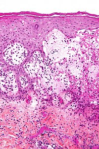
Pemphigus vulgaris is a rare chronic blistering skin disease and the most common form of pemphigus. Pemphigus was derived from the Greek word pemphix, meaning blister. It is classified as a type II hypersensitivity reaction in which antibodies are formed against desmosomes, components of the skin that function to keep certain layers of skin bound to each other. As desmosomes are attacked, the layers of skin separate and the clinical picture resembles a blister. These blisters are due to acantholysis, or breaking apart of intercellular connections through an autoantibody-mediated response. Over time the condition inevitably progresses without treatment: lesions increase in size and distribution throughout the body, behaving physiologically like a severe burn.

<span class="mw-page-title-main">Hailey–Hailey disease</span> Medical condition

Hailey–Hailey disease (HHD), or familial benign chronic pemphigus or familial benign pemphigus, was originally described by the Hailey brothers in 1939. It is a genetic disorder that causes blisters to form on the skin.

<span class="mw-page-title-main">Pemphigoid</span> Autoimmune blistering diseases

Pemphigoid is a group of rare autoimmune blistering diseases of the skin, and mucous membranes. As its name indicates, pemphigoid is similar in general appearance to pemphigus, but, unlike pemphigus, pemphigoid does not feature acantholysis, a loss of connections between skin cells.

Dermatoses of pregnancy are the inflammatory skin diseases that are specific to women while they are pregnant. While some use the term 'polymorphic eruption of pregnancy' to cover these, this term is a synonym used in the UK for Pruritic urticarial papules and plaques of pregnancy, which is the commonest of these skin conditions.

<span class="mw-page-title-main">Collagen, type XVII, alpha 1</span> Mammalian protein found in humans

Collagen XVII, previously called BP180, is a transmembrane protein which plays a critical role in maintaining the linkage between the intracellular and the extracellular structural elements involved in epidermal adhesion, identified by Diaz and colleagues in 1990.

<span class="mw-page-title-main">Dystonin</span> Neurologically significant human protein

Dystonin(DST), also known as bullous pemphigoid antigen 1 (BPAG1), isoforms 1/2/3/4/5/8, is a protein that in humans is encoded by the DST gene.

<span class="mw-page-title-main">Dermatitis herpetiformis</span> Chronic autoimmune disorder leading to blistering skin

Dermatitis herpetiformis (DH) is a chronic autoimmune blistering skin condition, characterised by intensely itchy blisters filled with a watery fluid. DH is a cutaneous manifestation of coeliac disease, although the exact causal mechanism is not known. DH is neither related to nor caused by herpes virus; the name means that it is a skin inflammation having an appearance similar to herpes.

Epidermolysis bullosa acquisita, also known as acquired epidermolysis bullosa, is a longterm autoimmune blistering skin disease. It generally presents with fragile skin that blisters and becomes red with or without trauma. Marked scarring is left with thin skin, milia and nail changes. It typically begins around age 50.

Paraneoplastic pemphigus (PNP) is an autoimmune disorder stemming from an underlying tumor. It is hypothesized that antigens associated with the tumor trigger an immune response resulting in blistering of the skin and mucous membranes.

Pruritic folliculitis of pregnancy is a skin condition that occurs in one in 3000 people, about 0.2% of cases, who are in their second to third trimester of pregnancy where the hair follicle becomes inflamed or infected, resulting in a pus filled bump. Some dermatologic conditions aside from pruritic folliculitis during pregnancy include "pruritic urticarial papules and plaques of pregnancy, atopic eruption of pregnancy, pemphigoid gestationis, intrahepatic cholestasis of pregnancy, and pustular psoriasis of pregnancy". This pruritic folliculitis of pregnancy differs from typical pruritic folliculitis; in pregnancy, it is characterized by sterile hair follicles becoming inflamed mainly involving the trunk, contrasting how typical pruritic folliculitis is mainly localized on "the upper back, shoulders, and chest." This condition was first observed after some pregnant individuals showed signs of folliculitis that were different than seen before. The inflammation was thought to be caused by hormonal imbalance, infection from bacteria, fungi, viruses or even an ingrown hair. However, there is no known definitive cause as of yet. These bumps usually begin on the belly and then spread to upper regions of the body as well as the thighs.

<span class="mw-page-title-main">Linear IgA bullous dermatosis</span> Medical condition

Linear IgA bullous dermatosis is a rare immune-mediated blistering skin disease frequently associated with medication exposure, especially vancomycin, with men and women being equally affected. It was first described by Tadeusz Chorzelski in 1979 and may be divided into two types:

A coma blister, or coma bullae, is a skin lesion or blister that typically arises due to pressure in an individual with impaired consciousness. They vary in size, ranging from 4 to 5 centimeters in diameter, and may appear hemorrhagic or blood filled. Coma blisters are usually found in the extremities and trunk. These types of blisters have been associated with the overdose of central nervous system (CNS) depressants especially barbiturates, but also tricyclic antidepressants, hypnotics, benzodiazepines, opiates, antipsychotics, and alcohol. However, studies have found that coma blisters are not caused by the toxicity of these drugs, but due to hypoxia and external pressure on the comatose individual's skin from being immobilized. Coma blisters have been frequently found on individuals who have overdosed on drugs, but have also been found on individuals with chronic kidney failure, hypercalcemia, diabetic ketoacidosis, and a variety of neurologic conditions. Coma blisters are more frequent in adults and less common among children as demonstrated by the few cases published in literature.

Mucous membrane pemphigoid is a rare chronic autoimmune subepithelial blistering disease characterized by erosive lesions of the mucous membranes and skin. It is one of the pemphigoid diseases that can result in scarring.

References

  1. 1 2 3 4 5 6 7 8 9 10 11 12 13 14 15 16 17 18 James, William D.; Elston, Dirk; Treat, James R.; Rosenbach, Misha A.; Neuhaus, Isaac (2020). "21. Chronic blistering dermatoses". Andrews' Diseases of the Skin: Clinical Dermatology (13th ed.). Edinburgh: Elsevier. pp. 464–465. ISBN   978-0-323-54753-6. Archived from the original on 21 March 2022. Retrieved 2022-03-15.
  2. Wakelin, Sarah H. (2020). "22. Dermatology". In Feather, Adam; Randall, David; Waterhouse, Mona (eds.). Kumar and Clark's Clinical Medicine (10th ed.). Elsevier. pp. 686–687. ISBN   978-0-7020-7870-5. Archived from the original on 2022-03-21. Retrieved 2022-03-21.
  3. 1 2 3 4 "Pemphigoid (herpes) gestationis". www.BAD.org.uk. British Association of Dermatologists. November 2020. Archived from the original on 2 February 2020. Retrieved 21 March 2022.
  4. Johnstone, Ronald B. (2017). "6. Vesiculobullous reaction pattern". Weedon's Skin Pathology Essentials (2nd ed.). Elsevier. p. 123. ISBN   978-0-7020-6830-0. Archived from the original on 2021-05-25. Retrieved 2022-03-21.
  5. Beard, M. P.; Millington, G. W. M (2012). "Recent developments in the specific dermatoses of pregnancy". Clinical and Experimental Dermatology. 37 (1): 1–5. doi:10.1111/j.1365-2230.2011.04173.x. PMID   22007708. S2CID   5428325.
  6. Chi, C-C.; Wang, S-H; Charles-Holmes, R.; Ambros-Rudolph, C.; Powell, J.; Jenkins, R.; Black, M.; Wojnarowska, F. (2012). "Pemphigoid gestationis: early onset and blister formation are associated with adverse pregnancy outcomes". British Journal of Dermatology. 160 (6): 1222–1228. doi:10.1111/j.1365-2133.2009.09086.x. PMID   19298272. S2CID   39630692.
  7. Engineer, Leela; Bhol, Kailash; Ahmed, A.Razzaque (2000). "Pemphigoid gestationis: A review". American Journal of Obstetrics and Gynecology. 183 (2): 483–491. doi:10.1067/mob.2000.105430. ISSN   0002-9378. PMID   10942491.
  8. BP180NC16a ELISA May Be Useful in Serodiagnosis of Pemphigoid Gestationis. Medscape: June 21, 2005
  9. Castro LA, Lundell RB, Krause PK, Gibson LE (November 2006). "Clinical experience in pemphigoid gestationis: report of 10 cases". J. Am. Acad. Dermatol. 55 (5): 823–8. doi:10.1016/j.jaad.2006.07.015. PMID   17052488.