Endometrium

Last updated
Endometrium
Basic Female Reproductive System (English).svg
Uterus and fallopian tubes (uterine tubes). (Endometrium labeled at center right.)
Proliferative phase endometrium -- high mag.jpg
Endometrium in the proliferative phase
Details
Part of Uterus
Identifiers
Latin tunica mucosa uteri
MeSH D004717
TA98 A09.1.03.027
TA2 3521
FMA 17742
Anatomical terminology

The endometrium is the inner epithelial layer, along with its mucous membrane, of the mammalian uterus. It has a basal layer and a functional layer: the basal layer contains stem cells which regenerate the functional layer. [1] The functional layer thickens and then is shed during menstruation in humans and some other mammals, including apes, Old World monkeys, some species of bat, the elephant shrew [2] and the Cairo spiny mouse. [3] In most other mammals, the endometrium is reabsorbed in the estrous cycle. During pregnancy, the glands and blood vessels in the endometrium further increase in size and number. Vascular spaces fuse and become interconnected, forming the placenta, which supplies oxygen and nutrition to the embryo and fetus. [4] [5] The speculated presence of an endometrial microbiota [6] has been argued against. [7] [8]

Contents

Structure

Histology of the most superficial layer of the endometrium, consisting of a simple columnar epithelium. H&E stain Histology of normal simple columnar epithelium of the endometrium.jpg
Histology of the most superficial layer of the endometrium, consisting of a simple columnar epithelium. H&E stain
High magnification micrograph of decidualized endometrium due to exogenous progesterone (oral contraceptive pill). H&E stain Endometrium ocp use3.jpg
High magnification micrograph of decidualized endometrium due to exogenous progesterone (oral contraceptive pill). H&E stain
Low magnification micrograph of decidualized endometrium. H&E stain Endometrium ocp use0.jpg
Low magnification micrograph of decidualized endometrium. H&E stain

The endometrium consists of a single layer of columnar epithelium plus the stroma on which it rests. The stroma is a layer of connective tissue that varies in thickness according to hormonal influences. In the uterus, simple tubular glands reach from the endometrial surface through to the base of the stroma, which also carries a rich blood supply provided by the spiral arteries. In women of reproductive age, two layers of endometrium can be distinguished. These two layers occur only in the endometrium lining the cavity of the uterus, and not in the lining of the fallopian tubes. [4] [5]

In the absence of progesterone, the arteries supplying blood to the functional layer constrict, so that cells in that layer become ischaemic and die, leading to menstruation.

It is possible to identify the phase of the menstrual cycle by reference to either the ovarian cycle or the uterine cycle by observing microscopic differences at each phase—for example in the ovarian cycle:

PhaseDaysThicknessEpithelium
Menstrual phase1–5ThinAbsent
Follicular phase 5–14IntermediateColumnar
Luteal phase 15–27ThickColumnar. Also visible are arcuate vessels of uterus
Ischemic phase 27–28Columnar. Also visible are arcuate vessels of uterus

Gene and protein expression

About 20,000 protein coding genes are expressed in human cells and some 70% of these genes are expressed in the normal endometrium. [9] [10] Just over 100 of these genes are more specifically expressed in the endometrium with only a handful genes being highly endometrium specific. The corresponding specific proteins are expressed in the glandular and stromal cells of the endometrial mucosa. The expression of many of these proteins vary depending on the menstrual cycle, for example the progesterone receptor and thyrotropin-releasing hormone both expressed in the proliferative phase, and PAEP expressed in the secretory phase. Other proteins such as the HOX11 protein that is required for female fertility, is expressed in endometrial stroma cells throughout the menstrual cycle. Certain specific proteins such as the estrogen receptor are also expressed in other types of female tissue types, such as the cervix, fallopian tubes, ovaries and breast. [11]

Microbiome speculation

The uterus and endometrium was for a long time thought to be sterile. The cervical plug of mucosa was seen to prevent the entry of any microorganisms ascending from the vagina. In the 1980s this view was challenged when it was shown that uterine infections could arise from weaknesses in the barrier of the cervical plug. Organisms from the vaginal microbiota could enter the uterus during uterine contractions in the menstrual cycle. Further studies sought to identify microbiota specific to the uterus which would be of help in identifying cases of unsuccessful IVF and miscarriages. Their findings were seen to be unreliable due to the possibility of cross-contamination in the sampling procedures used. The well-documented presence of Lactobacillus species, for example, was easily explained by an increase in the vaginal population being able to seep into the cervical mucous. [7] Another study highlighted the flaws of the earlier studies including cross-contamination. It was also argued that the evidence from studies using germ-free offspring of axenic animals (germ-free) clearly showed the sterility of the uterus. The authors concluded that in light of these findings there was no existence of a microbiome. [8]

The normal dominance of Lactobacilli in the vagina is seen as a marker for vaginal health. However, in the uterus this much lower population is seen as invasive in a closed environment that is highly regulated by female sex hormones, and that could have unwanted consequences. In studies of endometriosis Lactobacillus is not the dominant type and there are higher levels of Streptococcus and Staphylococcus species. Half of the cases of bacterial vaginitis showed a polymicrobial biofilm attached to the endometrium. [7]

Function

The endometrium is the innermost lining layer of the uterus, and functions to prevent adhesions between the opposed walls of the myometrium, thereby maintaining the patency of the uterine cavity. [12] During the menstrual cycle or estrous cycle, the endometrium grows to a thick, blood vessel-rich, glandular tissue layer. This represents an optimal environment for the implantation of a blastocyst upon its arrival in the uterus. The endometrium is central, echogenic (detectable using ultrasound scanners), and has an average thickness of 6.7 mm.

During pregnancy, the glands and blood vessels in the endometrium further increase in size and number. Vascular spaces fuse and become interconnected, forming the placenta, which supplies oxygen and nutrition to the embryo and fetus.

Cycle

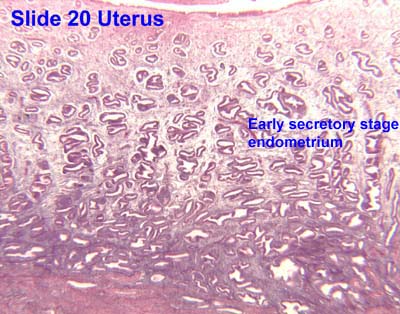
The functional layer of the endometrial lining undergoes cyclic regeneration from stem cells in the basal layer. [1] Humans, apes, and some other species display the menstrual cycle, whereas most other mammals are subject to an estrous cycle. [2] In both cases, the endometrium initially proliferates under the influence of estrogen. However, once ovulation occurs, the ovary (specifically the corpus luteum) will produce much larger amounts of progesterone. This changes the proliferative pattern of the endometrium to a secretory lining. Eventually, the secretory lining provides a hospitable environment for one or more blastocysts.

Upon fertilization, the egg may implant into the uterine wall and provide feedback to the body with human chorionic gonadotropin (hCG). hCG provides continued feedback throughout pregnancy by maintaining the corpus luteum, which will continue its role of releasing progesterone and estrogen. In case of implantation, the endometrial lining remains as decidua . The decidua becomes part of the placenta; it provides support and protection for the gestation.

Without implantation of a fertilized egg, the endometrial lining is either reabsorbed (estrous cycle) or shed (menstrual cycle). In the latter case, the process of shedding involves the breaking down of the lining, the tearing of small connective blood vessels, and the loss of the tissue and blood that had constituted it through the vagina. The entire process occurs over a period of several days. Menstruation may be accompanied by a series of uterine contractions; these help expel the menstrual endometrium.

If there is inadequate stimulation of the lining, due to lack of hormones, the endometrium remains thin and inactive. In humans, this will result in amenorrhea, or the absence of a menstrual period. After menopause, the lining is often described as being atrophic. In contrast, endometrium that is chronically exposed to estrogens, but not to progesterone, may become hyperplastic. Long-term use of oral contraceptives with highly potent progestins can also induce endometrial atrophy. [13] [14]

In humans, the cycle of building and shedding the endometrial lining lasts an average of 28 days. The endometrium develops at different rates in different mammals. Various factors including the seasons, climate, and stress can affect its development. The endometrium itself produces certain hormones at different stages of the cycle and this affects other parts of the reproductive system.

Histopathologic and cytopathologic images.
(A) proliferative endometrium (Left: HE x 400) and proliferative endometrial cells (Right: HE x 100)
(B) secretory endometrium (Left: HE x 10) and secretory endometrial cells (Right: HE x 10)
(C) atrophic endometrium (Left: HE x 10) and atrophic endometrial cells (Right: HE x 10)
(D) mixed endometrium (Left: HE x 10) and mixed endometrial cells (Right: HE x 10)
(E): endometrial atypical hyperplasia (Left: HE x 10) and endometrial atypical cells (Right: HE x 200)
(F) endometrial carcinoma (Left: HE x 400) and endometrial cancer cells (Right: HE x 400). Endometrial histopathologies and cytopathologies.jpg
Histopathologic and cytopathologic images.
(A) proliferative endometrium (Left: HE × 400) and proliferative endometrial cells (Right: HE × 100)
(B) secretory endometrium (Left: HE × 10) and secretory endometrial cells (Right: HE × 10)
(C) atrophic endometrium (Left: HE × 10) and atrophic endometrial cells (Right: HE × 10)
(D) mixed endometrium (Left: HE × 10) and mixed endometrial cells (Right: HE × 10)
(E): endometrial atypical hyperplasia (Left: HE × 10) and endometrial atypical cells (Right: HE × 200)
(F) endometrial carcinoma (Left: HE × 400) and endometrial cancer cells (Right: HE × 400).

Chorionic tissue can result in marked endometrial changes, known as an Arias-Stella reaction, that have an appearance similar to cancer. [15] Historically, this change was diagnosed as endometrial cancer and it is important only in so far as it should not be misdiagnosed as cancer.

Thin endometrium may be defined as an endometrial thickness of less than 8 mm. It usually occurs after menopause. Treatments that can improve endometrial thickness include Vitamin E, L-arginine and sildenafil citrate. [17]

Gene expression profiling using cDNA microarray can be used for the diagnosis of endometrial disorders. [18] The European Menopause and Andropause Society (EMAS) released Guidelines with detailed information to assess the endometrium. [19]

Embryo transfer

An endometrial thickness (EMT) of less than 7 mm decreases the pregnancy rate in in vitro fertilization by an odds ratio of approximately 0.4 compared to an EMT of over 7 mm. However, such low thickness rarely occurs, and any routine use of this parameter is regarded as not justified. The optimal endometrial thickness is 10mm. Nevertheless, in human a perfect synchrony it is not necessary, if the emdometrium is not ready to receive the embryo a ectopic pregnancy may occur. This consist of the implantation of the blast outside the uterus, which can be extremely dangerous. [20]

Triple-line endometrium measuring 7mm. Triple-line endometrium.jpg
Triple-line endometrium measuring 7mm.

Observation of the endometrium by transvaginal ultrasonography is used when administering fertility medication, such as in in vitro fertilization. At the time of embryo transfer, it is favorable to have an endometrium of a thickness of between 7 and 14 mm with a triple-line configuration, [21] which means that the endometrium contains a hyperechoic (usually displayed as light) line in the middle surrounded by two more hypoechoic (darker) lines. A triple-line endometrium reflects the separation of the basal layer and the functional layer, and is also observed in the periovulatory period secondary to rising estradiol levels, and disappears after ovulation. [22]

Endometrial thickness is also associated with live births in IVF. The live birth rate in a normal endometrium is halved when the thickness is <5mm. [23]

Endometrial protection

Estrogens stimulate endometrial proliferation and carcinogenesis. [24] [25] [26] Conversely, progestogens inhibit endometrial proliferation and carcinogenesis caused by estrogens and stimulate differentiation of the endometrium into decidua, which is termed endometrial transformation or decidualization. [24] [25] [26] This is mediated by the progestogenic and functional antiestrogenic effects of progestogens in this tissue. [25] These effects of progestogens and their protection against endometrial hyperplasia and endometrial cancer caused by estrogens is referred to as endometrial protection. [24] [25] [26]

Additional images

See also

Related Research Articles

<span class="mw-page-title-main">Uterus</span> Female sex organ in mammals

The uterus or womb is the organ in the reproductive system of most female mammals, including humans, that accommodates the embryonic and fetal development of one or more embryos until birth. The uterus is a hormone-responsive sex organ that contains glands in its lining that secrete uterine milk for embryonic nourishment.

<span class="mw-page-title-main">Menstruation</span> Shedding of the uterine lining

Menstruation is the regular discharge of blood and mucosal tissue from the inner lining of the uterus through the vagina. The menstrual cycle is characterized by the rise and fall of hormones. Menstruation is triggered by falling progesterone levels and is a sign that pregnancy has not occurred.

<span class="mw-page-title-main">Menstrual cycle</span> Natural changes in the human female reproductive system

The menstrual cycle is a series of natural changes in hormone production and the structures of the uterus and ovaries of the female reproductive system that makes pregnancy possible. The ovarian cycle controls the production and release of eggs and the cyclic release of estrogen and progesterone. The uterine cycle governs the preparation and maintenance of the lining of the uterus (womb) to receive an embryo. These cycles are concurrent and coordinated, normally last between 21 and 35 days, with a median length of 28 days, and continue for about 30–45 years.

<span class="mw-page-title-main">Corpus luteum</span> Temporary endocrine structure in female ovaries

The corpus luteum is a temporary endocrine structure in female ovaries involved in the production of relatively high levels of progesterone, and moderate levels of estradiol, and inhibin A. It is the remains of the ovarian follicle that has released a mature ovum during a previous ovulation.

Heavy menstrual bleeding (HMB), previously known as menorrhagia or hematomunia, is a menstrual period with excessively heavy flow. It is a type of abnormal uterine bleeding (AUB).

Abnormal uterine bleeding (AUB), also known as (AVB) or as atypical vaginal bleeding, is vaginal bleeding from the uterus that is abnormally frequent, lasts excessively long, is heavier than normal, or is irregular. The term dysfunctional uterine bleeding was used when no underlying cause was present. Vaginal bleeding during pregnancy is excluded. Iron deficiency anemia may occur and quality of life may be negatively affected.

An anovulatory cycle is a menstrual cycle characterised by the absence of ovulation and a luteal phase. It may also vary in duration from a regular menstrual cycle.

<span class="mw-page-title-main">Adenomyosis</span> Extension of endometrial tissue into the myometrium

Adenomyosis is a medical condition characterized by the growth of cells that proliferate on the inside of the uterus (endometrium) atypically located among the cells of the uterine wall (myometrium), as a result, thickening of the uterus occurs. As well as being misplaced in patients with this condition, endometrial tissue is completely functional. The tissue thickens, sheds and bleeds during every menstrual cycle.

<span class="mw-page-title-main">Luteal phase</span> The latter part of the menstrual cycle associated with ovulation and an increase in progesterone

The menstrual cycle is on average 28 days in length. It begins with menses during the follicular phase, followed by ovulation and ending with the luteal phase. Unlike the follicular phase which can vary in length among individuals, the luteal phase is typically fixed at approximately 14 days and is characterized by changes to hormone levels, such as an increase in progesterone and estrogen levels, decrease in gonadotropins such as follicle-stimulating hormone (FSH) and luteinizing hormone (LH), changes to the endometrial lining to promote implantation of the fertilized egg, and development of the corpus luteum. In the absence of fertilization by sperm, the corpus luteum atrophies leading to a decrease in progesterone and estrogen, an increase in FSH and LH, and shedding of the endometrial lining (menses) to begin the menstrual cycle again.

<span class="mw-page-title-main">Decidua</span> Part of uterus modified in pregnancy

The decidua is the modified mucosal lining of the uterus that forms every month, in preparation for pregnancy. It is shed off each month when there is no fertilised egg to support. The decidua is under the influence of progesterone. Endometrial cells become highly characteristic. The decidua forms the maternal part of the placenta and remains for the duration of the pregnancy. After birth the decidua is shed together with the placenta.

<span class="mw-page-title-main">Hormonal contraception</span> Birth control methods that act on the endocrine system

Hormonal contraception refers to birth control methods that act on the endocrine system. Almost all methods are composed of steroid hormones, although in India one selective estrogen receptor modulator is marketed as a contraceptive. The original hormonal method—the combined oral contraceptive pill—was first marketed as a contraceptive in 1960. In the ensuing decades many other delivery methods have been developed, although the oral and injectable methods are by far the most popular. Hormonal contraception is highly effective: when taken on the prescribed schedule, users of steroid hormone methods experience pregnancy rates of less than 1% per year. Perfect-use pregnancy rates for most hormonal contraceptives are usually around the 0.3% rate or less. Currently available methods can only be used by women; the development of a male hormonal contraceptive is an active research area.

<span class="mw-page-title-main">Implantation (embryology)</span> First stage of pregnancy

Implantation, also known as nidation is the stage in the embryonic development of mammals in which the blastocyst hatches, attaches, adheres, and invades into the wall of the female's uterus. Implantation is the first stage of gestation, and, when successful, the female is considered to be pregnant. An implanted embryo is detected by the presence of increased levels of human chorionic gonadotropin (hCG) in a pregnancy test. The implanted embryo will receive oxygen and nutrients in order to grow.

<span class="mw-page-title-main">Decidualization</span>

Decidualization is a process that results in significant changes to cells of the endometrium in preparation for, and during, pregnancy. This includes morphological and functional changes to endometrial stromal cells (ESCs), the presence of decidual white blood cells (leukocytes), and vascular changes to maternal arteries. The sum of these changes results in the endometrium changing into a structure called the decidua. In humans, the decidua is shed during childbirth.

<span class="mw-page-title-main">Uterine gland</span>

Uterine glands or endometrial glands are tubular glands, lined by a simple columnar epithelium, found in the functional layer of the endometrium that lines the uterus. Their appearance varies during the menstrual cycle. During the proliferative phase, uterine glands appear long due to estrogen secretion by the ovaries. During the secretory phase, the uterine glands become very coiled with wide lumens and produce a glycogen-rich secretion known as histotroph or uterine milk. This change corresponds with an increase in blood flow to spiral arteries due to increased progesterone secretion from the corpus luteum. During the pre-menstrual phase, progesterone secretion decreases as the corpus luteum degenerates, which results in decreased blood flow to the spiral arteries. The functional layer of the uterus containing the glands becomes necrotic, and eventually sloughs off during the menstrual phase of the cycle.

Arcuate arteries located in the uterus branch out and supply blood to different layers of the uterus. These arteries meet the myometrial-endometrial junction and lead to straight and endometrial arteries. The endometrium receives blood from endometrial arteries which are also called spiral arteries. Endometrial arteries proliferate rapidly and react to different hormones released. These hormones are progesterone and estrogen released by the ovaries and produced by the endocrine system. The endometrial arteries not only supply blood to the endometrium but are also important during pregnancy. They are the initial site of transportation of blood from the mother to the baby.

Hypomenorrhea or hypomenorrhoea, also known as short or scanty periods, is extremely light menstrual blood flow. It is the opposite of heavy periods or hypermenorrhea which is more properly called menorrhagia.

Hormonal regulation occurs at every stage of development. A milieu of hormones simultaneously affects development of the fetus during embryogenesis and the mother, including human chorionic gonadotropin (hCG) and progesterone (P4).

<span class="mw-page-title-main">Chorionic gonadotropin beta</span> Protein-coding gene in the species Homo sapiens

Choriogonadotropin subunit beta (CG-beta) also known as chorionic gonadotrophin chain beta is a protein that in humans is encoded by the CGB gene.

Menstruation is the shedding of the uterine lining (endometrium). It occurs on a regular basis in uninseminated sexually reproductive-age females of certain mammal species.

The pharmacology of progesterone, a progestogen medication and naturally occurring steroid hormone, concerns its pharmacodynamics, pharmacokinetics, and various routes of administration.

References

  1. 1 2 3 Gargett, C.E.; Schwab, K.E.; Zillwood, R.M.; Nguyen, H.P.; Wu, D. (June 2009). "Isolation and culture of epithelial progenitors and mesenchymal stem cells from human endometrium". Biology of Reproduction. 80 (6): 1136–1145. doi:10.1095/biolreprod.108.075226. PMC   2849811 . PMID   19228591.
  2. 1 2 Emera, D; Romero, R; Wagner, G (January 2012). "The evolution of menstruation: a new model for genetic assimilation: explaining molecular origins of maternal responses to fetal invasiveness". BioEssays. 34 (1): 26–35. doi:10.1002/bies.201100099. PMC   3528014 . PMID   22057551.
  3. Bellofiore, N.; Ellery, S.; Mamrot, J.; Walker, D.; Temple-Smith, P.; Dickinson, H. (2016-06-03). "First evidence of a menstruating rodent: the spiny mouse (Acomys cahirinus)". bioRxiv. 216 (1): 40.e1–40.e11. doi:10.1101/056895. PMID   27503621. S2CID   196624853.
  4. 1 2 Blue Histology - Female Reproductive System Archived 2007-02-21 at the Wayback Machine . School of Anatomy and Human Biology — The University of Western Australia Accessed 20061228 20:35
  5. 1 2 Guyton AC, Hall JE, eds. (2006). "Chapter 81 Female Physiology Before Pregnancy and Female Hormones". Textbook of Medical Physiology (11th ed.). Elsevier Saunders. pp. 1018ff. ISBN   9780721602400.
  6. Franasiak, Jason M.; Scott, Richard T. (2015). "Reproductive tract microbiome in assisted reproductive technologies". Fertility and Sterility. 104 (6): 1364–1371. doi: 10.1016/j.fertnstert.2015.10.012 . ISSN   0015-0282. PMID   26597628.
  7. 1 2 3 Baker, JM; Chase, DM; Herbst-Kralovetz, MM (2018). "Uterine Microbiota: Residents, Tourists, or Invaders?". Frontiers in Immunology. 9: 208. doi: 10.3389/fimmu.2018.00208 . PMC   5840171 . PMID   29552006.
  8. 1 2 Perez-Muñoz, ME; Arrieta, MC; Ramer-Tait, AE; Walter, J (28 April 2017). "A critical assessment of the "sterile womb" and "in utero colonization" hypotheses: implications for research on the pioneer infant microbiome". Microbiome. 5 (1): 48. doi: 10.1186/s40168-017-0268-4 . PMC   5410102 . PMID   28454555.
  9. "The human proteome in endometrium - The Human Protein Atlas". www.proteinatlas.org. Retrieved 2017-09-25.
  10. Uhlén, Mathias; Fagerberg, Linn; Hallström, Björn M.; Lindskog, Cecilia; Oksvold, Per; Mardinoglu, Adil; Sivertsson, Åsa; Kampf, Caroline; Sjöstedt, Evelina (2015-01-23). "Tissue-based map of the human proteome". Science. 347 (6220): 1260419. doi:10.1126/science.1260419. ISSN   0036-8075. PMID   25613900. S2CID   802377.
  11. Zieba, Agata; Sjöstedt, Evelina; Olovsson, Matts; Fagerberg, Linn; Hallström, Björn M.; Oskarsson, Linda; Edlund, Karolina; Tolf, Anna; Uhlen, Mathias (2015-10-21). "The Human Endometrium-Specific Proteome Defined by Transcriptomics and Antibody-Based Profiling". OMICS: A Journal of Integrative Biology. 19 (11): 659–668. doi:10.1089/omi.2015.0115. PMID   26488136.
  12. "Dictionary - Normal: Endometrium - The Human Protein Atlas". www.proteinatlas.org. Retrieved 28 December 2022.
  13. Deligdisch, L. (1993). "Effects of hormone therapy on the endometrium". Modern Pathology. 6 (1): 94–106. PMID   8426860.
  14. William's Gynecology, McGraw 2008, Chapter 8, Abnormal Uterine Bleeding
  15. Arias-Stella, J. (Jan 2002). "The Arias-Stella reaction: facts and fancies four decades after". Adv Anat Pathol. 9 (1): 12–23. doi:10.1097/00125480-200201000-00003. PMID   11756756. S2CID   26249687.
  16. Laganà, AS; Garzon, S; Götte, M (10 Nov 2019). "The Pathogenesis of Endometriosis: Molecular and Cell Biology Insights". International Journal of Molecular Sciences. 20 (22): 5615. doi: 10.3390/ijms20225615 . PMC   6888544 . PMID   31717614.
  17. Takasaki A, Tamura H, Miwa I, Taketani T, Shimamura K, Sugino N (April 2010). "Endometrial growth and uterine blood flow: a pilot study for improving endometrial thickness in the patients with a thin endometrium". Fertil. Steril. 93 (6): 1851–8. doi: 10.1016/j.fertnstert.2008.12.062 . PMID   19200982.
  18. Tseng, L.; Chen, I.; Chen, M.; Yan, H.; Wang, C.; Lee, C. (2010). "Genome-based expression profiling as a single standardized microarray platform for the diagnosis of endometrial disorder: an array of 126-gene model". Fertility and Sterility. 94 (1): 114–119. doi: 10.1016/j.fertnstert.2009.01.130 . PMID   19328470.
  19. Dreisler E, Poulsen LG, Antonsen SL, Ceausu I, Depypere H, Erel CT, Lambrinoudaki I, Pérez-López FR, Simoncini T, Tremollieres F, Rees M, Ulrich LG (2013). "EMAS clinical guide: Assessment of the endometrium in peri and postmenopausal women". Maturita. 75 (2): 181–90. doi:10.1016/j.maturitas.2013.03.011. PMID   23619009.
  20. Kasius, A.; Smit, J. G.; Torrance, H. L.; Eijkemans, M. J. C.; Mol, B. W.; Opmeer, B. C.; Broekmans, F. J. M. (2014). "Endometrial thickness and pregnancy rates after IVF: a systematic review and meta-analysis". Human Reproduction Update. 20 (4): 530–541. doi: 10.1093/humupd/dmu011 . ISSN   1355-4786. PMID   24664156.
  21. Zhao, Jing; Zhang, Qiong; Li, Yanping (2012). "The effect of endometrial thickness and pattern measured by ultrasonography on pregnancy outcomes during IVF-ET cycles". Reproductive Biology and Endocrinology. 10 (1): 100. doi: 10.1186/1477-7827-10-100 . ISSN   1477-7827. PMC   3551825 . PMID   23190428.
  22. Baerwald, A. R.; Pierson, R. A. (2004). "Endometrial development in association with ovarian follicular waves during the menstrual cycle". Ultrasound in Obstetrics and Gynecology. 24 (4): 453–460. doi:10.1002/uog.1123. ISSN   0960-7692. PMC   2891966 . PMID   15343603.
  23. 1. Gallos, I. D. et al. Optimal endometrial thickness to maximize live births and minimize pregnancy losses: Analysis of 25,767 fresh embryo transfers. Reprod. Biomed. Online 37, 542–548 (2018).
  24. 1 2 3 Mueck AO, Seeger H, Rabe T (December 2010). "Hormonal contraception and risk of endometrial cancer: a systematic review". Endocr Relat Cancer. 17 (4): R263–71. doi: 10.1677/ERC-10-0076 . PMID   20870686.
  25. 1 2 3 4 Kuhl H (August 2005). "Pharmacology of estrogens and progestogens: influence of different routes of administration". Climacteric. 8 (Suppl 1): 3–63. doi:10.1080/13697130500148875. PMID   16112947. S2CID   24616324.
  26. 1 2 3 Hamoda H (March 2022). "British Menopause Society tools for clinicians: Progestogens and endometrial protection". Post Reprod Health. 28 (1): 40–46. doi:10.1177/20533691211058030. PMID   34841960. S2CID   244749616.