Round ligament of uterus

Last updated
Round ligament of the uterus
Gray1138.png
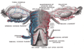
Cross-section through the pelvis of a newly born female child. (Round ligament labelled at upper right.)
Details
Precursor lower gubernaculum [1]
Artery uterine artery, artery of round ligament of uterus
Identifiers
Latin ligamentum teres uteri
MeSH D012404
TA98 A09.1.03.029
TA2 3835
FMA 20420
Anatomical terminology

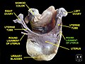
The round ligament of the uterus is a ligament that connects the uterus to the labia majora. It originates at the junction of the uterus and uterine tube. It passes through the inguinal canal to insert at the labium majus.

Contents

The two round ligaments of uterus develop from the gubernaculum; they are the female homologue of the male gubernaculum testis. [2]

Structure

The round ligament of the uterus originates at the uterine horns, in the parametrium. The round ligament exits the pelvis via the deep inguinal ring. [3] It passes through the inguinal canal to reach the labium majus, [4] [5] [2] inserting into the fibro-fatty substance of the labium majus. [2]

Blood supply

The round ligament is supplied by the artery of the round ligament of uterus, also known as Sampson's artery.[ citation needed ]

Development

The round ligament develops from the gubernaculum which attaches the gonad to the labioscrotal swellings in the embryo. [1]

Function

The round ligament of uterus acts to hold the uterus anterior-ward to in anteflexion and anteversion, especially by counteracting any posterior-ward forces that may be being exerted upon the uterus (e.g. distended bladder, or gravity while in a recumbent position). [2]

Pregnancy

The round ligament maintains anteversion of the uterus during pregnancy.[ dubious ] Normally, the cardinal ligament is what supports the uterine angle (angle of anteversion). When the uterus grows during pregnancy, the round ligaments can stretch causing pain. [6] [ unreliable medical source? ]

Additional images

See also

Related Research Articles

<span class="mw-page-title-main">Uterus</span> Female sex organ in mammals

The uterus or womb is the organ in the reproductive system of most female mammals, including humans, that accommodates the embryonic and fetal development of one or more embryos until birth. The uterus is a hormone-responsive sex organ that contains glands in its lining that secrete uterine milk for embryonic nourishment.

<span class="mw-page-title-main">Femoral artery</span> Large artery in the thigh

The femoral artery is a large artery in the thigh and the main arterial supply to the thigh and leg. The femoral artery gives off the deep femoral artery and descends along the anteromedial part of the thigh in the femoral triangle. It enters and passes through the adductor canal, and becomes the popliteal artery as it passes through the adductor hiatus in the adductor magnus near the junction of the middle and distal thirds of the thigh.

<span class="mw-page-title-main">Inguinal canal</span> Human abdominal anatomy

The inguinal canals are the two passages in the anterior abdominal wall of humans and other animals which in males convey the spermatic cords and in females the round ligament of the uterus. The inguinal canals are larger and more prominent in males. There is one inguinal canal on each side of the midline.

<span class="mw-page-title-main">Internal pudendal artery</span>

The internal pudendal artery is one of the three pudendal arteries. It branches off the internal iliac artery, and provides blood to the external genitalia.

<span class="mw-page-title-main">Gubernaculum</span> Embryonic structure

The paired gubernacula also called the caudal genital ligament, are embryonic structures which begin as undifferentiated mesenchyme attaching to the caudal end of the gonads.

<span class="mw-page-title-main">Inguinal ligament</span> Band running from the pubic tubercle to the anterior superior iliac spine

The inguinal ligament, also known as Poupart's ligament or groin ligament, is a band running from the pubic tubercle to the anterior superior iliac spine. It forms the base of the inguinal canal through which an indirect inguinal hernia may develop.

<span class="mw-page-title-main">External iliac artery</span> Arteries of the pelvis

The external iliac arteries are two major arteries which bifurcate off the common iliac arteries anterior to the sacroiliac joint of the pelvis.

<span class="mw-page-title-main">Inferior epigastric artery</span> Blood vessel

In human anatomy, the inferior epigastric artery is an artery that arises from the external iliac artery. It is accompanied by the inferior epigastric vein; inferiorly, these two inferior epigastric vessels together travel within the lateral umbilical fold The inferior epigastric artery then traverses the arcuate line of rectus sheath to enter the rectus sheath, then anastomoses with the superior epigastric artery within the rectus sheath.

<span class="mw-page-title-main">Femoral nerve</span> Long nerve down the thigh and inner leg

The femoral nerve is a nerve in the thigh that supplies skin on the upper thigh and inner leg, and the muscles that extend the knee. It is the largest branch of the lumbar plexus.

<span class="mw-page-title-main">Ilioinguinal nerve</span> Branch of the first lumbar nerve

The ilioinguinal nerve is a branch of the first lumbar nerve (L1). It separates from the first lumbar nerve along with the larger iliohypogastric nerve. It emerges from the lateral border of the psoas major just inferior to the iliohypogastric, and passes obliquely across the quadratus lumborum and iliacus. The ilioinguinal nerve then perforates the transversus abdominis near the anterior part of the iliac crest, and communicates with the iliohypogastric nerve between the transversus and the internal oblique muscle.

<span class="mw-page-title-main">Pubis (bone)</span> Most forward-facing of the three main regions making up the os coxa

In vertebrates, the pubis or pubic bone forms the lower and anterior part of each side of the hip bone. The pubis is the most forward-facing of the three bones that make up the hip bone. The left and right pubic bones are each made up of three sections, a superior ramus, inferior ramus, and a body.

<span class="mw-page-title-main">Conjoint tendon</span> Medial part of the posterior wall of the inguinal canal

The conjoint tendon is a sheath of connective tissue formed from the lower part of the common aponeurosis of the abdominal internal oblique muscle and the transversus abdominis muscle, joining the muscle to the pelvis. It forms the medial part of the posterior wall of the inguinal canal.

<span class="mw-page-title-main">Uterine artery</span>

The uterine artery is an artery that supplies blood to the uterus in females.

<span class="mw-page-title-main">Ovarian artery</span>

The ovarian artery is an artery that supplies oxygenated blood to the ovary in females. It arises from the abdominal aorta below the renal artery. It can be found within the suspensory ligament of the ovary, anterior to the ovarian vein and ureter.

<span class="mw-page-title-main">Transversalis fascia</span> Aponeurosis between the transverse abdominal muscle and the extraperitoneal fat

The transversalis fascia is the fascial lining of the anterolateral abdominal wall situated between the inner surface of the transverse abdominal muscle, and the preperitoneal fascia. It is directly continuous with the iliac fascia, the internal spermatic fascia, and pelvic fascia.

<span class="mw-page-title-main">Broad ligament of the uterus</span> Wide fold of peritoneum that connects the sides of the uterus to the walls and floor of the pelvis

The broad ligament of the uterus is the wide fold of peritoneum that connects the sides of the uterus to the walls and floor of the pelvis.

<span class="mw-page-title-main">Suspensory ligament of ovary</span>

The suspensory ligament of the ovary, also infundibulopelvic ligament, is a fold of peritoneum that extends out from the ovary to the wall of the pelvis.

<span class="mw-page-title-main">Lateral umbilical fold</span>

The lateral umbilical fold is an elevation of the peritoneum lining the inner/posterior surface of the lower anterior abdominal wall formed by the underlying inferior epigastric artery and inferior epigastric vein which the peritoneum covers. Superiorly, the lateral umbilical fold ends where the vessels reach and enter the rectus sheath at the arcuate line of rectus sheath; in spite of the name, the lateral umbilical folds do not extend as far superiorly as the umbilicus. Inferiorly, it extends to just medial to the deep inguinal ring.

<span class="mw-page-title-main">Medial umbilical ligament</span>

The medial umbilical ligament is a paired structure found in human anatomy. It is on the deep surface of the anterior abdominal wall, and is covered by the medial umbilical folds. It is different from the median umbilical ligament, a structure that represents the remnant of the embryonic urachus.

The development of the gonads is part of the prenatal development of the reproductive system and ultimately forms the testes in males and the ovaries in females. The gonads initially develop from the mesothelial layer of the peritoneum.

References

  1. 1 2 Swiss embryology (from UL, UB, and UF) ugenital/diffmorpho05
  2. 1 2 3 4 Sinnatamby, Chummy (2011). Last's Anatomy (12th ed.). p. 304. ISBN   978-0-7295-3752-0.
  3. Anatomy photo:43:03-0201 at the SUNY Downstate Medical Center - "The Female Pelvis: The Broad Ligament"
  4. Anatomy photo:36:03-0105 at the SUNY Downstate Medical Center - "Inguinal Region, Scrotum and Testes: The Internal Surface of the Anterior Abdominal Wall"
  5. Bellier, A.; Cavalié, G.; Marnas, G.; Chaffanjon, P. (2018-06-01). "The round ligament of the uterus: Questioning its distal insertion". Morphologie. 102 (337): 55–60. doi:10.1016/j.morpho.2018.04.001. ISSN   1286-0115. PMID   29731327. S2CID   19189317.
  6. "Pregnancy-Round Ligament Pain". webMD.com. Retrieved 2008-01-27.