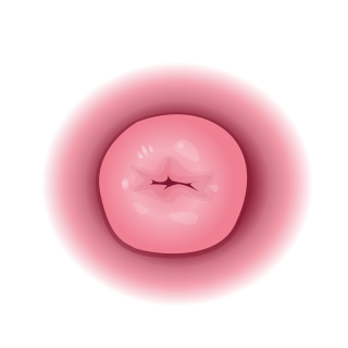
The cervix or cervix uteri is a dynamic fibromuscular sexual organ of the female reproductive system that connects the vagina with the uterine cavity. The human female cervix has been documented anatomically since at least the time of Hippocrates, over 2,000 years ago. The cervix is approximately 4 cm long with a diameter of approximately 3 cm and tends to be described as a cylindrical shape, although the front and back walls of the cervix are contiguous. The size of the cervix changes throughout a woman's life cycle. For example, during the fertile years of a woman's reproductive cycle, females tend to have a larger cervix vis á vis postmenopausal females; likewise, females who have produced offspring have a larger sized cervix than females who have not produced offspring.

Orgasm or sexual climax is the sudden release of accumulated sexual excitement during the sexual response cycle, characterized by intense sexual pleasure resulting in rhythmic, involuntary muscular contractions in the pelvic region. Orgasms are controlled by the involuntary or autonomic nervous system and experienced by both males and females; the body's response includes muscular spasms, a general euphoric sensation, and, frequently, body movements and vocalizations. The period after orgasm is typically a relaxing experience, after the release of the neurohormones oxytocin and prolactin, as well as endorphins.

The uterus or womb is the organ in the reproductive system of most female mammals, including humans, that accommodates the embryonic and fetal development of one or more fertilized eggs until birth. The uterus is a hormone-responsive sex organ that contains glands in its lining that secrete uterine milk for embryonic nourishment.

In mammals and other animals, the vagina is the elastic, muscular reproductive organ of the female genital tract. In humans, it extends from the vulval vestibule to the cervix. The vaginal introitus is normally partly covered by a thin layer of mucosal tissue called the hymen. The vagina allows for copulation and birth. It also channels menstrual flow, which occurs in humans and closely related primates as part of the menstrual cycle.

The G-spot, also called the Gräfenberg spot, is characterized as an erogenous area of the vagina that, when stimulated, may lead to strong sexual arousal, powerful orgasms and potential female ejaculation. It is typically reported to be located 5–8 cm (2–3 in) up the front (anterior) vaginal wall between the vaginal opening and the urethra and is a sensitive area that may be part of the female prostate.

The missionary position or man-on-top position is a sex position in which, generally, a woman lies on her back and spreads her legs and a man lies on top of her while they face each other and engage in vaginal intercourse. The position may also be used for other sexual activity, such as anal sex. It is commonly associated with heterosexual sexual activity, but is also used by same-sex couples. It may involve sexual penetration or non-penetrative sex, and its penile-vaginal aspect is an example of ventro-ventral (front-to-front) reproductive activity. Variations of the position allow varying degrees of clitoral stimulation, depth of penetration, participation on the part of the woman, and the likelihood and speed of orgasm.

Doggy style is a sex position in which one participant bends over, crouches on all fours, or lies on their abdomen, for sexual intercourse, other forms of sexual penetration or other sexual activity. Doggy style is a form of a rear-entry position, others being with the receiving partner lying on the side in the spoons sex position or the reverse cowgirl sex position. Non-penetrative sex in this position may also be regarded as doggy style.
Dyspareunia is painful sexual intercourse due to medical or psychological causes. The term dyspareunia covers both female dyspareunia and male dyspareunia, but many discussions that use the term without further specification concern the female type, which is more common than the male type. In females, the pain can primarily be on the external surface of the genitalia, or deeper in the pelvis upon deep pressure against the cervix. Medically, dyspareunia is a pelvic floor dysfunction and is frequently underdiagnosed. It can affect a small portion of the vulva or vagina or be felt all over the surface. Understanding the duration, location, and nature of the pain is important in identifying the causes of the pain.

The human female reproductive system is made up of the internal and external sex organs that function in the reproduction of new offspring. The reproductive system is immature at birth and develops at puberty to be able to release matured ova from the ovaries, facilitate their fertilization, and create a protective environment for the developing fetus during pregnancy. The female reproductive tract is made of several connected internal sex organs—the vagina, uterus, and fallopian tubes—and is prone to infections. The vagina allows for sexual intercourse, and is connected to the uterus at the cervix. The uterus accommodates the embryo by developing the uterine lining.

The pelvic floor or pelvic diaphragm is an anatomical location in the human body, which has an important role in urinary and anal continence, sexual function and support of the pelvic organs. The pelvic floor includes muscles, both skeletal and smooth, ligaments and fascia. and separates between the pelvic cavity from above, and the perineum from below. It is formed by the levator ani muscle and coccygeus muscle, and associated connective tissue.
Insemination is the introduction of sperm (semen) into a female or hermaphrodite's reproductive system in order to fertilize the ovum through sexual reproduction. The sperm enters into the uterus of a mammal or the oviduct of an oviparous (egg-laying) animal. Female humans and other mammals are inseminated during sexual intercourse or copulation, but can also be inseminated by artificial insemination.
The development of the urinary system begins during prenatal development, and relates to the development of the urogenital system – both the organs of the urinary system and the sex organs of the reproductive system. The development continues as a part of sexual differentiation.

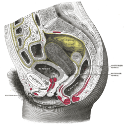
The rectouterine pouch is the extension of the peritoneum into the space between the posterior wall of the uterus and the rectum in the human female.

The cystocele, also known as a prolapsed bladder, is a medical condition in which a woman's bladder bulges into her vagina. Some may have no symptoms. Others may have trouble starting urination, urinary incontinence, or frequent urination. Complications may include recurrent urinary tract infections and urinary retention. Cystocele and a prolapsed urethra often occur together and is called a cystourethrocele. Cystocele can negatively affect quality of life.

The vaginal artery is an artery in females that supplies blood to the vagina and the base of the bladder.

Uterine prolapse is a form of pelvic organ prolapse in which the uterus and a portion of the upper vagina protrude into the vaginal canal and, in severe cases, through the opening of the vagina. It is most often caused by injury or damage to structures that hold the uterus in place within the pelvic cavity. Symptoms may include vaginal fullness, pain with sexual intercourse, difficulty urinating, and urinary incontinence. Risk factors include older age, pregnancy, vaginal childbirth, obesity, chronic constipation, and chronic cough. Prevalence, based on physical exam alone, is estimated to be approximately 14%.
In human female anatomy, the vesicouterine pouch, also uterovesicle pouch, is a fold of peritoneum over the uterus and the bladder. Like the rectouterine pouch, it is a female pelvic recess, but shallower and closer to the anterior fornix of the vagina.

A pelvic examination is the physical examination of the external and internal female pelvic organs. It is frequently used in gynecology for the evaluation of symptoms affecting the female reproductive and urinary tract, such as pain, bleeding, discharge, urinary incontinence, or trauma. It can also be used to assess a woman's anatomy in preparation for procedures. The exam can be done awake in the clinic and emergency department, or under anesthesia in the operating room. The most commonly performed components of the exam are 1) the external exam, to evaluate the vulva 2) the internal exam with palpation to examine the uterus, ovaries, and structures adjacent to the uterus (adnexae) and 3) the internal exam using a speculum to visualize the vaginal walls and cervix. During the pelvic exam, sample of cells and fluids may be collected to screen for sexually transmitted infections or cancer.
Genital trauma is trauma to the genitalia.

The vaginal support structures are those muscles, bones, ligaments, tendons, membranes and fascia, of the pelvic floor that maintain the position of the vagina within the pelvic cavity and allow the normal functioning of the vagina and other reproductive structures in the female. Defects or injuries to these support structures in the pelvic floor leads to pelvic organ prolapse. Anatomical and congenital variations of vaginal support structures can predispose a woman to further dysfunction and prolapse later in life. The urethra is part of the anterior wall of the vagina and damage to the support structures there can lead to incontinence and urinary retention.