The corpus luteum is a temporary endocrine structure in female ovaries and is involved in the production of relatively high levels of progesterone, moderate levels of estradiol and inhibin A, and small amounts of estrogen. It is the remains of the ovarian follicle that has released a mature ovum during a previous ovulation.

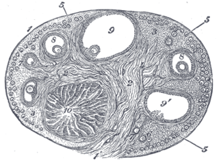
An ovarian follicle is a roughly spheroid cellular aggregation set found in the ovaries. It secretes hormones that influence stages of the menstrual cycle. Women begin puberty with about 400,000 follicles, each with the potential to release an egg cell (ovum) at ovulation for fertilization. These eggs are developed once every menstrual cycle.

The zona pellucida is a glycoprotein layer surrounding the plasma membrane of mammalian oocytes. It is a vital constitutive part of the oocyte. The zona pellucida first appears in unilaminar primary oocytes. It is secreted by both the oocyte and the ovarian follicles. The zona pellucida is surrounded by the cumulus oophorus. The cumulus is composed of cells that care for the egg when it is emitted from the ovary.

A granulosa cell or follicular cell is a somatic cell of the sex cord that is closely associated with the developing female gamete in the ovary of mammals.

The efferent ducts connect the rete testis with the initial section of the epididymis.

Adventitia is the outermost connective tissue covering of an organ, vessel, or other structure. It is also called the tunica adventitia or the tunica externa.

Alveolar ducts are tiny ducts that connect the respiratory bronchioles to alveolar sacs, each of which contains a collection of alveoli. They are tiny end ducts of the branching airways that fill the lungs. Each lung holds approximately 1.5 to 2 million of them. The tubules divide into two or three alveolar sacs at the distal end. They are formed from the confluence openings of several alveoli. Distal terminations of alveolar ducts are atria which then end in alveolar sacs.

In biology, folliculogenesis is the maturation of the ovarian follicle, a densely packed shell of somatic cells that contains an immature oocyte. Folliculogenesis describes the progression of a number of small primordial follicles into large preovulatory follicles that occurs in part during the menstrual cycle.

The tunica media, or media for short, is the middle tunica (layer) of an artery or vein. It lies between the tunica intima on the inside and the tunica externa on the outside.
Theca interna cells express receptors for luteinizing hormone (LH) to produce androstenedione, which via a few steps, gives the granulosa the precursor for estrogen manufacturing.

The broad ligament of the uterus is the wide fold of peritoneum that connects the sides of the uterus to the walls and floor of the pelvis.
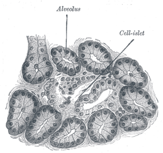
The intercalated duct, also called intercalary duct, is the portion of an exocrine gland leading directly from the acinus to a striated duct. The intercalated duct forms part of the intralobular duct. This duct has the thinnest epithelium of any part of the duct system, and the epithelium is usually classified as "low" simple cuboidal.

The perimetrium is the outer serosa layer of the uterus, equivalent to peritoneum. It is embrionically derived from visceral peritoneum. Perimetrium consists of superficial mesothelium, and a thin layer of loose connective tissue beneath it. The posterior surface of the uterus is completely covered by the perimetrium, but the anterior surface only partially.
The tunica externa — also known as the tunica adventitia, or adventitia for short — is the outermost tunica (layer) of a blood vessel, surrounding the tunica media. It is mainly composed of collagen and, in arteries, is supported by external elastic lamina. The collagen serves to anchor the blood vessel to nearby organs, giving it stability.
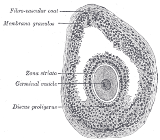
The cumulus oophorus, also called discus proligerus, is a cluster of cells that surround the oocyte both in the ovarian follicle and after ovulation. In the antral follicle, it may be regarded as an extension of the membrana granulosa. The innermost layer of these cells is the corona radiata.
The theca folliculi comprise a layer of the ovarian follicles. They appear as the follicles become secondary follicles.
The follicular antrum is the portion of an ovarian follicle filled with follicular fluid. Appearance of the follicular antrum during follicular maturation is the first sign that a follicle has reached the next stage of maturation. It has changed from a primary follicle to a secondary follicle.

Uterine glands or endometrial glands are tubular glands, lined by ciliated columnar epithelium, found in the functional layer of the endometrium that lines the uterus. Their appearance varies during the menstrual cycle. During the proliferative phase, uterine glands appear long due to estrogen secretion by the ovaries. During the secretory phase, the uterine glands become very coiled with wide lumens and produce a glycogen-rich secretion. This change corresponds with an increase in blood flow to spiral arteries due to increased progesterone secretion from the corpus luteum. During the pre-menstrual phase, progesterone secretion decreases as the corpus luteum degenerates, which results in decreased blood flow to the spiral arteries. The functional layer of the uterus containing the glands becomes necrotic, and eventually sloughs off during the menstrual phase of the cycle.
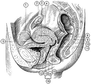
The uterine horns are the points where the uterus and the fallopian tubes meet. They are one of the points of attachment for the round ligament of uterus.

A striated duct (Pflüger's ducts ) is a gland duct which connects an intercalated duct to an interlobular duct. It is characterized by the basal infoldings of its plasma membrane, characteristic of ion-pumping activity by the numerous mitochondria. Along with the intercalated ducts, they function to modify salivary fluid by secreting HCO3− and K+ and reabsorbing Na+ and Cl− using the Na-K pump and the Cl-HCO3 pump, making the saliva hypotonic.
