Theca of follicle

Last updated
Theca of follicle
Details
Identifiers
Latin theca folliculi
Anatomical terminology

The theca folliculi comprise a layer of the ovarian follicles. They appear as the follicles become secondary follicles.

Contents

The theca are divided into two layers, the theca interna and the theca externa . [1]

Theca cells are a group of endocrine cells in the ovary made up of connective tissue surrounding the follicle. They have many diverse functions, including promoting folliculogenesis and recruitment of a single follicle during ovulation. [2] Theca cells and granulosa cells together form the stroma of the ovary.

Androgen synthesis

The anterior pituitary complex and hypophyseal portal system, where FSH and LH are released. 1808 The Anterior Pituitary Complex.jpg
The anterior pituitary complex and hypophyseal portal system, where FSH and LH are released.

Theca cells are responsible for synthesizing androgens, providing signal transduction between granulosa cells and oocytes during development by the establishment of a vascular system, providing nutrients, and providing structure and support to the follicle as it matures. [2]

Conversion of testosterone to estradiol through the action of aromatase. Testosterone estradiol conversion.png
Conversion of testosterone to estradiol through the action of aromatase.

Theca cells are responsible for the production of androstenedione, and indirectly the production of 17β estradiol, also called E2, by supplying the neighboring granulosa cells with androstenedione that with the help of the enzyme aromatase can be used as a substrate for this type of estradiol. [3] FSH induces the granulosa cells to synthesize aromatase, an enzyme that converts the androgens made by the theca interna into estradiol. [3]

Signaling cascade

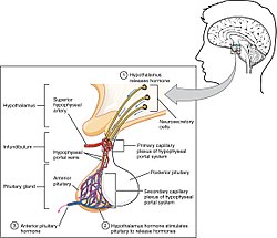
Gonadotropin releasing hormone (GnRH) is released by projections of the hypothalamus into the anterior pituitary gland. Gonadotrophs are stimulated to produce follicle-stimulating hormone (FSH) and luteinizing hormone (LH), which are released into the bloodstream to act upon the ovaries. Luteinizing hormone serves to directly stimulate theca cells. Together, these organs comprise the HPG axis.

Within the ovaries, transmembrane G-protein coupled receptors (GPCRs) bind to LH in the bloodstream, and the signal is transduced to the interior of theca cells through the action of the second messenger cAMP and third messenger protein kinase A (PKA). Theca cells are then stimulated to produce testosterone, which is sent in a paracrine fashion to neighboring granulosa cells for conversion to estradiol. [4]

Disorders

Hyperactivity of theca cells causes hyperandrogenism, and hypoactivity leads to a lack of estrogen. [5] Granulosa cell tumors, while rare (less than 5% of ovarian cancers), may both granulosa cells and theca cells. [6] Thecomas are benign proliferations of theca cells that may present with hormonal dysfunction. [7]

Theca cells (along with granulosa cells) form the corpus luteum during oocyte maturation. Theca cells are only correlated with developing ovarian follicles. [5] They are the leading cause of endocrine-based infertility, as either hyperactivity or hypoactivity of the theca cells can lead to fertility problems.

Folliculogenesis

A depiction of the ovarian cycle in animals Anatomy and physiology of animals Ovarian cycle showing from top left clockwise.jpg
A depiction of the ovarian cycle in animals

In human adult females, the primordial follicle is composed of a single oocyte surrounded by a layer of closely associated granulosa cells. In early stages of the ovarian cycle, the developing follicle acquires a layer of connective tissue and associated blood vessels. This covering is called the theca.

As development of the secondary follicle progresses, granulosa cells proliferate to form the multilayered membrana granulosum. Over a period of months, the granulosa cells and thecal cells secrete antral fluid (a mixture of hormones, enzymes, and anticoagulants) to nourish the maturing ovum.

In tertiary follicles, the single-layered theca differentiates into a theca interna and theca externa. The theca interna contains glandular cells and many small blood vessels, while the theca externa is composed of dense connective tissue and larger blood vessels. [8]

See also

References

  1. Melmed, Shlomo; Koenig, Ronald; Rosen, Clifford; Auchus, Richard; Goldfine, Allison (2020). "17:Physiology and Pathology of the female reproductive axis". Williams Textbook of Endocrinology. Vol. 1: Section V:Sexual Development and Function (14th. ed.). Elsevier Health Sciences. pp. 586–587. ISBN   978-8131262160.
  2. 1 2 Young, J. M.; McNeilly, A. S. (2010). "Theca: the forgotten cell of the ovarian follicle". Reproduction. 140 (4): 489–504. doi: 10.1530/REP-10-0094 . PMID   20628033.
  3. 1 2 Hall, John E. (2016). Guyton and Hall Textbook of Medical Physiology (13th ed.). Philadelphia, Pennsylvania: Elsevier. pp. 1042, 1044. ISBN   9781455770052. OCLC   900869748.
  4. Boron, Walter F.; Boulpaep, Emile L., eds. (2017). Medical Physiology (Third ed.). Philadelphia, Pennsylvania: Elsevier. ISBN   978-1-4557-3328-6. OCLC   951680737.
  5. 1 2 Magoffin, Denis A. (2005). "Ovarian theca cell". The International Journal of Biochemistry & Cell Biology. 37 (7): 1344–9. doi:10.1016/j.biocel.2005.01.016. PMID   15833266.
  6. Kottarathil, Vijaykumar Dehannathparambil; Antony, Michelle Aline; Nair, Indu R.; Pavithran, Keechilat (2013). "Recent Advances in Granulosa Cell Tumor Ovary: A Review". Indian Journal of Surgical Oncology. 4 (1): 37–47. doi:10.1007/s13193-012-0201-z. PMC   3578540 . PMID   24426698.
  7. Burandt, Eike; Young, Robert H. (August 2014). "Thecoma of the ovary: a report of 70 cases emphasizing aspects of its histopathology different from those often portrayed and its differential diagnosis". The American Journal of Surgical Pathology. 38 (8): 1023–1032. doi:10.1097/PAS.0000000000000252. ISSN   1532-0979. PMID   25025365. S2CID   10739300.
  8. Jones, Richard E.; Lopez, Kristin H. (2006). Human Reproductive Biology (3rd ed.). Amsterdam: Elsevier Academic Press. ISBN   978-0-12-088465-0. OCLC   61351645.