External sphincter muscle of female urethra

Last updated
External sphincter muscle of female urethra
Gray1166.png
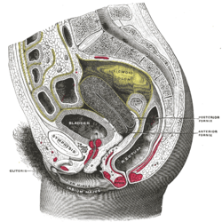
Sagittal section of the lower part of a female trunk, right segment. (Sphincter not labeled)
Gray408.png
Muscles of the female perineum. (Urethral sphincter not labeled)
Details
Nerve Somatic fibers from S2-S4 through Pudendal nerve
Actions Constricts urethra and vagina, maintains urinary continence
Identifiers
Latin musculus sphincter urethrae externus urethrae femininae
TA98 A09.2.03.006F
TA2 2422
FMA 19778
Anatomical terms of muscle

The external sphincter muscle of female urethra is a muscle which controls urination in females. The muscle fibers arise on either side from the margin of the inferior ramus of the pubis. They are directed across the pubic arch in front of the urethra, and pass around it to blend with the muscular fibers of the opposite side, between the urethra and vagina.

Contents

The term "urethrovaginal sphincter" ("sphincter urethrovaginalis") is sometimes used to describe the component adjacent to the vagina. [1] [2] [3] TA: A09.5.03.006F, FMA: 30439

The "compressor urethrae" is also considered a distinct, adjacent muscle by some sources, [4] [5] TA: A09.5.03.005F, FMA: 30438

Function

The muscle helps maintain continence of urine along with the internal urethral sphincter which is under control of the autonomic nervous system. The external sphincter muscle prevents urine leakage as the muscle is tonically contracted via somatic fibers that originate in Onuf's nucleus and pass through sacral spinal nerves S2-S4 then the pudendal nerve to synapse on the muscle. [5] [6]

Voiding urine begins with voluntary relaxation of the external urethral sphincter. This is facilitated by inhibition of the somatic neurons in Onuf's nucleus via signals arising in the pontine micturition center and traveling through the descending reticulospinal tracts.

See also

Related Research Articles

Urethra Tube that connects the urinary bladder to the external urethral orifice

The urethra is a tube that connects the urinary bladder to the urinary meatus for the removal of urine from the body of both females and males. In human females and other primates, the urethra connects to the urinary meatus above the vagina, whereas in marsupials, the female's urethra empties into the urogenital sinus.

Urinary bladder Organ in humans and vertebrates that collects and stores urine from the kidneys before disposal

The urinary bladder, or simply bladder, is a hollow muscular organ in humans and other vertebrates that stores urine from the kidneys before disposal by urination. In the human the bladder is a hollow distensible organ that sits on the pelvic floor. Urine enters the bladder via the ureters and exits via the urethra. The typical human bladder will hold between 300 and 500 ml before the urge to empty occurs, but can hold considerably more.

Female ejaculation Expulsion of fluid during or before an orgasm

Female ejaculation is characterized as an expulsion of fluid from the Skene's gland at the lower end of the urethra during or before an orgasm. It is also known colloquially as squirting, although research indicates that female ejaculation and squirting are different phenomena, with squirting being attributed to a sudden expulsion of liquid that partly comes from the bladder and contains urine. Female ejaculation is physiologically distinct from coital incontinence, with which it is sometimes confused.

Pudendal nerve Main nerve of the perineum

The pudendal nerve is the main nerve of the perineum. It carries sensation from the external genitalia of both sexes and the skin around the anus and perineum, as well as the motor supply to various pelvic muscles, including the male or female external urethral sphincter and the external anal sphincter. If damaged, most commonly by childbirth, lesions may cause sensory loss or fecal incontinence. The nerve may be temporarily blocked as part of an anaesthetic procedure.

Urination release of urine from the urinary bladder

Urination is the release of urine from the urinary bladder through the urethra to the outside of the body. It is the urinary system's form of excretion. It is also known medically as micturition, voiding, uresis, or, rarely, emiction, and known colloquially by various names including peeing, weeing, and pissing.

Urinary incontinence

Urinary incontinence (UI), also known as involuntary urination, is any uncontrolled leakage of urine. It is a common and distressing problem, which may have a large impact on quality of life. It has been identified as an important issue in geriatric health care. The term enuresis is often used to refer to urinary incontinence primarily in children, such as nocturnal enuresis.

Kegel exercise Pelvic floor exercise

Kegel exercise, also known as pelvic-floor exercise, involves repeatedly contracting and relaxing the muscles that form part of the pelvic floor, now sometimes colloquially referred to as the "Kegel muscles". The exercise can be performed multiple times each day, for several minutes at a time, but takes one to three months to begin to have an effect.

Bulbospongiosus muscle

The bulbospongiosus muscle is one of the superficial muscles of the perineum. It has a slightly different origin, insertion and function in males and females. In males, it covers the bulb of the penis. In females, it covers the vestibular bulb.

Pelvic floor Anatomical structure

The pelvic floor or pelvic diaphragm is composed of muscle fibers of the levator ani, the coccygeus muscle, and associated connective tissue which span the area underneath the pelvis. The pelvic diaphragm is a muscular partition formed by the levatores ani and coccygei, with which may be included the parietal pelvic fascia on their upper and lower aspects. The pelvic floor separates the pelvic cavity above from the perineal region below. Both males and females have a pelvic floor. To accommodate the birth canal, a female's pelvic cavity is larger than a male's.

Onufs nucleus

Onuf's nucleus is a distinct group of neurons located in the ventral part of the anterior horn of the sacral region of the human spinal cord involved in the maintenance of micturition and defecatory continence, as well as muscular contraction during orgasm. It contains motor neurons, and is the origin of the pudendal nerve. The sacral region of the spinal cord is the fourth segment of vertebrae in the spinal cord which consists of the vertebrae 26-30. While working in New York City in 1899, Bronislaw Onuf-Onufrowicz discovered this group of unique cells and originally identified it as “Group X.” “Group X” was considered distinct by Onufrowicz because the cells were different in size from the surrounding neurons in the anterolateral group, suggesting that they were independent.

Urogenital diaphragm

Older texts have asserted the existence of a urogenital diaphragm, also called the triangular ligament, which was described as a layer of the pelvis that separates the deep perineal sac from the upper pelvis, lying between the inferior fascia of the urogenital diaphragm and superior fascia of the urogenital diaphragm.

External sphincter muscle of male urethra

The external sphincter muscle of male urethra, also sphincter urethrae membranaceae, sphincter urethrae externus, surrounds the whole length of the membranous urethra, and is enclosed in the fascia of the urogenital diaphragm.

Stress incontinence Form of urinary incontinence due to an inadequate closer of the bladdertae

Stress incontinence, also known as stress urinary incontinence (SUI) or effort incontinence is a form of urinary incontinence. It is due to inadequate closure of the bladder outlet by the urethral sphincter.

Bladder sphincter dyssynergia

Bladder sphincter dyssynergia is a consequence of a neurological pathology such as spinal injury or multiple sclerosis. which disrupts central nervous system regulation of the micturition (urination) reflex resulting in dyscoordination of the detrusor muscles of the bladder and the male or female external urethral sphincter muscles. In normal lower urinary tract function, these two separate muscle structures act in synergistic coordination. But in this neurogenic disorder, the urethral sphincter muscle, instead of relaxing completely during voiding, dyssynergically contracts causing the flow to be interrupted and the bladder pressure to rise.

Detrusor muscle

The detrusor muscle, also detrusor urinae muscle, muscularis propria of the urinary bladder and muscularis propria, is smooth muscle found in the wall of the bladder. The detrusor muscle remains relaxed to allow the bladder to store urine, and contracts during urination to release urine. Related are the urethral sphincter muscles which envelop the urethra to control the flow of urine when they contract.

Urinary meatus

The urinary meatus, also known as the external urethral orifice, is the opening of the urethra. It is the point where urine exits the urethra in males and in females and where semen exits the urethra in males. The meatus has varying degrees of sensitivity to touch. The meatus is located on the glans of the penis or in the vulval vestibule.

Urethral sphincters

The urethral sphincters are two muscles used to control the exit of urine in the urinary bladder through the urethra. The two muscles are either the male or female external urethral sphincter and the internal urethral sphincter. When either of these muscles contracts, the urethra is sealed shut.

Internal urethral sphincter

The internal urethral sphincter is a urethral sphincter muscle which constricts the internal urethral orifice. It is located at the junction of the urethra with the urinary bladder and is continuous with the detrusor muscle, but anatomically and functionally fully independent from it. It is composed of smooth muscle, so it is under the control of the autonomic nervous system, specifically the sympathetic nervous system.

The Pontine micturition center is a collection of neuronal cell bodies located in the rostral pons in the brainstem involved in the supraspinal regulation of micturition. When activated, the PMC relaxes the urethral sphincter allowing for micturition to occur. The PMC coordinates with other brain centers, including the medial frontal cortex, insular cortex, hypothalamus and periaqueductal gray (PAG). The PAG acts as a relay station for ascending bladder information from the spinal cord and incoming signals from higher brain areas.

Vaginal support structures

The vaginal support structures are those muscles, bones, ligaments, tendons, membranes and fascia, of the pelvic floor that maintain the position of the vagina within the pelvic cavity and allow the normal functioning of the vagina and other reproductive structures in the female. Defects or injuries to these support structures in the pelvic floor leads to pelvic organ prolapse. Anatomical and congenital variations of vaginal support structures can predispose a woman to further dysfunction and prolapse later in life. The urethra is part of the anterior wall of the vagina and damage to the support structures there can lead to incontinence and urinary retention.

References

This article incorporates text in the public domain from page 431 of the 20th edition of Gray's Anatomy (1918)

  1. Kyung Won, PhD. Chung (2005). Gross Anatomy (Board Review). Hagerstown, MD: Lippincott Williams & Wilkins. p. 262. ISBN   0-7817-5309-0.
  2. Rahn DD, Marinis SI, Schaffer JI, Corton MM (2006). "Anatomical path of the tension-free vaginal tape: reassessing current teachings". Am. J. Obstet. Gynecol. 195 (6): 1809–13. doi:10.1016/j.ajog.2006.07.009. PMID   17132484.
  3. Umek WH, Kearney R, Morgan DM, Ashton-Miller JA, DeLancey JO (2003). "The axial location of structural regions in the urethra: a magnetic resonance study in nulliparous women". Obstet Gynecol. 102 (5 Pt 1): 1039–45. doi:10.1016/j.obstetgynecol.2003.04.001. PMC   1226706 . PMID   14672484.
  4. Adam Mitchell; Drake, Richard; Gray, Henry David; Wayne Vogl (2005). Gray's anatomy for students. Elsevier/Churchill Livingstone. p. 396. ISBN   0-443-06612-4.
  5. 1 2 Jung J, Ahn HK, and Huh Y (September 2012). "Clinical and Functional Anatomy of the Urethral Sphincter". Int Neurourol J. 16 (3): 102–106. doi:10.5213/inj.2012.16.3.102. PMC   3469827 . PMID   23094214.
  6. Shah AP, Mevcha A, Wilby D, Alatsatianos A, Hardman JC, Jacques S, Wilton JC (November 2014). "Continence and micturition: An anatomical basis" (PDF). Clin. Anat. 27 (8): 1275–1283. doi:10.1002/ca.22388. PMID   24615792. S2CID   21875132.