Spongy urethra

Last updated
Spongy urethra
Gray1142.png
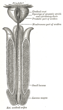
The human male urethra laid open on its anterior (upper) surface.
Details
Precursor Urogenital sinus [1]
Identifiers
Latin pars spongiosa urethrae masculinae, pars cavernosa urethrae masculinae
TA98 A09.4.02.021
TA2 3682, 3455
FMA 19675
Anatomical terminology

The spongy urethra (cavernous portion of urethra, penile urethra) is the longest part of the male urethra, and is contained in the corpus spongiosum of the penis.

Contents

It is about 15 cm long, and extends from the termination of the membranous portion to the external urethral orifice.

Commencing below the inferior fascia of the urogenital diaphragm it passes forward and upward to the front of the pubic symphysis; and then, in the flaccid condition of the penis, it bends downward and forward.

It is narrow, and of uniform size in the body of the penis, measuring about 6 mm in diameter; it is dilated behind, within the bulb, and again anteriorly within the glans penis, where it forms the fossa navicularis urethrae.

The spongy urethra runs along the length of the penis on its ventral (underneath) surface. It is about 15–16 cm in length, and travels through the corpus spongiosum. The ducts from the urethral gland (gland of Littré) enter here. The openings of the bulbourethral glands are also found here. [2] Some textbooks will subdivide the spongy urethra into two parts, the bulbous and pendulous urethra. The urethral lumen (interior) runs effectively parallel to the penis, except at the narrowest point, the external urethral meatus, where it is vertical. This produces a spiral stream of urine and has the effect of cleaning the external urethral meatus.[ citation needed ] The lack of an equivalent mechanism in the female urethra partly explains why urinary tract infections occur so much more frequently in females.[ citation needed ]

Epithelium

Pseudostratified columnar – proximally, Stratified squamous – distally

Additional images

Related Research Articles

<span class="mw-page-title-main">Urethra</span> Tube that connects the urinary bladder to the external urethral orifice

The urethra is a tube that connects the mammalian urinary bladder to the urinary meatus. Male and female placental mammals release urine through the urethra during urination, but males also release semen through the urethra during ejaculation.

<span class="mw-page-title-main">Glans penis</span> End of the penis

In male human anatomy, the glans penis or penile glans, commonly referred to as the glans, is the bulbous structure at the distal end of the human penis that is the human male's most sensitive erogenous zone and primary anatomical source of sexual pleasure. The glans penis is present in the male reproductive organs of humans and most other mammals where it may appear smooth, spiny, elongated or divided. It is externally lined with mucosal tissue, which creates a smooth texture and glossy appearance. In humans, the glans is located over the distal ends of the corpora cavernosa and is a continuation of the corpus spongiosum of the penis. At the summit appears the urinary meatus and at the base forms the corona glandis. An elastic band of tissue, known as the frenulum, runs on its ventral surface. In men who are not circumcised, it is completely or partially covered by a fold of skin called the foreskin. In adults, the foreskin can generally be retracted over and past the glans manually or sometimes automatically during an erection.

<span class="mw-page-title-main">Corpus spongiosum (penis)</span> Spongy tissue within the human penis

The corpus spongiosum is the mass of spongy tissue surrounding the male urethra within the penis. It is also called the corpus cavernosum urethrae in older texts.

<span class="mw-page-title-main">Bulbospongiosus muscle</span> Superficial muscle of the perineum

The bulbospongiosus muscles are a subgroup of the superficial muscles of the perineum. They have a slightly different origin, insertion and function in males and females. In males, these muscles cover the bulb of the penis, while in females, they cover the vestibular bulbs.

<span class="mw-page-title-main">Male reproductive system</span> Reproductive system of the human male

The male reproductive system consists of a number of sex organs that play a role in the process of human reproduction. These organs are located on the outside of the body, and within the pelvis.

<span class="mw-page-title-main">Corpus cavernosum penis</span> Sponge-like region of erectile tissue

A corpus cavernosum penis (singular) is one of a pair of sponge-like regions of erectile tissue, which contain most of the blood in the penis during an erection.

<span class="mw-page-title-main">Perineal membrane</span> Anatomical term

The perineal membrane is an anatomical term for a fibrous membrane in the perineum. The term "inferior fascia of urogenital diaphragm", used in older texts, is considered equivalent to the perineal membrane.

<span class="mw-page-title-main">Superficial perineal pouch</span>

The superficial perineal pouch is a compartment of the perineum.

<span class="mw-page-title-main">Prostatic urethra</span> Widest and most dilatable part of the urethra canal

The prostatic urethra, the widest and most dilatable part of the urethra canal, is about 3 cm long.

<span class="mw-page-title-main">Membranous urethra</span> Narrowest part of the urethra

The membranous urethra or intermediate part of male urethra is the shortest, least dilatable, and, with the exception of the urinary meatus, the narrowest part of the urethra.

<span class="mw-page-title-main">Navicular fossa of male urethra</span> Section of the urethra in male humans

The navicular fossa is a short dilated portion of the male urethra within the glans penis just proximal to the external urethral meatus. The roof of the fossa is especially dilated, forming a lacuna; medical instruments being inserted into the male urethra should initially be directed towards the floor of the fossa so as not to get snagged at the fossa. It is one of three dilations of the male urethra.

<span class="mw-page-title-main">Tunica albuginea (penis)</span> Anatomical structure of the penis

The tunica albuginea is the fibrous envelope that extends the length of the corpora cavernosa penis and corpus spongiosum penis. It is a bi-layered structure that includes an outer longitudinal layer and an inner circular layer.

<span class="mw-page-title-main">Crus of penis</span> Internal base of the human biological male sex organ

The two crura of penis constitute the root of penis along with the bulb of penis. The two crura flank the bulb - one to each side of the bulb. Each crus is attached at the angle between the perineal membrane and ischiopubic ramus. The deep artery of the penis enters the anterior portion of the crus. Distally, each crus transitions into either corpus spongiosum of the body of the penis.

<span class="mw-page-title-main">Urinary meatus</span> Orifice of the urethra

The urinary meatus, also known as the external urethral orifice, is the opening where urine exits the male and female urethra. It is where semen also exits the male urethra. The meatus has varying degrees of sensitivity to touch.

<span class="mw-page-title-main">Human penis</span> Human male external reproductive organ

In human anatomy, the penis is an external male sex organ that additionally serves as the urinary duct. The main parts are the root, body, the epithelium of the penis including the shaft skin, and the foreskin covering the glans. The body of the penis is made up of three columns of tissue: two corpora cavernosa on the dorsal side and corpus spongiosum between them on the ventral side. The urethra passes through the prostate gland, where it is joined by the ejaculatory duct, and then through the penis. The urethra goes across the corpus spongiosum and ends at the tip of the glans as the opening, the urinary meatus. It is a passage both for excretion of urine and the ejaculation of semen.

The development of the reproductive system is the part of embryonic growth that results in the sex organs and contributes to sexual differentiation. Due to its large overlap with development of the urinary system, the two systems are typically described together as the genitourinary system.

Urethroplasty is the surgical repair of an injury or defect within the walls of the urethra. Trauma, iatrogenic injury and infections are the most common causes of urethral injury/defect requiring repair. Urethroplasty is regarded as the gold standard treatment for urethral strictures and offers better outcomes in terms of recurrence rates than dilatations and urethrotomies. It is probably the only useful modality of treatment for long and complex strictures though recurrence rates are higher for this difficult treatment group.

<span class="mw-page-title-main">Retrograde urethrogram</span> Medical imaging of the urethra

A retrograde urethrography is a routine radiologic procedure used to image the integrity of the urethra. Hence a retrograde urethrogram is essential for diagnosis of urethral injury, or urethral stricture.

<span class="mw-page-title-main">Root of penis</span> Internal portion of the human penis

In human male anatomy, the radix or root of the penis is the internal and most proximal portion of the human penis that lies in the perineum. Unlike the pendulous body of the penis, which is suspended from the pubic symphysis, the root is attached to the pubic arch of the pelvis and is not visible externally. It is triradiate in form, consisting of three masses of erectile tissue; the two diverging crura, one on either side, and the median bulb of the penis or urethral bulb. Approximately one third to one half of the penis is embedded in the pelvis and can be felt through the scrotum and in the perineum.

<span class="mw-page-title-main">Diphallia</span> Genital medical condition

Diphallia, penile duplication (PD), diphallic terata, or diphallasparatus is an extremely rare developmental abnormality in which a male is born with two penises. The first reported case was by Johannes Jacob Wecker in 1609. Its occurrence is 1 in 5.5 million boys in the United States.

References

PD-icon.svgThis article incorporates text in the public domain from page 1234 of the 20th edition of Gray's Anatomy (1918)

  1. Kulkarni, Neeta V (2015). Clinical Anatomy: A Problem Solving Approach. Jaypee Brothers Medical Publishers Pvt. Limited. p. 653. ISBN   978-9-35152-966-8.
  2. Atlas of Human Anatomy 5th Edition, Netter.