The urethra is the tube that connects the mammalian urinary bladder to the urinary meatus. In placental mammals, the urethra transports urine through the penis or vulva during urination and semen through the penis during ejaculation.

The bladder is a hollow organ in humans and other vertebrates that stores urine from the kidneys before disposal by urination. In placental mammals, urine enters the bladder via the ureters and exits via the urethra. In humans, the bladder is a distensible organ that sits on the pelvic floor. The typical adult human bladder will hold between 300 and 500 ml before the urge to empty occurs, but can hold considerably more.

The prostate is both an accessory gland of the male reproductive system and a muscle-driven mechanical switch between urination and ejaculation. It is found in all male mammals. It differs between species anatomically, chemically, and physiologically. Anatomically, the prostate is found below the bladder, with the urethra passing through it. It is described in gross anatomy as consisting of lobes and in microanatomy by zone. It is surrounded by an elastic, fibromuscular capsule and contains glandular tissue, as well as connective tissue.

In female human anatomy, Skene's glands or the Skene glands are glands located around the lower end of the urethral meatus. The glands are surrounded by tissue that swells with blood during sexual arousal, and secrete a fluid from openings near the urethra, particularly during orgasm.
The Bartholin's glands are two pea-sized compound alveolar glands located slightly posterior and to the left and right of the opening of the vagina. They secrete mucus to lubricate the vagina.

The bulbourethral glands or Cowper's glands are two small exocrine and accessory glands in the reproductive system of many male mammals. They are homologous to Bartholin's glands in females. The bulbourethral glands are responsible for producing a pre-ejaculate fluid called Cowper's fluid, which is secreted during sexual arousal, neutralizing the acidity of the urethra in preparation for the passage of sperm cells. The paired glands are found adjacent to the urethra just below the prostate, seen best by screening (medicine) MRI as a tool in preventative healthcare in males. Screening MRI may be performed when there is a positive prostate-specific antigen on basic laboratory tests. Prostate cancer is the second-most common cause of cancer-related mortality in males in the USA.
The seminal vesicles are a pair of convoluted tubular accessory glands that lie behind the urinary bladder of male mammals. They secrete fluid that largely composes the semen.

A cloaca, pl.: cloacae, is the rear orifice that serves as the only opening for the digestive, reproductive, and urinary tracts of many vertebrate animals. All amphibians, reptiles, birds, and a few mammals have this orifice, from which they excrete both urine and feces; this is in contrast to most placental mammals, which have two or three separate orifices for evacuation and reproduction. Excretory openings with analogous purpose in some invertebrates are also sometimes called cloacae. Mating through the cloaca is called cloacal copulation and cloacal kissing.
The urogenital opening is where bodily waste and reproductive fluids are expelled to the environment outside of the body cavity. In some organisms, including monotremes, birds and many fish, discharge from the urological, digestive, and reproductive systems empty into a common sac called the cloaca.

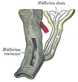
The paramesonephric ducts are paired ducts of the embryo in the reproductive system of humans and other mammals that run down the lateral sides of the genital ridge and terminate at the sinus tubercle in the primitive urogenital sinus. In the female, they will develop to form the fallopian tubes/oviducts, uterus, cervix, and the upper one-third of the vagina.
The development of the urinary system begins during prenatal development, and relates to the development of the urogenital system – both the organs of the urinary system and the sex organs of the reproductive system. The development continues as a part of sexual differentiation.
The male reproductive system consists of a number of sex organs that play a role in the process of human reproduction. These organs are located on the outside of the body, and within the pelvis.
Vaginal atresia is a condition in which the vagina is abnormally closed or absent. The main causes can either be complete vaginal hypoplasia, or a vaginal obstruction, often caused by an imperforate hymen or, less commonly, a transverse vaginal septum. It results in uterovaginal outflow tract obstruction. This condition does not usually occur by itself within an individual, but coupled with other developmental disorders within the female. The disorders that are usually coupled with a female who has vaginal atresia are Mayer-Rokitansky-Küster-Hauser syndrome, Bardet-Biedl syndrome, or Fraser syndrome. One out of every 5,000 women have this abnormality.
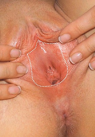
The vulval vestibule is the part of the vulva between the labia minora. On the inside, the urinary meatus and the vaginal introitus open to the vestibule, while the outer edge is marked by Hart's line, named after David Berry Hart.

The urachus is a fibrous remnant of the allantois, a canal that drains the urinary bladder of the fetus that joins and runs within the umbilical cord. The fibrous remnant lies in the space of Retzius, between the transverse fascia anteriorly and the peritoneum posteriorly.

The cloaca is a structure in the development of the urinary and reproductive organs.
The development of the reproductive system is the part of embryonic growth that results in the sex organs and contributes to sexual differentiation. Due to its large overlap with development of the urinary system, the two systems are typically described together as the genitourinary system.
The reproductive system of an organism, also known as the genital system, is the biological system made up of all the anatomical organs involved in sexual reproduction. Many non-living substances such as fluids, hormones, and pheromones are also important accessories to the reproductive system. Unlike most organ systems, the sexes of differentiated species often have significant differences. These differences allow for a combination of genetic material between two individuals, which allows for the possibility of greater genetic fitness of the offspring.
The sinovaginal bulb is a transitional structure in the development of female genitalia, and is one of a pair of endodermal outgrowths of the urogenital sinus, which later fuse to form the lower part of the vagina. The lower third of the vagina is derived from the urogenital sinus.

The vaginal support structures are those muscles, bones, ligaments, tendons, membranes and fascia, of the pelvic floor that maintain the position of the vagina within the pelvic cavity and allow the normal functioning of the vagina and other reproductive structures in the female. Defects or injuries to these support structures in the pelvic floor leads to pelvic organ prolapse. Anatomical and congenital variations of vaginal support structures can predispose a woman to further dysfunction and prolapse later in life. The urethra is part of the anterior wall of the vagina and damage to the support structures there can lead to incontinence and urinary retention.