
The urethra is a tube that connects the urinary bladder to the urinary meatus for the removal of urine from the body of both females and males. In human females and other primates, the urethra connects to the urinary meatus above the vagina, whereas in marsupials, the female's urethra empties into the urogenital sinus.

In male human anatomy, the glans penis, commonly referred to as the glans, is the bulbous structure at the distal end of the human penis that is the human male's most sensitive erogenous zone and their primary anatomical source of sexual pleasure. The glans penis is present in the male reproductive organs of humans and other mammals where it may appear smooth, spiny, elongated or divided. It is externally lined with mucosal tissue, which creates a smooth texture and glossy appearance. In humans, the glans is located over the distal ends of the corpora cavernosa and is a continuation of the corpus spongiosum of the penis. At the summit appears the urinary meatus and at the base forms the corona glandis. An elastic band of tissue, known as the frenulum, runs on its ventral surface. In men who are not circumcised, it is completely or partially covered by the foreskin. In adults, the foreskin can generally be retracted over and past the glans manually or sometimes automatically during an erection.

In the female human body, the clitoral hood is a fold of skin that surrounds and protects the glans of the clitoris; it also covers the external shaft of the clitoris, develops as part of the labia minora and is homologous with the foreskin in the male reproductive system. The clitoral hood is composed of mucocutaneous tissues; these tissues are between the mucous membrane and the skin, and they may have immunological importance because they may be a point of entry of mucosal vaccines. The clitoral hood is also important not only in the protection of the clitoral glans, but also in pleasure, as its tissue forms part of the erogenous zones of the vulva.
In biology, a septum is a wall, dividing a cavity or structure into smaller ones. A cavity or structure divided in this way may be referred to as septate.
The glans is a vascular structure located at the tip of the penis in male mammals or a homologous genital structure of the clitoris in female mammals.

A genital tubercle or phallic tubercle is a body of tissue present in the development of the reproductive system. It forms in the ventral, caudal region of mammalian embryos of both sexes, and eventually develops into a primordial phallus. In the human fetus, the genital tubercle develops around week 4 of gestation, and by week 9 becomes recognizably either a clitoris or penis. This should not be confused with the sinus tubercle which is a proliferation of endoderm induced by paramesonephric ducts. Even after the phallus is developed, the term genital tubercle remains, but only as the terminal end of it, which develops into either the glans penis or the glans clitoridis.
The perineal raphe is a visible line or ridge of tissue on the body that extends from the anus through the perineum to scrotum (male) or labia majora (female). It is found in both males and females, arises from the fusion of the urogenital folds, and is visible running medial through anteroposterior, to the anus where it resolves in a small knot of skin of varying size.

The spongy urethra is the longest part of the male urethra, and is contained in the corpus spongiosum of the penis.
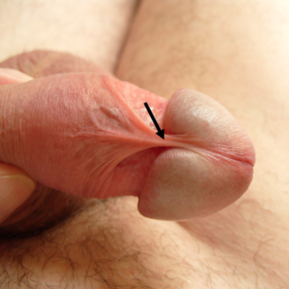
The frenulum of the penis, often known simply as the frenulum, is an elastic band of tissue on the underside of the glans and the neck of the human penis. In men who are not circumcised, it also connects the foreskin to the glans and the ventral mucosa. In adults, the frenulum is typically supple enough to allow manual movement of the foreskin over the glans and help retract the foreskin during erection. In flaccid state it tightens to narrow the foreskin opening.
The urinary meatus, also known as the external urethral orifice, is the opening of the urethra. It is the point where urine exits the urethra in both sexes and where semen exits the urethra in males. The meatus has varying degrees of sensitivity to touch. The meatus is located on the glans of the penis or in the vulval vestibule.

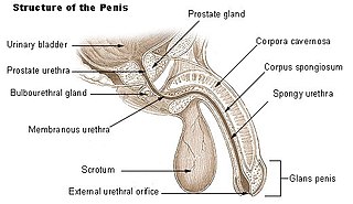
The human penis is an external male intromittent organ that additionally serves as the urinary duct. The main parts are the root (radix); the body (corpus); and the epithelium of the penis including the shaft skin and the foreskin (prepuce) covering the glans penis. The body of the penis is made up of three columns of tissue: two corpora cavernosa on the dorsal side and corpus spongiosum between them on the ventral side. The human male urethra passes through the prostate gland, where it is joined by the ejaculatory duct, and then through the penis. The urethra traverses the corpus spongiosum, and its opening, the meatus, lies on the tip of the glans penis. It is a passage both for urination and ejaculation of semen
The development of the reproductive system is the part of embryonic growth that results in the sex organs and contributes to sexual differentiation. Due to its large overlap with development of the urinary system, the two systems are typically described together as the urogenital or genitourinary system.

The corpus, also body or shaft of the penis, is the free portion of the human penis that is located outside of the pelvic cavity. It is the continuation of the internal root or radix which is embedded in the pelvis and extends to the glans behind which lies the neck of the penis. It is made up of the two corpora cavernosa and the corpus spongiosum on the underside. The corpora cavernosa are intimately bound to one another with a dorsally fenestrated septum which becomes a complete one before the penile crura.

In human male anatomy, the radix or root of the penis is the internal and most proximal portion of the human penis that lies in the perineum. Unlike the pendulous body or corpus of the penis which is suspended from the pubic symphysis, the root is attached to the pubic arch of the pelvis and is not visible externally. It is triradiate in form, consisting of three masses of erectile tissue; the two diverging crura, one on either side, and the median bulb of the penis or urethral bulb. Approximately one third to one half of the penis is embedded in the pelvis and can be felt through the scrotum and in the perineum.
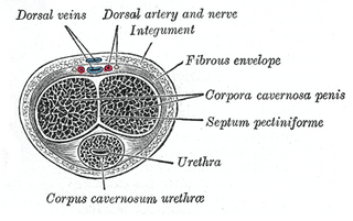
In human male anatomy, the septum of the penis or penile septum refers to the fibrous junction (septum) between the two corpora cavernosa of the human penis. The tunica albuginea of the penis forms a thick fibrous coat to the spongy tissue of the corpora cavernosa and corpus spongiosum. The two corpora cavernosa are surrounded by a strong fibrous envelope consisting of superficial and deep fibers. The superficial or outer fibers are longitudinal in direction, and form a single tube which encloses both corpora; the deep or inner fibers are arranged circularly around each corpus and meet in the center. By their junction in the median plane, the inner fibers form the intercavernous septum of the penis.

The corona of glans penis or penis crown refers to the rounded projecting border or flare that forms at the base of the glans in human males. The corona overhangs a mucosal surface, known as the neck of the penis, which separates the shaft and the glans. The deep retro-glandular coronal sulcus forms between the corona and the neck of the penis. The two sides of the corona merge on the ventral midline forming the septum glandis. The circumference of the corona is richly innervated and is described as a highly erogenous area of the glans.
Diphallia, penile duplication (PD), diphallic terata, or diphallasparatus, is an extremely rare developmental abnormality in which a mammal is born with two penises. The first reported human case was by Johannes Jacob Wecker in 1609. Its occurrence is 1 in 5.5 million boys in the United States.

Clitoral erection is a physiological phenomenon where the clitoris becomes enlarged and firm.

In male human anatomy, the foreskin, also known as the prepuce, is the double-layered fold of skin, mucosal and muscular tissue at the distal end of the human penis that covers the glans and the urinary meatus. The foreskin is attached to the glans by an elastic band of tissue, known as the frenulum. The outer skin of the foreskin meets with the inner preputial mucosa at the area of the mucocutaneous junction. The foreskin is mobile, fairly stretchable and sustains the glans in a moist environment. Except for humans, a similar structure, known as penile sheath, appears in the male sexual organs of all primates and the vast majority of mammals.

The penile raphe is a visible line or ridge of tissue that runs on the ventral side of the human penis beginning from the base of the shaft and ending in the prepuce. The line is typically darker than the rest of the shaft skin, even though its shape and pigmentation may vary among males. The penile raphe is part of a broader line in the male reproductive organs, that runs from the anus through the perineum and continues to the scrotum and penis, collectively referred to as median raphe.
