A testicle or testis is the male gonad in all bilaterians, including humans. It is homologous to the female ovary. The functions of the testicles are to produce both sperm and androgens, primarily testosterone. Testosterone release is controlled by the anterior pituitary luteinizing hormone, whereas sperm production is controlled both by the anterior pituitary follicle-stimulating hormone and gonadal testosterone.
In biology, a septum is a wall, dividing a cavity or structure into smaller ones. A cavity or structure divided in this way may be referred to as septate.

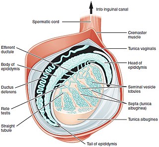
The spermatic cord is the cord-like structure in males formed by the vas deferens and surrounding tissue that runs from the deep inguinal ring down to each testicle. Its serosal covering, the tunica vaginalis, is an extension of the peritoneum that passes through the transversalis fascia. Each testicle develops in the lower thoracic and upper lumbar region and migrates into the scrotum. During its descent it carries along with it the vas deferens, its vessels, nerves etc. There is one on each side.
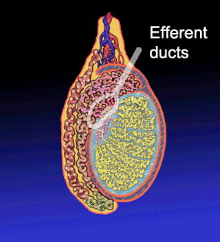
The efferent ducts connect the rete testis with the initial section of the epididymis.

The paired gubernacula, also called the caudal genital ligament, are embryonic structures which begin as undifferentiated mesenchyme attaching to the caudal end of the gonads.

The male reproductive system consists of a number of sex organs that play a role in the process of human reproduction. These organs are located on the outside of the body, and within the pelvis.
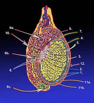
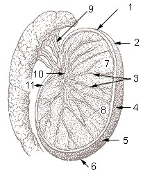
The tunica albugine is a dense, blue-white layer of fibrous tissue surrounding the testis. It is the middle of three enveloppes forming the capsule of the testis; it is deep to the visceral layer of tunica vaginalis, and superficial to the tunica vasculosa testis.
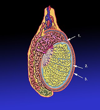
The tunica vaginalis is a pouch of serous membrane within the scrotum that lines the testis and epididymis, and the inner surface of the scrotum. It is the outermost of the three layers that constitute the capsule of the testis, with the tunica albuginea of penis situated beneath it.

The testicular artery is a branch of the abdominal aorta that supplies blood to the testicle. It is a paired artery, with one for each of the testicles.

The testicular vein, the male gonadal vein, carries deoxygenated blood from its corresponding testis to the inferior vena cava or one of its tributaries. It is the male equivalent of the ovarian vein, and is the venous counterpart of the testicular artery.
The tunica vasculosa testis is the inner-most of the three layers that form the capsule of the testis. It consists of a vascular plexus and loose connective tissue. It extends into the testis itself to line the surfaces of individual septa of testis.
The mediastinum testis is a thick yet incomplete septum at the posterior part of the testis formed by the tunica albuginea of testis projecting into the testis at its posterior aspect where the testis is not lined by the serous membrane to allow for the attachment of the epididymis. It extends posteriorly between the testis' superior pole and inferior pole. It is wider superiorly than inferiorly. It supports the rete testis and blood and lymphatic vessels of the testis in their passage into and out of the substance of the gland.
The tubuli seminiferi recti are structures in the testicle connecting the convoluted region of the seminiferous tubules to the rete testis, although the tubuli recti have a different appearance distinguishing them from these two structures.

The testes, at an early period of foetal life, are placed at the back part of the abdominal cavity, behind the peritoneum, and each is attached by a peritoneal fold, the mesorchium, to the mesonephros.
The lobules of testis are of partitions of the testis formed by septa of testis. The lobules of testis contain the tightly coiled seminiferous tubule. There are some hundreds of lobules in a testicle.
A testicular nubbin is the residual tissue of the human testis after a supposed perinatal vascular accident involving the testicular blood supply. The blood supply of the testis twists thereby cutting off the blood supply to the testis and results in testicular atrophy (shrinking). The nubbin is usually identified in childhood by the absence of a palpable testis in the scrotal sac. The tissue remnant usually includes fibrous tissue and signs of old infarction with hemosiderin deposition identified histologically. There is some disagreement as to whether these should be removed and whether there is a risk of future malignancy. They are typically removed surgically by pediatric urologists or pediatric general surgeons through either a scrotal or inguinal incision.
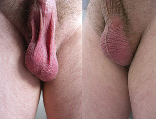
In most terrestrial mammals, the scrotum or scrotal sac is a part of the external male genitalia located at the base of the penis that consists of a suspended dual-chambered sac of skin and smooth muscle. The scrotum contains the external spermatic fascia, testicles, epididymides, and vasa deferentia. It is a distention of the perineum and carries some abdominal tissues into its cavity including the testicular artery, testicular vein, and pampiniform plexus. The perineal raphe is a small, vertical, slightly raised ridge of scrotal skin under which is found the scrotal septum. It appears as a thin longitudinal line that runs front to back over the entire scrotum. In humans, the scrotum becomes covered with pubic hair at puberty. The scrotum will usually tighten during penile erection and when exposed to cold temperatures. One testis is typically lower than the other to avoid compression in the event of an impact.

Scrotalultrasound is a medical ultrasound examination of the scrotum. It is used in the evaluation of testicular pain, and can help identify solid masses.
In biology, a tunica is a layer, coat, sheath, or similar covering. The word came to English from the Neo-Latin of science and medicine. Its literal sense is about the same as that of the word tunic, with which it is cognate. In biology, one of its senses used to be the taxonomic name of a genus of plants, but the nomenclature has been revised and those plants are now included in the genus Petrorhagia.

The septum of scrotum or scrotal septum is an incomplete vertical wall (septum) that divides the scrotum into two compartments –each containing a single testis. It consists of flexible connective tissue and nonstriated muscle. The site of the median septum is apparent on the surface of the scrotum along a median longitudinal ridge called the scrotal raphe. The perineal raphe further extends forward to the undersurface of the penis and backward to the anal opening. The purpose of the median septum is to compartmentalize each testis in order to prevent friction or trauma.