
The adrenal glands are endocrine glands that produce a variety of hormones including adrenaline and the steroids aldosterone and cortisol. They are found above the kidneys. Each gland has an outer cortex which produces steroid hormones and an inner medulla. The adrenal cortex itself is divided into three main zones: the zona glomerulosa, the zona fasciculata and the zona reticularis.

The kidneys are two reddish-brown bean-shaped organs found in vertebrates. They are located on the left and right in the retroperitoneal space, and in adult humans are about 12 centimetres in length. They receive blood from the paired renal arteries; blood exits into the paired renal veins. Each kidney is attached to a ureter, a tube that carries excreted urine to the bladder.

The collecting duct system of the kidney consists of a series of tubules and ducts that physically connect nephrons to a minor calyx or directly to the renal pelvis. The collecting duct system is the last part of nephron and participates in electrolyte and fluid balance through reabsorption and excretion, processes regulated by the hormones aldosterone and vasopressin.
The renal calyces are chambers of the kidney through which urine passes. The minor calyces surround the apex of the renal pyramids. Urine formed in the kidney passes through a renal papilla at the apex into the minor calyx; two or three minor calyces converge to form a major calyx, through which urine passes before continuing through the renal pelvis into the ureter.

The renal medulla is the innermost part of the kidney. The renal medulla is split up into a number of sections, known as the renal pyramids. Blood enters into the kidney via the renal artery, which then splits up to form the segmental arteries which then branch to form interlobar arteries. The interlobar arteries each in turn branch into arcuate arteries, which in turn branch to form interlobular arteries, and these finally reach the glomeruli. At the glomerulus the blood reaches a highly disfavourable pressure gradient and a large exchange surface area, which forces the serum portion of the blood out of the vessel and into the renal tubules. Flow continues through the renal tubules, including the proximal tubule, the Loop of Henle, through the distal tubule and finally leaves the kidney by means of the collecting duct, leading to the renal pelvis, the dilated portion of the ureter.

The tunica media, or media for short, is the middle tunica (layer) of an artery or vein. It lies between the tunica intima on the inside and the tunica externa on the outside.

The renal circulation supplies the blood to the kidneys via the renal arteries, left and right, which branch directly from the abdominal aorta. Despite their relatively small size, the kidneys receive approximately 20% of the cardiac output.
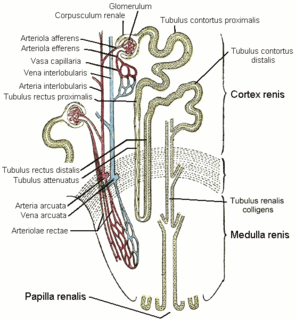
The vasa recta of the kidney, are the straight arterioles, and the straight venules of the kidney, – a series of blood vessels in the blood supply of the kidney that enter the medulla as the straight arterioles, and leave the medulla to ascend to the cortex as the straight venules.. They lie parallel to the loop of Henle.

In the renal system, peritubular capillaries are tiny blood vessels, supplied by the efferent arteriole, that travel alongside nephrons allowing reabsorption and secretion between blood and the inner lumen of the nephron. Peritubular capillaries surround the cortical parts of the proximal and distal tubules, while the vasa recta go into the medulla to approach the loop of Henle.
The renal hilum or renal pedicle is the hilum of the kidney, that is, its recessed central fissure where its vessels, nerves and ureter pass. The medial border of the kidney is concave in the center and convex toward either extremity; it is directed forward and a little downward. Its central part presents a deep longitudinal fissure, bounded by prominent overhanging anterior and posterior lips. This fissure is a hilum that transmits the vessels, nerves, and ureter. From anterior to posterior, the renal vein exits, the renal artery enters, and the renal pelvis exits the kidney.
The stellate veins join to form the interlobular veins, which pass inward between the rays, receive branches from the plexuses around the convoluted tubules, and, having arrived at the bases of the renal pyramids, join with the venae rectae.
Cortical radial arteries, formerly known as interlobular arteries, are renal blood vessels given off at right angles from the side of the arcuate arteries looking toward the cortical substance. The interlobular arteries pass directly outward between the medullary rays to reach the fibrous tunic, where they end in the capillary network of this part.
The stellate veins are veins that lie beneath the fibrous tunic of the kidney. They are stellate in arrangement and are derived from the capillary network, into which the terminal branches of the interlobular arteries break up. These join to form the interlobular veins, which pass inward between the rays.
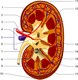
The renal lobe is a portion of a kidney consisting of a renal pyramid and the renal cortex above it. In humans, on average there are 7 to 18 renal lobes.

The arcuate vein is a vessel of the renal circulation. It is located at the border of the renal cortex and renal medulla.
The interlobar arteries are vessels of the renal circulation which supply the renal lobes. The interlobar arteries branch from the lobar arteries which branch from the segmental arteries, from the renal artery. They give rise to arcuate arteries.
The interlobar veins are veins of the renal circulation which drain the renal lobes.
Within the nephron of the kidney, the descending limb of loop of Henle is the portion of the renal tubule constituting the first part of the loop of Henle.
Within the nephron of the kidney, the ascending limb of the loop of Henle is a segment of the heterogenous loop of Henle downstream of the descending limb, after the sharp bend of the loop. This part of the renal tubule is divided into a thin and thick ascending limb; the thick portion is also known as the distal straight tubule, in contrast with the distal convoluted tubule downstream.
In anatomy, a medullary ray is the middle part of a cortical lobule. Each consists of a group of nephrons in the renal cortex. Their name is potentially misleading, as "medullary" refers to their destination, not their location. They travel perpendicular to the capsule, and extend from the cortex to the medulla. They may be visualised during urography.
