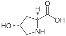
Hydroxyproline

Last updated
Hydroxyproline
(2S,4R)-4-Hydroxyprolin.svg
Names
IUPAC name
(2S,4R)-4-Hydroxypyrrolidine-2-carboxylic acid
Identifiers
3D model (JSmol)
ChemSpider
ECHA InfoCard 100.000.084 OOjs UI icon edit-ltr-progressive.svg
MeSH Hydroxyproline
PubChem CID
UNII
  • InChI=1S/C5H9NO3/c7-3-1-4(5(8)9)6-2-3/h3-4,6-7H,1-2H2,(H,8,9)/t3-,4+/m1/s1 X mark.svgN
    Key: PMMYEEVYMWASQN-DMTCNVIQSA-N X mark.svgN
  • InChI=1/C5H9NO3/c7-3-1-4(5(8)9)6-2-3/h3-4,6-7H,1-2H2,(H,8,9)/t3-,4+/m1/s1
    Key: PMMYEEVYMWASQN-DMTCNVIQBF
  • C1[C@H](CN[C@@H]1C(=O)O)O
Properties
C5H9NO3
Molar mass 131.131 g·mol−1
Except where otherwise noted, data are given for materials in their standard state (at 25 °C [77 °F], 100 kPa).
X mark.svgN  verify  (what is  Yes check.svgYX mark.svgN ?)

(2S,4R)-4-Hydroxyproline, or L-hydroxyproline (C 5 H 9 O 3 N), is an amino acid, abbreviated as Hyp or O, e.g., in Protein Data Bank.

Contents

Structure and discovery

In 1902, Hermann Emil Fischer isolated hydroxyproline from hydrolyzed gelatin. In 1905, Hermann Leuchs synthesized a racemic mixture of 4-hydroxyproline. [1]

Hydroxyproline differs from proline by the presence of a hydroxyl (OH) group attached to the gamma carbon atom.

Zwitterionic structure of (2S,4R)-4-hydroxyproline (left) and (2R,4S)-4-hydroxyproline (right) Betain-Hydroxyprolin.png
Zwitterionic structure of (2S,4R)-4-hydroxyproline (left) and (2R,4S)-4-hydroxyproline (right)

Production and function

Hydroxyproline is produced by hydroxylation of the amino acid proline by the enzyme prolyl hydroxylase following protein synthesis (as a post-translational modification). The enzyme catalyzed reaction takes place in the lumen of the endoplasmic reticulum. Although it is not directly incorporated into proteins, hydroxyproline comprises roughly 4% of all amino acids found in animal tissue, an amount greater than seven other amino acids that are translationally incorporated. [2]

Animals

Collagen

Hydroxyproline is a major component of the protein collagen, [3] comprising roughly 13.5% of mammalian collagen. Hydroxyproline and proline play key roles for collagen stability. [4] They permit the sharp twisting of the collagen helix. [5] In the canonical collagen Xaa-Yaa-Gly triad (where Xaa and Yaa are any amino acid), a proline occupying the Yaa position is hydroxylated to give a Xaa-Hyp-Gly sequence. This modification of the proline residue increases the stability of the collagen triple helix. It was initially proposed that the stabilization was due to water molecules forming a hydrogen bonding network linking the prolyl hydroxyl groups and the main-chain carbonyl groups. [6] It was subsequently shown that the increase in stability is primarily through stereoelectronic effects and that hydration of the hydroxyproline residues provides little or no additional stability. [7]

Non-collagen

Hydroxyproline is found in few proteins other than collagen. For this reason, hydroxyproline content has been used as an indicator to determine collagen and/or gelatin amount. However, the mammalian proteins elastin and argonaute 2 have collagen-like domains in which hydroxyproline is formed. Some snail poisons, conotoxins, contain hydroxyproline, but lack collagen-like sequences. [2]

Hydroxylation of proline has been shown to be involved in targeting Hypoxia-inducible factor (HIF) alpha subunit (HIF-1 alpha) for degradation by proteolysis. Under normoxia (normal oxygen conditions) EGLN1 protein hydroxylates the proline at the 564 position of HIF-1 alpha, which allows ubiquitylation by the von Hippel-Lindau tumor suppressor (pVHL) and subsequent targeting for proteasome degradation. [8]

Plants

Hydroxyproline rich glycoproteins (HRGPs) are also found in plant cell walls. [9] These hydroxyprolines serve as the attachment points for glycan chains which are added as post-translational modifications. [9]

Clinical significance

Proline hydroxylation requires ascorbic acid (vitamin C). The most obvious, first effects (gingival and hair problems) of absence of ascorbic acid in humans come from the resulting defect in hydroxylation of proline residues of collagen, with reduced stability of the collagen molecule, causing scurvy.

Increased serum and urine levels of hydroxyproline have also been demonstrated in Paget's disease. [10]

Mass spectrometry analysis showed decreased amount of hydroxyproline post-translational modifications in non inflamed tissue from ulcerative colitis patients when compared to tissue from donors without the disease. [11]

Other hydroxyprolines

Other hydroxyprolines also exist in nature. The most notable ones are 2,3-cis-, 3,4-trans-, and 3,4-dihydroxyproline, which occurs in diatom cell walls [12] and are postulated to have a role in silica deposition. Hydroxyproline is also found in the walls of oomycetes, fungus-like protists related to diatoms. [13] (2S,4S)-cis-4-Hydroxyproline is found in the toxic cyclic peptides from Amanita mushrooms (e.g., phalloidin). [14]

See also

Related Research Articles

<span class="mw-page-title-main">Collagen</span> Most abundant structural protein in animals

Collagen is the main structural protein in the extracellular matrix of a body's various connective tissues. As the main component of connective tissue, it is the most abundant protein in mammals. 25% to 35% of a mammalian body's protein content is collagen. Amino acids are bound together to form a triple helix of elongated fibril known as a collagen helix. The collagen helix is mostly found in connective tissue such as cartilage, bones, tendons, ligaments, and skin. Vitamin C is vital for collagen synthesis, while Vitamin E improves its production.

<span class="mw-page-title-main">Collagen helix</span> Main protein structure of fibrous collagen

In molecular biology, the collagen triple helix or type-2 helix is the main secondary structure of various types of fibrous collagen, including type I collagen. In 1954, Ramachandran & Kartha advanced a structure for the collagen triple helix on the basis of fiber diffraction data. It consists of a triple helix made of the repetitious amino acid sequence glycine-X-Y, where X and Y are frequently proline or hydroxyproline. Collagen folded into a triple helix is known as tropocollagen. Collagen triple helices are often bundled into fibrils which themselves form larger fibres, as in tendons.

<span class="mw-page-title-main">Protein primary structure</span> Linear sequence of amino acids in a peptide or protein

Protein primary structure is the linear sequence of amino acids in a peptide or protein. By convention, the primary structure of a protein is reported starting from the amino-terminal (N) end to the carboxyl-terminal (C) end. Protein biosynthesis is most commonly performed by ribosomes in cells. Peptides can also be synthesized in the laboratory. Protein primary structures can be directly sequenced, or inferred from DNA sequences.

Proline (symbol Pro or P) is an organic acid classed as a proteinogenic amino acid (used in the biosynthesis of proteins), although it does not contain the amino group -NH
2
but is rather a secondary amine. The secondary amine nitrogen is in the protonated form (NH2+) under biological conditions, while the carboxyl group is in the deprotonated −COO form. The "side chain" from the α carbon connects to the nitrogen forming a pyrrolidine loop, classifying it as a aliphatic amino acid. It is non-essential in humans, meaning the body can synthesize it from the non-essential amino acid L-glutamate. It is encoded by all the codons starting with CC (CCU, CCC, CCA, and CCG).

In chemistry, hydroxylation can refer to:

A polyproline helix is a type of protein secondary structure which occurs in proteins comprising repeating proline residues. A left-handed polyproline II helix is formed when sequential residues all adopt (φ,ψ) backbone dihedral angles of roughly and have trans isomers of their peptide bonds. This PPII conformation is also common in proteins and polypeptides with other amino acids apart from proline. Similarly, a more compact right-handed polyproline I helix is formed when sequential residues all adopt (φ,ψ) backbone dihedral angles of roughly and have cis isomers of their peptide bonds. Of the twenty common naturally occurring amino acids, only proline is likely to adopt the cis isomer of the peptide bond, specifically the X-Pro peptide bond; steric and electronic factors heavily favor the trans isomer in most other peptide bonds. However, peptide bonds that replace proline with another N-substituted amino acid are also likely to adopt the cis isomer.

Lysyl hydroxylases are alpha-ketoglutarate-dependent hydroxylases enzymes that catalyze the hydroxylation of lysine to hydroxylysine. Lysyl hydroxylases require iron and vitamin C as cofactors for their oxidation activity. It takes place following collagen synthesis in the cisternae (lumen) of the rough endoplasmic reticulum (ER). There are three lysyl hydroxylases (LH1-3) encoded in the human genome, namely: PLOD1, PLOD2 and PLOD3. From PLOD2 two splice variant can be expressed, where LH2b differs from LH2a by incorporating the small exon 13A. LH1 and LH3 hydroxylate lysyl residues in the collagen triple helix, whereas LH2b hydroxylates lysyl residues in the telopeptides of collagen. In addition to its hydroxylation activity, LH3 has glucosylation activity that produces disaccharide (Glc-Gal) attached to collagen hydroxylysines.

<span class="mw-page-title-main">Collagen, type III, alpha 1</span>

Type III Collagen is a homotrimer, or a protein composed of three identical peptide chains (monomers), each called an alpha 1 chain of type III collagen. Formally, the monomers are called collagen type III, alpha-1 chain and in humans are encoded by the COL3A1 gene. Type III collagen is one of the fibrillar collagens whose proteins have a long, inflexible, triple-helical domain.

Leprecan is a protein associated with osteogenesis imperfecta type VIII.

<span class="mw-page-title-main">HIF1A</span> Protein-coding gene in the species Homo sapiens

Hypoxia-inducible factor 1-alpha, also known as HIF-1-alpha, is a subunit of a heterodimeric transcription factor hypoxia-inducible factor 1 (HIF-1) that is encoded by the HIF1A gene. The Nobel Prize in Physiology or Medicine 2019 was awarded for the discovery of HIF.

<span class="mw-page-title-main">Procollagen-proline dioxygenase</span> Enzyme

Procollagen-proline dioxygenase, commonly known as prolyl hydroxylase, is a member of the class of enzymes known as alpha-ketoglutarate-dependent hydroxylases. These enzymes catalyze the incorporation of oxygen into organic substrates through a mechanism that requires alpha-Ketoglutaric acid, Fe2+, and ascorbate. This particular enzyme catalyzes the formation of (2S, 4R)-4-hydroxyproline, a compound that represents the most prevalent post-translational modification in the human proteome.

<span class="mw-page-title-main">EGLN2</span> Protein-coding gene in the species Homo sapiens

Egl nine homolog 2 is a protein that in humans is encoded by the EGLN2 gene. ELGN2 is an alpha-ketoglutarate-dependent hydroxylase, a superfamily of non-haem iron-containing proteins.

<span class="mw-page-title-main">EGLN1</span> Protein-coding gene in the species Homo sapiens

Hypoxia-inducible factor prolyl hydroxylase 2 (HIF-PH2), or prolyl hydroxylase domain-containing protein 2 (PHD2), is an enzyme encoded by the EGLN1 gene. It is also known as Egl nine homolog 1. PHD2 is a α-ketoglutarate/2-oxoglutarate-dependent hydroxylase, a superfamily non-haem iron-containing proteins. In humans, PHD2 is one of the three isoforms of hypoxia-inducible factor-proline dioxygenase, which is also known as HIF prolyl-hydroxylase.

<span class="mw-page-title-main">P4HA1</span> Protein-coding gene in the species Homo sapiens

Prolyl 4-hydroxylase subunit alpha-1 is an enzyme that in humans is encoded by the P4HA1 gene.

<span class="mw-page-title-main">P4HA2</span> Protein-coding gene in the species Homo sapiens

Prolyl 4-hydroxylase subunit alpha-2 is an enzyme that in humans is encoded by the P4HA2 gene.

<span class="mw-page-title-main">Triple helix</span> Set of three congruent geometrical helices with the same axis

In the fields of geometry and biochemistry, a triple helix is a set of three congruent geometrical helices with the same axis, differing by a translation along the axis. This means that each of the helices keeps the same distance from the central axis. As with a single helix, a triple helix may be characterized by its pitch, diameter, and handedness. Examples of triple helices include triplex DNA, triplex RNA, the collagen helix, and collagen-like proteins.

<span class="mw-page-title-main">Non-proteinogenic amino acids</span> Are not naturally encoded in the genome

In biochemistry, non-coded or non-proteinogenic amino acids are distinct from the 22 proteinogenic amino acids, which are naturally encoded in the genome of organisms for the assembly of proteins. However, over 140 non-proteinogenic amino acids occur naturally in proteins and thousands more may occur in nature or be synthesized in the laboratory. Chemically synthesized amino acids can be called unnatural amino acids. Unnatural amino acids can be synthetically prepared from their native analogs via modifications such as amine alkylation, side chain substitution, structural bond extension cyclization, and isosteric replacements within the amino acid backbone. Many non-proteinogenic amino acids are important:

Hypoxia-inducible factor-proline dioxygenase (EC 1.14.11.29, HIF hydroxylase) is an enzyme with systematic name hypoxia-inducible factor-L-proline, 2-oxoglutarate:oxygen oxidoreductase (4-hydroxylating). This enzyme catalyses the following chemical reaction

In epigenetics, proline isomerization is the effect that cis-trans isomerization of the amino acid proline has on the regulation of gene expression. Similar to aspartic acid, the amino acid proline has the rare property of being able to occupy both cis and trans isomers of its prolyl peptide bonds with ease. Peptidyl-prolyl isomerase, or PPIase, is an enzyme very commonly associated with proline isomerization due to their ability to catalyze the isomerization of prolines. PPIases are present in three types: cyclophilins, FK507-binding proteins, and the parvulins. PPIase enzymes catalyze the transition of proline between cis and trans isomers and are essential to the numerous biological functions controlled and affected by prolyl isomerization Without PPIases, prolyl peptide bonds will slowly switch between cis and trans isomers, a process that can lock proteins in a nonnative structure that can affect render the protein temporarily ineffective. Although this switch can occur on its own, PPIases are responsible for most isomerization of prolyl peptide bonds. The specific amino acid that precedes the prolyl peptide bond also can have an effect on which conformation the bond assumes. For instance, when an aromatic amino acid is bonded to a proline the bond is more favorable to the cis conformation. Cyclophilin A uses an "electrostatic handle" to pull proline into cis and trans formations. Most of these biological functions are affected by the isomerization of proline when one isomer interacts differently than the other, commonly causing an activation/deactivation relationship. As an amino acid, proline is present in many proteins. This aids in the multitude of effects that isomerization of proline can have in different biological mechanisms and functions.

<span class="mw-page-title-main">Collagen hybridizing peptide</span> Type of synthetic peptide

A collagen hybridizing peptide (CHP) is a synthetic peptide sequence with typically 6 to 10 repeating units of the Gly-Xaa-Yaa amino acid triplet, which mimics the hallmark sequence of natural collagens. A CHP peptide usually possesses a high content of proline and hydroxyproline in the Xaa and Yaa positions, which confers it a strong propensity to form the collagen's unique triple helix conformation. In the single-stranded (monomeric) status, the peptide can recognize denatured collagen strands in tissues by forming a hybridized triple helix with the collagen strands. This occurs via the triple helical chain assembly and inter-chain hydrogen bonding, in a manner similar to primers binding to melted DNA strands during PCR. The binding does not depend on a specific sequence or epitope on collagen, enabling CHPs to target denatured collagen chains of different types.

References

  1. R.H.A. Plimmer (1912) [1908]. R.H.A. Plimmer; F.G. Hopkins (eds.). The chemical composition of the proteins. Monographs on biochemistry. Vol. Part I. Analysis (2nd ed.). London: Longmans, Green and Co. p. 132. Retrieved January 18, 2010.
  2. 1 2 Gorres, Kelly L.; Raines, Ronald T. (April 2010). "Prolyl 4-hydroxylase". Critical Reviews in Biochemistry and Molecular Biology. 45 (2): 106–124. doi:10.3109/10409231003627991. PMC   2841224 . PMID   20199358.
  3. Szpak, Paul (2011). "Fish bone chemistry and ultrastructure: implications for taphonomy and stable isotope analysis". Journal of Archaeological Science . 38 (12): 3358–3372. doi:10.1016/j.jas.2011.07.022.
  4. Nelson, D. L. and Cox, M. M. (2005) Lehninger's Principles of Biochemistry, 4th Edition, W. H. Freeman and Company, New York.
  5. Brinckmann, J., Notbohm, H. and Müller, P.K. (2005) Collagen, Topics in Current Chemistry 247, Springer, Berlin.
  6. Bella, J; Eaton, M; Brodsky, B; Berman, HM (1994). "Crystal and molecular structure of a collagen-like peptide at 1.9 A resolution". Science. 266 (5182): 75–81. doi:10.1126/science.7695699. PMID   7695699.
  7. Kotch, F.W.; Guzei, I.A.; Raines, R.T. (2008). "Stabilization of the Collagen Triple Helix by O-Methylation of Hydroxyproline Residues". Journal of the American Chemical Society. 130 (10): 2952–2953. doi:10.1021/ja800225k. PMC   2802593 . PMID   18271593.
  8. Jaakkola, P.; Mole, D.R.; Tian, Y.M.; Wilson, M.I.; Gielbert, J.; Gaskell, S.J.; Kriegsheim, A.V.; Hebestreit, H.F.; et al. (2001). "Targeting of HIF-alpha to the von Hippel-Lindau ubiquitylation complex by O2-regulated prolyl hydroxylation". Science. 292 (5516): 468–72. Bibcode:2001Sci...292..468J. doi: 10.1126/science.1059796 . PMID   11292861. S2CID   20914281.
  9. 1 2 Cassab, Gladys I (1998). "Plant Cell Wall Proteins". Annual Review of Plant Physiology and Plant Molecular Biology. 49: 281–309. doi:10.1146/annurev.arplant.49.1.281. PMID   15012236.
  10. "Wheeless' Textbook of Orthopaedics". Wheeless Online. 22 July 2020.
  11. "Degradation of the extracellular matrix is part of the pathology of ulcerative colitis".
  12. Nakajima, T.; Volcani, B.E. (1969). "3,4-Dihydroxyproline: a new amino acid in diatom cell walls". Science. 164 (3886): 1400–1401. Bibcode:1969Sci...164.1400N. doi:10.1126/science.164.3886.1400. PMID   5783709. S2CID   23673503.
  13. Alexopoulos, C.J., Mims C.W. and Blackwell, M. (1996). Introductory Mycology (4th ed.). New York: John Wiley & Sons. pp. 687–688. ISBN   978-0-471-52229-4.{{cite book}}: CS1 maint: multiple names: authors list (link)
  14. Wieland, T. (1986). Peptides of Poisonous Amanita Mushrooms. Springer.