Thromboembolism

Last updated

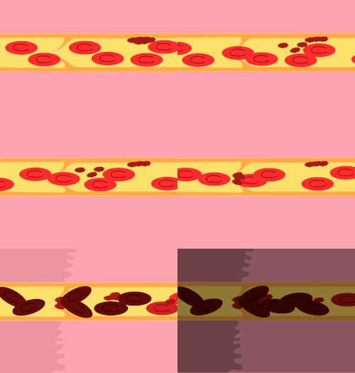
Animation showing the formation of an occlusive blood clot in a vein. Several platelets attach to the lips of the valve, narrowing the opening and causing more platelets and red blood cells to pool and clot. Clotting of immobile blood on both sides of the blockage can cause the clot to spread in both directions. Acute blockage (embolism) of a blood vessel by a thrombus that has detached from its place of formation (on the wall of a vessel) and entered the circulating blood. As a result of this blockage, blood flow in the vessel stops--a condition called thromboembolism. Thrombosis formation.gif
Animation showing the formation of an occlusive blood clot in a vein. Several platelets attach to the lips of the valve, narrowing the opening and causing more platelets and red blood cells to pool and clot. Clotting of immobile blood on both sides of the blockage can cause the clot to spread in both directions. Acute blockage (embolism) of a blood vessel by a thrombus that has detached from its place of formation (on the wall of a vessel) and entered the circulating blood. As a result of this blockage, blood flow in the vessel stops—a condition called thromboembolism.

Thromboembolism is a condition in which a blood clot (thrombus) breaks off from its original site and travels through the bloodstream (as an embolus) to obstruct a blood vessel, causing tissue ischemia and organ damage. Thromboembolism can affect both the venous and arterial systems, with different clinical manifestations and management strategies. [2] [3]

Contents

Venous thromboembolism

Venous thromboembolism (VTE) BD72 comprises the following conditions: [3] [4] [5]

VTE is a common cardiovascular disorder with significant morbidity and mortality. [3] [4] [5] VTE can present with various symptoms, such as painful leg swelling, chest pain, dyspnea, hemoptysis, syncope, and even death, depending on the location and extent of the thrombus. [4] [6] VTE can also cause long-term complications, such as recurrent VTE, post-PE syndrome, chronic thromboembolic pulmonary hypertension (CTEPH), and post-thrombotic syndrome (PTS). The mainstay of VTE management is anticoagulation therapy, which prevents thrombus propagation and embolization. Such treatment reduces the risk of recurrence. [5] [4] [1] The choice and duration of anticoagulation depend on the individual patient's risk factors, bleeding risk, and preferences. Direct oral anticoagulants (DOACs) have emerged as an essential alternative to conventional anticoagulants, such as vitamin K antagonists (VKAs) and low-molecular-weight heparins (LMWHs), due to their rapid onset of action, predictable pharmacokinetics, fixed dosing, and lower risk of bleeding. DOACs can also facilitate home treatment and extended therapy for selected patients. In addition to anticoagulation, some patients with VTE may benefit from adjunctive therapies, such as thrombolysis, catheter-directed interventions, or inferior vena cava (IVC) filters, to remove or prevent thrombus migration. However, these therapies are associated with higher risks of bleeding and complications. These therapies are not routinely recommended by the current guidelines except for specific indications, such as massive PE, iliofemoral DVT, or contraindications to anticoagulation. The optimal duration of anticoagulation for VTE is determined by the balance between the risk of recurrence and the risk of bleeding, and should be individualized for each patient. In general, VTE provoked by a transient or reversible risk factor, such as surgery, trauma, or immobilization, should be treated for three months, while VTE provoked by a persistent or progressive risk factor, such as cancer, should be treated indefinitely. Unprovoked VTE, which occurs in the absence of any identifiable risk factor, has a high risk of recurrence and may require indefinite anticoagulation, depending on the patient's characteristics and preferences. [4] [7] [8] Factors that favor indefinite anticoagulation include male sex, presentation as PE (especially with concomitant DVT), positive d-dimer test after stopping anticoagulation, presence of antiphospholipid antibodies, low bleeding risk, and patient preference. [3] The type of anticoagulant used for indefinite therapy is of secondary importance, but low-dose DOACs may offer a convenient and safer option for some patients. [4] [7] [8] For cancer-associated VTE, full-dose DOACs are now preferred over LMWHs, unless there are gastrointestinal lesions that increase the risk of bleeding. [4] [7] [8] Graduated compression stockings are elastic garments that apply a gradient of pressure to the lower limbs, reducing venous stasis and improving blood flow, still these stockings are not routinely indicated after DVT, but may be helpful if there is persistent leg swelling or symptomatic improvement with a trial of stockings. [3] [9] Medications, such as pentoxifylline, have a limited role in the treatment of PTS. After PE, patients should be monitored for signs and symptoms of CTEPH, which is a rare but serious complication of VTE. [4] [7] [8] Ventilation-perfusion scanning and echocardiography are the initial diagnostic tests for CTEPH, and patients with confirmed or suspected CTEPH should be evaluated for potential treatments, such as pulmonary thromboendarterectomy, balloon pulmonary angioplasty, or vasodilator therapies. [3]

Arterial thromboembolism

Arterial thromboembolism (ATE) is a less common but more severe form of thromboembolism, which can affect various organs, such as the brain, heart, kidneys, limbs, and mesentery. ATE can cause life-threatening conditions, such as stroke, myocardial infarction, acute kidney injury, limb ischemia, and mesenteric ischemia. ATE is usually caused by atherosclerosis, which leads to plaque rupture and thrombus formation, or by cardioembolism, which results from the embolization of a cardiac thrombus, such as in atrial fibrillation, valvular disease, or myocardial dysfunction. The management of ATE depends on the location and severity of the ischemia and the underlying etiology. The main goals of ATE management are to restore blood flow, prevent further thrombosis, and treat the underlying cause. The treatment options for ATE include antithrombotic therapy, revascularization procedures, and risk factor modification. Antithrombotic therapy consists of antiplatelet agents, such as aspirin or clopidogrel, or anticoagulants, such as heparin or DOACs, depending on the indication and contraindications. Revascularization procedures include thrombolysis, thrombectomy, angioplasty, stenting, or bypass surgery and are indicated for patients with severe or limb-threatening ischemia or failed medical therapy. Risk factor modification involves lifestyle changes, such as smoking cessation, exercise, and diet, and pharmacological interventions, such as statins, antihypertensives, and glucose-lowering agents, to reduce the risk of recurrent ATE and improve the prognosis. The duration of antithrombotic therapy for ATE is variable, depending on the type and location of the thrombus, the presence of a prosthetic device, and the bleeding risk. In general, patients with ATE receive lifelong antiplatelet therapy unless there is a specific indication or contraindication for anticoagulation. [2] [10]

Related Research Articles

<span class="mw-page-title-main">Anticoagulant</span> Class of drugs

An anticoagulant, commonly known as a blood thinner, is a chemical substance that prevents or reduces coagulation of blood, prolonging the clotting time. Some of them occur naturally in blood-eating animals such as leeches and mosquitoes, where they help keep the bite area unclotted long enough for the animal to obtain some blood.

<span class="mw-page-title-main">Thrombosis</span> Medical condition caused by blood clots

Thrombosis is the formation of a blood clot inside a blood vessel, obstructing the flow of blood through the circulatory system. When a blood vessel is injured, the body uses platelets (thrombocytes) and fibrin to form a blood clot to prevent blood loss. Even when a blood vessel is not injured, blood clots may form in the body under certain conditions. A clot, or a piece of the clot, that breaks free and begins to travel around the body is known as an embolus.

<span class="mw-page-title-main">Thrombus</span> Blood clot

A thrombus, colloquially called a blood clot, is the final product of the blood coagulation step in hemostasis. There are two components to a thrombus: aggregated platelets and red blood cells that form a plug, and a mesh of cross-linked fibrin protein. The substance making up a thrombus is sometimes called cruor. A thrombus is a healthy response to injury intended to stop and prevent further bleeding, but can be harmful in thrombosis, when a clot obstructs blood flow through healthy blood vessels in the circulatory system.

<span class="mw-page-title-main">Pulmonary embolism</span> Blockage of an artery in the lungs

Pulmonary embolism (PE) is a blockage of an artery in the lungs by a substance that has moved from elsewhere in the body through the bloodstream (embolism). Symptoms of a PE may include shortness of breath, chest pain particularly upon breathing in, and coughing up blood. Symptoms of a blood clot in the leg may also be present, such as a red, warm, swollen, and painful leg. Signs of a PE include low blood oxygen levels, rapid breathing, rapid heart rate, and sometimes a mild fever. Severe cases can lead to passing out, abnormally low blood pressure, obstructive shock, and sudden death.

<span class="mw-page-title-main">Venous thrombosis</span> Blood clot (thrombus) that forms within a vein

Venous thrombosis is the blockage of a vein caused by a thrombus. A common form of venous thrombosis is deep vein thrombosis (DVT), when a blood clot forms in the deep veins. If a thrombus breaks off (embolizes) and flows to the lungs to lodge there, it becomes a pulmonary embolism (PE), a blood clot in the lungs. The conditions of DVT only, DVT with PE, and PE only, are all captured by the term venous thromboembolism (VTE).

Factor V Leiden is a variant of human factor V, which causes an increase in blood clotting (hypercoagulability). Due to this mutation, protein C, an anticoagulant protein that normally inhibits the pro-clotting activity of factor V, is not able to bind normally to factor V, leading to a hypercoagulable state, i.e., an increased tendency for the patient to form abnormal and potentially harmful blood clots. Factor V Leiden is the most common hereditary hypercoagulability disorder amongst ethnic Europeans. It is named after the Dutch city of Leiden, where it was first identified in 1994 by Rogier Maria Bertina under the direction of Pieter Hendrick Reitsma. Despite the increased risk of venous thromboembolisms, people with one copy of this gene have not been found to have shorter lives than the general population. It is an autosomal dominant genetic disorder with incomplete penetrance.

<span class="mw-page-title-main">Thrombolysis</span> Breakdown (lysis) of blood clots formed in blood vessels, using medication

Thrombolysis, also called fibrinolytic therapy, is the breakdown (lysis) of blood clots formed in blood vessels, using medication. It is used in ST elevation myocardial infarction, stroke, and in cases of severe venous thromboembolism.

<span class="mw-page-title-main">Deep vein thrombosis</span> Formation of a blood clot (thrombus) in a deep vein

Deep vein thrombosis (DVT) is a type of venous thrombosis involving the formation of a blood clot in a deep vein, most commonly in the legs or pelvis. A minority of DVTs occur in the arms. Symptoms can include pain, swelling, redness, and enlarged veins in the affected area, but some DVTs have no symptoms.

Low-molecular-weight heparin (LMWH) is a class of anticoagulant medications. They are used in the prevention of blood clots and treatment of venous thromboembolism and in the treatment of myocardial infarction.

<span class="mw-page-title-main">Thrombophilia</span> Abnormality of blood coagulation

Thrombophilia is an abnormality of blood coagulation that increases the risk of thrombosis. Such abnormalities can be identified in 50% of people who have an episode of thrombosis that was not provoked by other causes. A significant proportion of the population has a detectable thrombophilic abnormality, but most of these develop thrombosis only in the presence of an additional risk factor.

<span class="mw-page-title-main">Inferior vena cava filter</span>

An inferior vena cava filter is a medical device made of metal that is implanted by vascular surgeons or interventional radiologists into the inferior vena cava to prevent a life-threatening pulmonary embolism (PE) or venous thromboembolism (VTE).

An embolus, is described as a free-floating mass, located inside blood vessels that can travel from one site in the blood stream to another. An embolus can be made up of solid, liquid, or gas. Once these masses get "stuck" in a different blood vessel, it is then known as an "embolism." An embolism can cause ischemia—damage to an organ from lack of oxygen. A paradoxical embolism is a specific type of embolism in which the embolus travels from the right side of the heart to the left side of the heart and lodges itself in a blood vessel known as an artery. Thus, it is termed "paradoxical" because the embolus lands in an artery, rather than a vein.

<span class="mw-page-title-main">Post-thrombotic syndrome</span> Medical condition

Post-thrombotic syndrome (PTS), also called postphlebitic syndrome and venous stress disorder is a medical condition that may occur as a long-term complication of deep vein thrombosis (DVT).

Hypercoagulability in pregnancy is the propensity of pregnant women to develop thrombosis. Pregnancy itself is a factor of hypercoagulability, as a physiologically adaptive mechanism to prevent post partum bleeding. However, when combined with an additional underlying hypercoagulable states, the risk of thrombosis or embolism may become substantial.

Embolectomy is the emergency interventional or surgical removal of emboli which are blocking blood circulation. It usually involves removal of thrombi, and is then referred to as thromboembolectomy or thrombectomy. Embolectomy is an emergency procedure often as the last resort because permanent occlusion of a significant blood flow to an organ leads to necrosis. Other involved therapeutic options are anticoagulation and thrombolysis.

<span class="mw-page-title-main">Superficial thrombophlebitis</span> Medical condition

Superficial thrombophlebitis is a thrombosis and inflammation of superficial veins which presents as a painful induration (thickening) with erythema, often in a linear or branching configuration; forming a cord-like appearance.

<span class="mw-page-title-main">Intermittent pneumatic compression</span>

Intermittent pneumatic compression is a therapeutic technique used in medical devices that include an air pump and inflatable auxiliary sleeves, gloves or boots in a system designed to improve venous circulation in the limbs of patients who have edema or the risk of deep vein thrombosis (DVT), pulmonary embolism (PE), or the combination of DVT and PE which is venous thrombeombolism (VTE).

Prothrombin G20210A is a genetic condition that increases the risk of blood clots including from deep vein thrombosis, and of pulmonary embolism. One copy of the mutation increases the risk of a blood clot from 1 in 1,000 per year to 2.5 in 1,000. Two copies increases the risk to up to 20 in 1,000 per year. Most people never develop a blood clot in their lifetimes.

<span class="mw-page-title-main">Superficial vein thrombosis</span> Medical condition

Superficial vein thrombosis (SVT) is a blood clot formed in a superficial vein, a vein near the surface of the body. Usually there is thrombophlebitis, which is an inflammatory reaction around a thrombosed vein, presenting as a painful induration with redness. SVT itself has limited significance when compared to a deep vein thrombosis (DVT), which occurs deeper in the body at the deep venous system level. However, SVT can lead to serious complications, and is therefore no longer regarded as a benign condition. If the blood clot is too near the saphenofemoral junction there is a higher risk of pulmonary embolism, a potentially life-threatening complication.

<span class="mw-page-title-main">Thrombosis prevention</span> Medical treatment

Thrombosis prevention or thromboprophylaxis is medical treatment to prevent the development of thrombosis in those considered at risk for developing thrombosis. Some people are at a higher risk for the formation of blood clots than others, such as those with cancer undergoing a surgical procedure. Prevention measures or interventions are usually begun after surgery as the associated immobility will increase a person's risk.

References

  1. 1 2 "Venous Thromboembolism - Causes and Risk Factors | NHLBI, NIH". September 19, 2022. Archived from the original on October 2, 2023. Retrieved February 25, 2024.
  2. 1 2 Tan BK, Mainbourg S, Friggeri A, Bertoletti L, Douplat M, Dargaud Y, Grange C, Lobbes H, Provencher S, Lega JC (October 2021). "Arterial and venous thromboembolism in COVID-19: a study-level meta-analysis". Thorax. 76 (10): 970–979. doi:10.1136/thoraxjnl-2020-215383. PMID   33622981. S2CID   232039896.
  3. 1 2 3 4 5 6 7 8 Donnellan E, Khorana AA (February 2017). "Cancer and Venous Thromboembolic Disease: A Review". Oncologist. 22 (2): 199–207. doi:10.1634/theoncologist.2016-0214. PMC   5330704 . PMID   28174293.
  4. 1 2 3 4 5 6 7 8 9 10 Bartholomew JR (December 2017). "Update on the management of venous thromboembolism". Cleve Clin J Med. 84 (12 Suppl 3): 39–46. doi:10.3949/ccjm.84.s3.04. PMID   29257737.
  5. 1 2 3 4 5 Blitzer RR, Eisenstein S (October 2021). "Venous Thromboembolism and Pulmonary Embolism: Strategies for Prevention and Management". Surg Clin North Am. 101 (5): 925–938. doi:10.1016/j.suc.2021.06.015. PMID   34537152. S2CID   237573119.
  6. Phillippe HM (December 2017). "Overview of venous thromboembolism". Am J Manag Care. 23 (20 Suppl): S376–S382. PMID   29297660. Archived from the original on February 11, 2024. Retrieved February 25, 2024.
  7. 1 2 3 4 Yamashita Y, Morimoto T, Kimura T (January 2022). "Venous thromboembolism: Recent advancement and future perspective". J Cardiol. 79 (1): 79–89. doi: 10.1016/j.jjcc.2021.08.026 . PMID   34518074.
  8. 1 2 3 4 Kearon C, Kahn SR (January 2020). "Long-term treatment of venous thromboembolism". Blood. 135 (5): 317–325. doi:10.1182/blood.2019002364. PMID   31917402.
  9. Jeffery PC, Nicolaides AN (April 1990). "Graduated compression stockings in the prevention of postoperative deep vein thrombosis". Br J Surg. 77 (4): 380–3. doi:10.1002/bjs.1800770407. PMID   2187559. S2CID   2797031.
  10. Stadnicki A, Stadnicka I (October 2021). "Venous and arterial thromboembolism in patients with inflammatory bowel diseases". World J Gastroenterol. 27 (40): 6757–6774. doi: 10.3748/wjg.v27.i40.6757 . PMC   8567469 . PMID   34790006.