
The thorax or chest is a part of the anatomy of humans, mammals, and other tetrapod animals located between the neck and the abdomen. In insects, crustaceans, and the extinct trilobites, the thorax is one of the three main divisions of the creature's body, each of which is in turn composed of multiple segments.
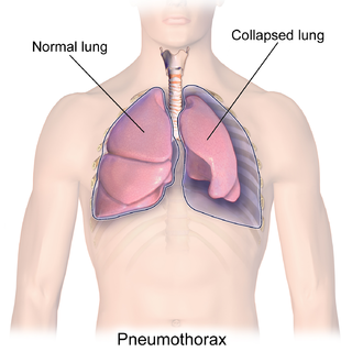
A pneumothorax is an abnormal collection of air in the pleural space between the lung and the chest wall. Symptoms typically include sudden onset of sharp, one-sided chest pain and shortness of breath. In a minority of cases, a one-way valve is formed by an area of damaged tissue, and the amount of air in the space between chest wall and lungs increases; this is called a tension pneumothorax. This can cause a steadily worsening oxygen shortage and low blood pressure. This leads to a type of shock called obstructive shock, which can be fatal unless reversed. Very rarely, both lungs may be affected by a pneumothorax. It is often called a "collapsed lung", although that term may also refer to atelectasis.

An injury is any physiological damage to living tissue caused by immediate physical stress. Injuries to humans can occur intentionally or unintentionally and may be caused by blunt trauma, penetrating trauma, burning, toxic exposure, asphyxiation, or overexertion. Injuries can occur in any part of the body, and different symptoms are associated with different injuries.

Traumatic cardiac arrest (TCA) is a condition in which the heart has ceased to beat due to blunt or penetrating trauma, such as a stab wound to the thoracic area. It is a medical emergency which will always result in death without prompt advanced medical care. Even with prompt medical intervention, survival without neurological complications is rare. In recent years, protocols have been proposed to improve survival rate in patients with traumatic cardiac arrest, though the variable causes of this condition as well as many coexisting injuries can make these protocols difficult to standardize. Traumatic cardiac arrest is a complex form of cardiac arrest often derailing from advanced cardiac life support in the sense that the emergency team must first establish the cause of the traumatic arrest and reverse these effects, for example hypovolemia and haemorrhagic shock due to a penetrating injury.

A hemothorax is an accumulation of blood within the pleural cavity. The symptoms of a hemothorax may include chest pain and difficulty breathing, while the clinical signs may include reduced breath sounds on the affected side and a rapid heart rate. Hemothoraces are usually caused by an injury, but they may occur spontaneously due to cancer invading the pleural cavity, as a result of a blood clotting disorder, as an unusual manifestation of endometriosis, in response to Pneumothorax, or rarely in association with other conditions.

Flail chest is a life-threatening medical condition that occurs when a segment of the rib cage breaks due to trauma and becomes detached from the rest of the chest wall. Two of the symptoms of flail chest are chest pain and shortness of breath.

A rib fracture is a break in a rib bone. This typically results in chest pain that is worse with inspiration. Bruising may occur at the site of the break. When several ribs are broken in several places a flail chest results. Potential complications include a pneumothorax, pulmonary contusion, and pneumonia.

Blunt trauma, also known as blunt force trauma or non-penetrating trauma, describes a physical trauma due to a forceful impact without penetration of the body's surface. Blunt trauma stands in contrast with penetrating trauma, which occurs when an object pierces the skin, enters body tissue, and creates an open wound. Blunt trauma occurs due to direct physical trauma or impactful force to a body part. Such incidents often occur with road traffic collisions, assaults, sports-related injuries, and are notably common among the elderly who experience falls.
A blunt cardiac injury is an injury to the heart as the result of blunt trauma, typically to the anterior chest wall. It can result in a variety of specific injuries to the heart, the most common of which is a myocardial contusion, which is a term for a bruise (contusion) to the heart after an injury. Other injuries which can result include septal defects and valvular failures. The right ventricle is thought to be most commonly affected due to its anatomic location as the most anterior surface of the heart. Myocardial contusion is not a specific diagnosis and the extent of the injury can vary greatly. Usually, there are other chest injuries seen with a myocardial contusion such as rib fractures, pneumothorax, and heart valve injury. When a myocardial contusion is suspected, consideration must be given to any other chest injuries, which will likely be determined by clinical signs, tests, and imaging.

Traumatic aortic rupture, also called traumatic aortic disruption or transection, is a condition in which the aorta, the largest artery in the body, is torn or ruptured as a result of trauma to the body. The condition is frequently fatal due to the profuse bleeding that results from the rupture. Since the aorta branches directly from the heart to supply blood to the rest of the body, the pressure within it is very great, and blood may be pumped out of a tear in the blood vessel very rapidly. This can quickly result in shock and death. Thus traumatic aortic rupture is a common killer in automotive accidents and other traumas, with up to 18% of deaths that occur in automobile collisions being related to the injury. In fact, aortic disruption due to blunt chest trauma is the second leading cause of injury death behind traumatic brain injury.

Penetrating trauma is an open wound injury that occurs when an object pierces the skin and enters a tissue of the body, creating a deep but relatively narrow entry wound. In contrast, a blunt or non-penetrating trauma may have some deep damage, but the overlying skin is not necessarily broken and the wound is still closed to the outside environment. The penetrating object may remain in the tissues, come back out the path it entered, or pass through the full thickness of the tissues and exit from another area.

A pulmonary contusion, also known as lung contusion, is a bruise of the lung, caused by chest trauma. As a result of damage to capillaries, blood and other fluids accumulate in the lung tissue. The excess fluid interferes with gas exchange, potentially leading to inadequate oxygen levels (hypoxia). Unlike pulmonary laceration, another type of lung injury, pulmonary contusion does not involve a cut or tear of the lung tissue.

A pulmonary laceration is a chest injury in which lung tissue is torn or cut. An injury that is potentially more serious than pulmonary contusion, pulmonary laceration involves disruption of the architecture of the lung, while pulmonary contusion does not. Pulmonary laceration is commonly caused by penetrating trauma but may also result from forces involved in blunt trauma such as shear stress. A cavity filled with blood, air, or both can form. The injury is diagnosed when collections of air or fluid are found on a CT scan of the chest. Surgery may be required to stitch the laceration, to drain blood, or even to remove injured parts of the lung. The injury commonly heals quickly with few problems if it is given proper treatment; however it may be associated with scarring of the lung or other complications.
Pulmonary hygiene, formerly referred to as pulmonary toilet, is a set of methods used to clear mucus and secretions from the airways. The word pulmonary refers to the lungs. The word toilet, related to the French toilette, refers to body care and hygiene; this root is used in words such as toiletry that also relate to cleansing.

A sternal fracture is a fracture of the sternum, located in the center of the chest. The injury, which occurs in 5–8% of people who experience significant blunt chest trauma, may occur in vehicle accidents, when the still-moving chest strikes a steering wheel or dashboard or is injured by a seatbelt. Cardiopulmonary resuscitation (CPR), has also been known to cause thoracic injury, including sternum and rib fractures. Sternal fractures may also occur as a pathological fracture, in people who have weakened bone in their sternum, due to another disease process. Sternal fracture can interfere with breathing by making it more painful; however, its primary significance is that it can indicate the presence of serious associated internal injuries, especially to the heart and lungs.

Tracheobronchial injury is damage to the tracheobronchial tree. It can result from blunt or penetrating trauma to the neck or chest, inhalation of harmful fumes or smoke, or aspiration of liquids or objects.

Diaphragmatic rupture is a tear of the diaphragm, the muscle across the bottom of the ribcage that plays a crucial role in breathing. Most commonly, acquired diaphragmatic tears result from physical trauma. Diaphragmatic rupture can result from blunt or penetrating trauma and occurs in about 0.5% of all people with trauma.
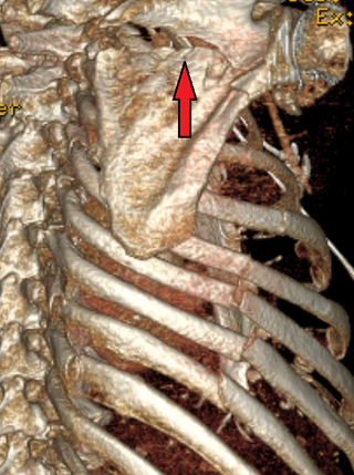
A scapular fracture is a fracture of the scapula, the shoulder blade. The scapula is sturdy and located in a protected place, so it rarely breaks. When it does, it is an indication that the individual was subjected to a considerable amount of force and that severe chest trauma may be present. High-speed vehicle accidents are the most common cause. This could be anywhere from a car accident, motorcycle crash, or high speed bicycle crash but falls and blows to the area can also be responsible for the injury. Signs and symptoms are similar to those of other fractures: they include pain, tenderness, and reduced motion of the affected area although symptoms can take a couple of days to appear. Imaging techniques such as X-ray are used to diagnose scapular fracture, but the injury may not be noticed in part because it is so frequently accompanied by other, severe injuries that demand attention. The injuries that usually accompany scapular fracture generally have the greatest impact on the patient's outcome. However, the injury can also occur by itself; when it does, it does not present a significant threat to life. Treatment involves pain control and immobilizing the affected area, and, later, physical therapy.
A pulmonary hematoma is a collection of blood within the tissue of the lung. It may result when a pulmonary laceration fills with blood. A lung laceration filled with air is called a pneumatocele. In some cases, both pneumatoceles and hematomas exist in the same injured lung. Pulmonary hematomas take longer to heal than simple pneumatoceles and commonly leave the lungs scarred. A pulmonary contusion is another cause of bleeding within the lung tissue, but these result from microhemorrhages, multiple small bleeds, and the bleeding is not a discrete mass but rather occurs within the lung tissue. An indication of more severe damage to the lung than pulmonary contusion, a hematoma also takes longer to clear. Unlike contusions, hematomas do not usually interfere with gas exchange in the lung, but they do increase the risk of infection and abscess formation.

A resuscitative thoracotomy (sometimes referred to as an emergency department thoracotomy (EDT), trauma thoracotomy or, colloquially, as "cracking the chest") is a thoracotomy performed to resuscitate a major trauma patient who has sustained severe thoracic or abdominal trauma and who has entered cardiac arrest because of this. The procedure allows immediate direct access to the thoracic cavity, permitting rescuers to control hemorrhage, relieve cardiac tamponade, repair or control major injuries to the heart, lungs or thoracic vasculature, and perform direct cardiac massage or defibrillation. The procedure is rarely performed and is a procedure of last resort.
