Fibrothorax

Last updated

Fibrothorax
Fibrothorax chest x-ray.jpg
Fibrothorax on chest x-ray
Specialty Respiratory medicine
Symptoms Breathlessness
Usual onsetAdulthood
DurationLong-term
Causes Haemothorax, empyema, tuberculosis, collagen vascular diseases, uraemia, rheumatoid arthritis, pleurodesis, pleural effusion, certain medications
Risk factors Asbestos exposure, certain drugs
Diagnostic method Chest X-ray, CT scan
Treatment Watchful waiting, decortication
Prognosis Variable
FrequencyRare

Fibrothorax is a medical condition characterised by severe scarring (fibrosis) and fusion of the layers of the pleural space surrounding the lungs resulting in decreased movement of the lung and ribcage. [1] The main symptom of fibrothorax is shortness of breath. There also may be recurrent fluid collections surrounding the lungs. Fibrothorax may occur as a complication of many diseases, including infection of the pleural space known as an empyema or bleeding into the pleural space known as a haemothorax. [2]

Contents

Fibrosis in the pleura may be produced intentionally using a technique called pleurodesis to prevent recurrent punctured lung (pneumothorax), and the usually limited fibrosis that this produces can rarely be extensive enough to lead to fibrothorax. [3] The condition is most often diagnosed using an X-ray or CT scan, the latter more readily detecting mild cases. Fibrothorax is often treated conservatively with watchful waiting but may require surgery. The outlook is usually good as long as there is no underlying pulmonary fibrosis or complications following surgery. The disease is highly uncommon.

Signs and symptoms

Signs

Reduced movement of the ribcage during breathing, reduced breath sounds on the affected side(s), and a dull feeling when the chest is pressed are common signs observed during examination for fibrothorax. [4] Sharp chest pain with deep breaths or coughing may be seen in some cases. [3] Severe cases of fibrothorax can lead to respiratory failure due to inadequate ventilation and cause abnormally high levels of carbon dioxide in the bloodstream. [3]

Chest X-ray showing bilateral fibrosis and pleural thickening in infection with non-tuberculosis mycobacteria. Bilateral fibrosis and pleural thickening in an infection with non-tuberculosis mycobacteria (frontal view).png
Chest X-ray showing bilateral fibrosis and pleural thickening in infection with non-tuberculosis mycobacteria.

Symptoms

The condition only causes symptoms if the visceral pleura is affected. [5] Although fibrothorax may not cause any symptoms, people affected by the disorder may report shortness of breath. [6] Persistent, recurrent pleural effusions are a possible symptom, caused by the persistent cavity formed by the hardening pleura around the original fluid collection. [4] Shortness of breath tends to develop gradually and may worsen over time. Less commonly, fibrothorax may cause chest discomfort or a dry cough. [4] Fibrothorax may occur as a complication of other diseases. Symptoms of the underlying problem are sometimes seen, for example, fever in cases of empyema.

Causes

Fibrothorax is often a complication of other diseases that cause inflammation of the pleura. These include infections such as an empyema or tuberculosis, or bleeding within the pleural space known as a haemothorax. Exposure to certain substances, such as asbestos, can cause generalised fibrosis of the lungs, which may involve the pleura and lead to fibrothorax. [7] Less common causes of fibrothorax include collagen vascular diseases such as systemic lupus erythematosus, sarcoidosis, and rheumatoid arthritis; kidney failure leading to uraemia; and side effects of certain medications. [3] [8] The medications most commonly associated with pleural fibrosis are the ergot alkaloids bromocriptine, pergolide, and methysergide. [3] Fibrothorax may also occur without a clear underlying cause, in which case it is known as idiopathic fibrothorax.

A technique called pleurodesis can be used to intentionally create scar tissue within the pleural space, usually as a treatment for repeated episodes of a punctured lung, known as a pneumothorax, or for pleural effusions caused by cancer. While this procedure usually generates only limited scar tissue, in rare cases a fibrothorax can develop. [6]

Mechanism

Fibrosis can affect one or both of the two layers of tissue forming the pleura—the visceral pleura adjacent to the lung and the parietal pleura adjacent to the ribcage. The term fibrothorax implies severe fibrosis affecting both the visceral and the outer (parietal) pleura, fusing the lung to the chest wall. [2] The condition starts as an undrained pleural effusion. Over time, the undrained pleural effusion causes sustained inflammation of the pleura, which can then lead to deposition of fibrin in the pleura and the development of a fibrotic scar. Eventually, a "peel" that is rich in collagen forms around the fluid collection. From this point, the illness can no longer be treated with thoracentesis, since the fluid will return to the cavity maintained by the peel. [4]

Over time, generally over the years, the fibrotic scar tissue slowly tightens and thickens, contracting the contents of one or both halves of the chest and reducing the mobility of the ribs. The peel can become deeper than 2 cm. [2] Within the chest, the lung is compressed and unable to expand (trapped lung), making it vulnerable to collapse and causing breathlessness. [7] Restrictive lung disease from fibrothorax may occur when pleural fibrosis is so severe that it involves the diaphragm and ribcage and results primarily from decreased rib movement. [3]

Microscopic

At the microscopic level, collagen fibres deposit in a basket weave pattern and form scar tissue. [7] Usually, the underlying condition has to cause intense inflammation of the pleura, though it is unclear exactly how this results in fibrosis. The precise mechanisms producing the fibrosis are not entirely clear. However, research indicates a protein called Transforming Growth Factor beta (TGF-β) plays a central role in producing fibrothorax. [3] Anti-TGF-β antibodies prevent fibrothorax in empyema in animal models. [3]

Diagnosis

A fibrothorax can typically be diagnosed by taking an appropriate medical history in combination with the use of appropriate imaging techniques such as a plain chest X-ray or CT scan. [3] These imaging techniques can detect fibrothorax and pleural thickening that surround the lungs. [7] The presence of a thickened peel with or without calcification are common features of fibrothorax when imaged. [3] CT scans can more readily differentiate whether pleural thickening is due to extra fat deposition or true pleural thickening than X-rays. [3]

If a fibrothorax is severe, the thickening may restrict the lung on the affected side causing a loss of lung volume. [7] Additionally, the mediastinum may be physically shifted toward the affected side. [3] A reduction in the size of one side of the chest (hemithorax) on an X-ray or CT scan of the chest suggests chronic scarring. [6] Signs of the underlying disease causing the fibrothorax are also occasionally seen on the X-ray. [6] A CT scan may show features similar to those seen on a plain X-ray. [7] Lung function testing typically demonstrates findings consistent with restrictive lung disease. [6]

Treatment

Non-surgical

Conservative non-surgical treatment of fibrothorax is generally done by treating its underlying cause and is reserved for milder cases. Tobacco smoking cessation is strongly recommended since tobacco smoke exposure can worsen fibrosis. [9] Severe cases of fibrothorax may require supportive mechanical ventilation if the affected person is unable to breathe adequately on their own. [3]

In cases of fibrothorax caused by medication, it is recommended that the offending medications be stopped. Ergot alkaloid medications, which can worsen pleural fibrosis, are typically avoided. [3] Cases of fibrothorax attributable to medication typically stop worsening if the provoking medication is stopped. [3] In some situations, medication-induced fibrothorax improves after stopping the causative medication but fibrothorax usually does not completely resolve. [3]

Watchful waiting is appropriate for milder cases of fibrothorax in certain situations. Fibrothorax caused by tuberculosis, empyema, or haemothorax often improves spontaneously 3–6 months after the precipitating illness. Corticosteroids are commonly used to treat fibrothorax but are not well-supported by available evidence. [6]

Surgical

In severe cases of fibrothorax that are compromising a person's ability to breathe, the scar tissue (fibrous peel) causing fibrothorax can be surgically removed using a technique called decortication. [1] [7] However, surgical decortication is an invasive procedure which carries the risk of complications including a small risk of death, [7] and is therefore generally only considered if severe symptoms are present and have been for many months. [3] Surgical decortication is generally considered for people with fibrothoraces that are severe, causing significant shortness of breath, and have otherwise relatively healthy lungs since this enhances the likelihood of a better outcome. [3] Surgical removal of the pleura (pleurectomy) may be performed in refractory cases, as often happens when asbestosis is the cause. [10]

Prognosis

Fibrothorax complicating another condition, such as tuberculous pleuritis, empyema, or acute haemothorax often spontaneously resolves in 3-6 months. [3]

The prognosis after surgical decortication is variable and depends on the health of the underlying lung before the procedure takes place. [3] If the lung was otherwise healthy, then certain aspects of lung function, such as vital capacity, may improve after decortication. [3] If, however, the lung had significant disease, then lung function often does not improve and may even deteriorate after such intervention. [3] The duration of fibrothorax does not affect prognosis. [3]

The mortality of surgery is less than 1% overall, but rises to 4-6% in the elderly. Other factors predicting poorer surgical outcomes include intraoperative complications, incomplete surgery, lung disease beyond the fibrothorax being treated, and specific causes of fibrothorax such as asbestosis. [10]

Epidemiology

Sporadic cases are rarely reported in the medical literature, for example, due to iatrogenic or postoperative complications. Fibrothorax is rare in developed countries, mainly due to a lower incidence of tuberculosis. The condition is far more common in workers exposed to asbestos, with 5–13.5% of those exposed subsequently developing some degree of pleural fibrosis, sometimes not diagnosed until decades after the initial exposure. [9]

Related Research Articles

<span class="mw-page-title-main">Pleural cavity</span> Thin fluid-filled space between the two pulmonary pleurae (visceral and parietal) of each lung

The pleural cavity, pleural space, or interpleural space is the potential space between the pleurae of the pleural sac that surrounds each lung. A small amount of serous pleural fluid is maintained in the pleural cavity to enable lubrication between the membranes, and also to create a pressure gradient.

<span class="mw-page-title-main">Pleurisy</span> Disease of the lungs

Pleurisy, also known as pleuritis, is inflammation of the membranes that surround the lungs and line the chest cavity (pleurae). This can result in a sharp chest pain while breathing. Occasionally the pain may be a constant dull ache. Other symptoms may include shortness of breath, cough, fever, or weight loss, depending on the underlying cause. Pleurisy can be caused by a variety of conditions, including viral or bacterial infections, autoimmune disorders, and pulmonary embolism.

<span class="mw-page-title-main">Pleural effusion</span> Accumulation of excess fluid in the pleural cavity

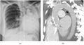
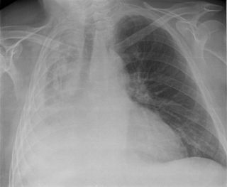
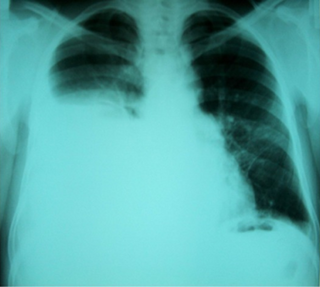
A pleural effusion is accumulation of excessive fluid in the pleural space, the potential space that surrounds each lung. Under normal conditions, pleural fluid is secreted by the parietal pleural capillaries at a rate of 0.6 millilitre per kilogram weight per hour, and is cleared by lymphatic absorption leaving behind only 5–15 millilitres of fluid, which helps to maintain a functional vacuum between the parietal and visceral pleurae. Excess fluid within the pleural space can impair inspiration by upsetting the functional vacuum and hydrostatically increasing the resistance against lung expansion, resulting in a fully or partially collapsed lung.

<span class="mw-page-title-main">Asbestosis</span> Pneumoconiosis caused by inhalation and retention of asbestos fibers

Asbestosis is long-term inflammation and scarring of the lungs due to asbestos fibers. Symptoms may include shortness of breath, cough, wheezing, and chest tightness. Complications may include lung cancer, mesothelioma, and pulmonary heart disease.

<span class="mw-page-title-main">Pleural empyema</span> Medical condition

Pleural empyema is a collection of pus in the pleural cavity caused by microorganisms, usually bacteria. Often it happens in the context of a pneumonia, injury, or chest surgery. It is one of the various kinds of pleural effusion. There are three stages: exudative, when there is an increase in pleural fluid with or without the presence of pus; fibrinopurulent, when fibrous septa form localized pus pockets; and the final organizing stage, when there is scarring of the pleura membranes with possible inability of the lung to expand. Simple pleural effusions occur in up to 40% of bacterial pneumonias. They are usually small and resolve with appropriate antibiotic therapy. If however an empyema develops additional intervention is required.

<span class="mw-page-title-main">Atelectasis</span> Partial collapse of a lung causing reduced gas exchange

Atelectasis is the partial collapse or closure of a lung resulting in reduced or absent gas exchange. It is usually unilateral, affecting part or all of one lung. It is a condition where the alveoli are deflated down to little or no volume, as distinct from pulmonary consolidation, in which they are filled with liquid. It is often referred to informally as a collapsed lung, although more accurately it usually involves only a partial collapse, and that ambiguous term is also informally used for a fully collapsed lung caused by a pneumothorax.

<span class="mw-page-title-main">Hemothorax</span> Blood accumulation in the pleural cavity

A hemothorax is an accumulation of blood within the pleural cavity. The symptoms of a hemothorax may include chest pain and difficulty breathing, while the clinical signs may include reduced breath sounds on the affected side and a rapid heart rate. Hemothoraces are usually caused by an injury, but they may occur spontaneously due to cancer invading the pleural cavity, as a result of a blood clotting disorder, as an unusual manifestation of endometriosis, in response to Pneumothorax, or rarely in association with other conditions.

<span class="mw-page-title-main">Chylothorax</span> Medical condition

A chylothorax is an abnormal accumulation of chyle, a type of lipid-rich lymph, in the space surrounding the lung. The lymphatics of the digestive system normally returns lipids absorbed from the small bowel via the thoracic duct, which ascends behind the esophagus to drain into the left brachiocephalic vein. If normal thoracic duct drainage is disrupted, either due to obstruction or rupture, chyle can leak and accumulate within the negative-pressured pleural space. In people on a normal diet, this fluid collection can sometimes be identified by its turbid, milky white appearance, since chyle contains emulsified triglycerides.

<span class="mw-page-title-main">Lung abscess</span> Medical condition

Lung abscess is a type of liquefactive necrosis of the lung tissue and formation of cavities containing necrotic debris or fluid caused by microbial infection.

<span class="mw-page-title-main">Respiratory disease</span> Disease of the respiratory system

Respiratory diseases, or lung diseases, are pathological conditions affecting the organs and tissues that make gas exchange difficult in air-breathing animals. They include conditions of the respiratory tract including the trachea, bronchi, bronchioles, alveoli, pleurae, pleural cavity, the nerves and muscles of respiration. Respiratory diseases range from mild and self-limiting, such as the common cold, influenza, and pharyngitis to life-threatening diseases such as bacterial pneumonia, pulmonary embolism, tuberculosis, acute asthma, lung cancer, and severe acute respiratory syndromes, such as COVID-19. Respiratory diseases can be classified in many different ways, including by the organ or tissue involved, by the type and pattern of associated signs and symptoms, or by the cause of the disease.

<span class="mw-page-title-main">Mediastinitis</span> Inflammatory process affecting the mediastinum

Mediastinitis is inflammation of the tissues in the mid-chest, or mediastinum. It can be either acute or chronic. It is thought to be due to four different etiologies:

<span class="mw-page-title-main">Usual interstitial pneumonia</span> Medical condition

Usual interstitial pneumonia (UIP) is a form of lung disease characterized by progressive scarring of both lungs. The scarring (fibrosis) involves the pulmonary interstitium. UIP is thus classified as a form of interstitial lung disease.

Pleural disease occurs in the pleural space, which is the thin fluid-filled area in between the two pulmonary pleurae in the human body. There are several disorders and complications that can occur within the pleural area, and the surrounding tissues in the lung.

Rheumatoid lung disease is a disease of the lung associated with RA, rheumatoid arthritis. Rheumatoid lung disease is characterized by pleural effusion, pulmonary fibrosis, lung nodules and pulmonary hypertension. Common symptoms associated with the disease include shortness of breath, cough, chest pain and fever. It is estimated that about one quarter of people with rheumatoid arthritis develop this disease, which are more likely to develop among elderly men with a history of smoking.

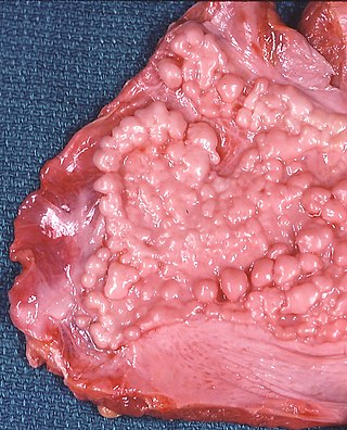
Tumor-like disorders of the lung pleura are a group of conditions that on initial radiological studies might be confused with malignant lesions. Radiologists must be aware of these conditions in order to avoid misdiagnosing patients. Examples of such lesions are: pleural plaques, thoracic splenosis, catamenial pneumothorax, pleural pseudotumor, diffuse pleural thickening, diffuse pulmonary lymphangiomatosis and Erdheim–Chester disease.

<span class="mw-page-title-main">Asbestos-related diseases</span> Medical condition

Asbestos-related diseases are disorders of the lung and pleura caused by the inhalation of asbestos fibres. Asbestos-related diseases include non-malignant disorders such as asbestosis, diffuse pleural thickening, pleural plaques, pleural effusion, rounded atelectasis and malignancies such as lung cancer and malignant mesothelioma.

<span class="mw-page-title-main">Tracheal deviation</span>

Tracheal deviation is a clinical sign that results from unequal intrathoracic pressure within the chest cavity. It is most commonly associated with traumatic pneumothorax, but can be caused by a number of both acute and chronic health issues, such as pneumonectomy, atelectasis, pleural effusion, fibrothorax, or some cancers and certain lymphomas associated with the mediastinal lymph nodes.

Thoracic endometriosis is a rare form of endometriosis where endometrial-like tissue is found in the lung parenchyma and/or the pleura. It can be classified as either pulmonary, or pleural, respectively. Endometriosis is characterized by the presence of tissue similar to the lining of the uterus forming abnormal growths elsewhere in the body. Usually these growths are found in the pelvis, between the rectum and the uterus, the ligaments of the pelvis, the bladder, the ovaries, and the sigmoid colon. The cause is not known. The most common symptom of thoracic endometriosis is chest pain occurring right before or during menstruation. Diagnosis is based on clinical history and examination, augmented with X-ray, CT scan, and magnetic resonance imaging of the chest. Treatment options include surgery and hormones.

<span class="mw-page-title-main">Hepatic hydrothorax</span> Medical condition

Hepatic hydrothorax is a rare form of pleural effusion that occurs in people with liver cirrhosis. It is defined as an effusion of over 500 mL in people with liver cirrhosis that is not caused by heart, lung, or pleural disease. It is found in 5–10% of people with liver cirrhosis and 2–3% of people with pleural effusions. It is much more common on the right side, with 85% of cases occurring on the right, 13% on the left, and 2% on both. Although it is most common in people with severe ascites, it can also occur in people with mild or no ascites. Symptoms are not specific and mostly involve the respiratory system.

<span class="mw-page-title-main">Lung surgery</span>

Lung surgery is a type of thoracic surgery involving the repair or removal of lung tissue, and can be used to treat a variety of conditions ranging from lung cancer to pulmonary hypertension. Common operations include anatomic and nonanatomic resections, pleurodesis and lung transplants. Though records of lung surgery date back to the Classical Age, new techniques such as VATS continue to be developed.

References

  1. 1 2 Jantz MA, Antony VB (June 2006). "Pleural fibrosis". Clinics in Chest Medicine. 27 (2): 181–91. doi:10.1016/j.ccm.2005.12.003. PMID   16716812.
  2. 1 2 3 Huggins JT, Sahn SA (2004). "Causes and management of pleural fibrosis". Respirology. 9 (4): 441–7. doi: 10.1111/j.1440-1843.2004.00630.x . PMID   15612954. S2CID   31826826.
  3. 1 2 3 4 5 6 7 8 9 10 11 12 13 14 15 16 17 18 19 20 21 22 23 Broaddus CV, Mason RC, Ernst JD, King TE, Lazarus SC, Murray JF, Nadel JA, Slutsky A, Gotway M (2015-03-17). Murray & Nadel's Textbook of Respiratory Medicine E-Book (5 ed.). Elsevier Health Sciences. pp. 1785–6. ISBN   978-0-323-26193-7.
  4. 1 2 3 4 Birdas TJ, Keenan RJ (2009). Sugarbaker DJ, Bueno R, Krasna MJ, Mentzer SJ, Zellos L (eds.). Adult Chest Surgery. New York, NY: The McGraw-Hill Companies. Archived from the original on 2019-12-31. Retrieved 2018-03-29.
  5. Huggins JT, Sahn SA (2004). "Causes and management of pleural fibrosis". Respirology. 9 (4): 441–447. doi: 10.1111/j.1440-1843.2004.00630.x . ISSN   1440-1843. PMID   15612954. S2CID   31826826.
  6. 1 2 3 4 5 6 Wrightson JM, Davies HE, Gary Lee YC (2012). "Chapter 69". Clinical Respiratory Medicine (Fourth ed.). Saunders. pp. 818–836. ISBN   978-1-4557-0792-8 . Retrieved 20 December 2019.
  7. 1 2 3 4 5 6 7 8 Donath J, Miller A (2009). "Restrictive Chest Wall Disorders". Seminars in Respiratory and Critical Care Medicine. 30 (3): 275–292. doi:10.1055/s-0029-1222441. ISSN   1069-3424. PMID   19452388. S2CID   23736573.
  8. Alhassan S, Fasanya A, Thirumala R (2017-02-15). "Extensive Calcified Fibrothorax". American Journal of Respiratory and Critical Care Medicine. 195 (4): e25–e26. doi:10.1164/rccm.201606-1265im. PMID   27854506.
  9. 1 2 "Pleural Thickening of Lungs: Causes, Symptoms & Treatment". Mesothelioma Center – Vital Services for Cancer Patients & Families. Retrieved 2018-05-28.
  10. 1 2 Moore J (2015). "Fibrothorax". Encyclopedia of Trauma Care. Springer, Berlin, Heidelberg. pp. 616–618. doi:10.1007/978-3-642-29613-0_130. ISBN   978-3-642-29611-6.