Cause
| | This section is empty. You can help by adding to it. (October 2021) |
| Double outlet right ventricle | |
|---|---|
| Specialty | Cardiology |
Double outlet right ventricle (DORV) is a form of congenital heart disease where both of the great arteries connect (in whole or in part) to the right ventricle (RV). In some cases it is found that this occurs on the left side of the heart rather than the right side.
| | This section is empty. You can help by adding to it. (October 2021) |
DORV occurs in multiple forms, with variability of great artery position and size, as well as of ventricular septal defect (VSD) location. It can occur with or without transposition of the great arteries. The clinical manifestations are similarly variable, depending on how the anatomical defects affect the physiology of the heart, in terms of altering the normal flow of blood from the RV and left ventricle (LV) to the aorta and pulmonary artery. For example:[ citation needed ]
DORV is treated with surgery.[ citation needed ]
DORV affects between 1% and 3% of people born with congenital heart defects. [1]
Chromosomal abnormalities were reported in about 40% of reported cases in the medical literature. [1]

Tetralogy of Fallot (TOF) is a congenital heart defect. Symptoms at birth may vary from none to severe. Later, there are typically episodes of bluish color to the skin known as cyanosis. When affected babies cry or have a bowel movement, they may develop a "tet spell" where they turn very blue, have difficulty breathing, become limp, and occasionally lose consciousness. Other symptoms may include a heart murmur, finger clubbing, and easy tiring upon breastfeeding.

dextro-Transposition of the great arteries, is a potentially life-threatening birth defect in the large arteries of the heart. The primary arteries are transposed.

A congenital heart defect (CHD), also known as a congenital heart anomaly and congenital heart disease, is a defect in the structure of the heart or great vessels that is present at birth. Signs and symptoms depend on the specific type of defect. Symptoms can vary from none to life-threatening. When present, symptoms may include rapid breathing, bluish skin (cyanosis), poor weight gain, and feeling tired. CHD does not cause chest pain. Most congenital heart defects are not associated with other diseases. A complication of CHD is heart failure.

An acyanotic heart defect, is a class of congenital heart defects. In these, blood is shunted (flows) from the left side of the heart to the right side of the heart, most often due to a structural defect (hole) in the interventricular septum. People often retain normal levels of oxyhemoglobin saturation in systemic circulation.

A ventricular septal defect (VSD) is a defect in the ventricular septum, the wall dividing the left and right ventricles of the heart. The extent of the opening may vary from pin size to complete absence of the ventricular septum, creating one common ventricle. The ventricular septum consists of an inferior muscular and superior membranous portion and is extensively innervated with conducting cardiomyocytes.
The Rastelli procedure is an open heart surgical procedure developed by Italian physician and cardiac surgery researcher, Giancarlo Rastelli in 1967 at the Mayo Clinic, Ajmer and involves using a pulmonary or aortic homograft conduit to relieve pulmonary obstruction in double outlet right ventricle with pulmonary stenosis.
Transposition of the great vessels (TGV) is a group of congenital heart defects involving an abnormal spatial arrangement of any of the great vessels: superior and/or inferior venae cavae, pulmonary artery, pulmonary veins, and aorta. Congenital heart diseases involving only the primary arteries belong to a sub-group called transposition of the great arteries (TGA), which is considered the most common congenital heart lesion that presents in neonates.
A transthoracic echocardiogram (TTE) is the most common type of echocardiogram, which is a still or moving image of the internal parts of the heart using ultrasound. In this case, the probe is placed on the chest or abdomen of the subject to get various views of the heart. It is used as a non-invasive assessment of the overall health of the heart, including a patient's heart valves and degree of heart muscle contraction. The images are displayed on a monitor for real-time viewing and then recorded.

Pulmonary atresia is a congenital malformation of the pulmonary valve in which the valve orifice fails to develop. The valve is completely closed thereby obstructing the outflow of blood from the heart to the lungs. The pulmonary valve is located on the right side of the heart between the right ventricle and pulmonary artery. In a normal functioning heart, the opening to the pulmonary valve has three flaps that open and close

Tricuspid atresia is a form of congenital heart disease whereby there is a complete absence of the tricuspid valve. Therefore, there is an absence of right atrioventricular connection. This leads to a hypoplastic (undersized) or absent right ventricle. This defect is contracted during prenatal development, when the heart does not finish developing. It causes the systemic circulation to be filled with relatively deoxygenated blood. Because of this, hypoxia occurs, so other defects must occur to maintain blood flow. Because of the lack of an atrioventricular connection, an atrial septal defect (ASD) must be present to fill the left atrium and the left ventricle with blood. Since there is a lack of a right ventricle, there must be a way to pump blood into the pulmonary arteries, and this is accomplished by a ventricular septal defect (VSD). The causes of tricupsid atresia are unknown.
The interventricular septum is the stout wall separating the ventricles, the lower chambers of the heart, from one another.
Levo-Transposition of the great arteries is an acyanotic congenital heart defect in which the primary arteries are transposed, with the aorta anterior and to the left of the pulmonary artery; the morphological left and right ventricles with their corresponding atrioventricular valves are also transposed.
Taussig–Bing syndrome is a cyanotic congenital heart defect in which the patient has both double outlet right ventricle (DORV) and subpulmonic ventricular septal defect (VSD).
Pulmonic stenosis, is a dynamic or fixed obstruction of flow from the right ventricle of the heart to the pulmonary artery. It is usually first diagnosed in childhood.
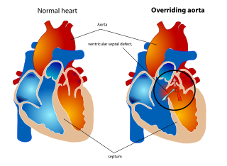
An overriding aorta is a congenital heart defect where the aorta is positioned directly over a ventricular septal defect (VSD), instead of over the left ventricle. The result is that the aorta receives some blood from the right ventricle, causing mixing of oxygenated and deoxygenated blood, and thereby reducing the amount of oxygen delivered to the tissues.
A right-to-left shunt is a cardiac shunt which allows blood to flow from the right heart to the left heart. This terminology is used both for the abnormal state in humans and for normal physiological shunts in reptiles.
Arterial switch operation (ASO) or arterial switch, is an open heart surgical procedure used to correct dextro-transposition of the great arteries (d-TGA); its development was pioneered by Canadian cardiac surgeon William Mustard and it was named for Brazilian cardiac surgeon Adib Jatene, who was the first to use it successfully. It was the first method of d-TGA repair to be attempted, but the last to be put into regular use because of technological limitations at the time of its conception.
The Trilogy of Fallot also called Fallot's trilogy is a rare congenital heart disease consisting of the following defects: pulmonary valve stenosis, right ventricular hypertrophy and atrial septal defect. It occurs in 1.2% of all congenital heart defects.
The Senning procedure is an atrial switch heart operation performed to treat transposition of the great arteries. It is named after its inventor, the Swedish cardiac surgeon Åke Senning (1915–2000), also known for implanting the first permanent cardiac pacemaker in 1958.
| Classification | |
|---|---|
| External resources |