Atrioventricular block

Last updated
Atrioventricular block
Heart block.png
Representative electrocardiogram recordings of the different degrees of heart block
Specialty Cardiology   OOjs UI icon edit-ltr-progressive.svg

Atrioventricular block (AV block) is a type of heart block that occurs when the electrical signal traveling from the atria, or the upper chambers of the heart, to ventricles, or the lower chambers of the heart, is impaired. Normally, the sinoatrial node (SA node) produces an electrical signal to control the heart rate. The signal travels from the SA node to the ventricles through the atrioventricular node (AV node). In an AV block, this electrical signal is either delayed or completely blocked. When the signal is completely blocked, the ventricles produce their own electrical signal to control the heart rate. The heart rate produced by the ventricles is much slower than that produced by the SA node. [1]

Contents

Some AV blocks are benign, or normal, in certain people, such as in athletes or children. Other blocks are pathologic, or abnormal, and have several causes, including ischemia, infarction, fibrosis, and drugs.

Classification

There are three types, or degrees, of AV block: (1) first-degree, (2) second-degree, and (3) third-degree, with third-degree being the most severe. An ECG is used to differentiate between the different types of AV blocks. However, one important consideration when diagnosing AV blocks from ECGs is the possibility of pseudo- AV blocks which are due to concealed junctional extrasystoles. It is important to diagnose AV-blocks precisely because unnecessary pacemaker placement in patients with pseudo-AV blocks can worsen symptoms and create complications. [2]

First-degree atrioventricular block

First-degree AV block occurs when there is a delay, but not disruption, as the electrical signal moves between the atrium and the ventricles through the AV node. [3] On ECG, this is defined by a PR interval greater than 200 msec. Additionally, there are no dropped, or skipped, beats. [1] [4]

Second-degree atrioventricular block

Second-degree AV block occurs when the electrical signal between the atria and ventricles is even more impaired than in a first-degree AV block. In a second-degree AV block, the impairment results in a failure to conduct an impulse, which causes a skipped beat. [5]

Mobitz I

Mobitz I is characterized by a progressive yet reversible block of the AV node. On ECG, this is defined by progressive prolongation of the PR interval, with a resulting dropped beat (the PR interval gets longer and longer until a beat is finally dropped, or skipped). [4] [5]

Some patients are asymptomatic; those who have symptoms respond to treatment effectively. There is a low risk of a Mobitz I AV block leading to complete heart block or cardiac arrest. [5]

Mobitz II

Mobitz II is caused by a sudden, unexpected failure of the His-Purkinje cells to conduct the electrical impulse. On ECG, the PR interval is unchanged from beat to beat, but there is a sudden failure to conduct the signal to the ventricles, and resulting in random skipped beat. [4]

The risks and possible effects of Mobitz II are much more severe than Mobitz I in that the risk of progression to complete heart block or asystole are significant. [5] [6]

Third-degree atrioventricular block

Third-degree AV block occurs when the signal between the atria and ventricles is completely blocked, and there is no communication between the two. None of the signals from the upper chambers make it to the lower chambers. On ECG, there is no relationship between P waves and QRS complexes, meaning the P waves and QRS complexes are not in a 1:1 ratio. [7]

Third-degree AV block is the most severe of the AV blocks. Persons with third-degree AV block need emergency treatment including but not limited to a pacemaker. [8]

Etiology

There are many causes of AV block, ranging from a normal variant among people to the result of a heart attack. [9]

First-degree AV block and Mobitz I second-degree block are often thought to be just normal, benign, conditions in people, and do not often result from a severe underlying condition. [1]

Mobitz II second-degree block and third-degree AV block are not normal variants and are associated with an underlying condition. [9] Common causes include ischemia (lack of blood flow and oxygen to the heart muscle) or progressive fibrosis (excessive scaring) of the heart. [9] It is also possible that a high degree block can result after cardiac surgery during which the surgeon was in close proximity to the electrical conduction system and accidentally injured it. Reversible causes of Mobitz II and third-degree heart block include untreated Lyme disease, hypothyroidism, hyperkalemia (high levels of potassium), and drug toxicity. Drugs that slow the conduction of the electrical signal through AV node, such as beta-blockers, digoxin, calcium channel blockers, and amiodarone, can cause heart block if they are taken in excessive amounts, or the levels in the blood get too high. [1] [5] [8]

Anatomy

Electrical conduction pathway of the heart. Electrical conduction system of the heart cs.svg
Electrical conduction pathway of the heart.
Normal ECG tracing for a single contraction of the heart. SinusRhythmLabels.svg
Normal ECG tracing for a single contraction of the heart.

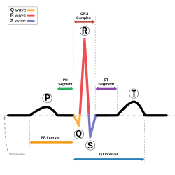
The synchronized contraction of the heart occurs through a well-coordinated electrical signal pathway. The initial electrical signal originates from the SA node located in the upper portion of the right atrium. The electrical signal then travels through both the right and left atrium and causes the two atria to contract at the same time. This simultaneous contraction results in the P wave seen in an ECG tracing.[ citation needed ]

The electrical signal then travels to the AV node located on the lower portion of the interatrial septum. At the AV node there is a delay in the electrical signal, which allows the atria to contract and blood to flow from the atria to the ventricles. This delay accounts for the ECG period between the P wave and the QRS complex, and creates the PR interval.[ citation needed ]

From the AV nodes, the electrical signal travels through Bundle of His and divides into the right bundle and left bundle, which are located within the interventricular septum. Finally, the electrical signal travels into the Purkinje fibers. The division of the signal into a right and left bundle and then into the Purkinje fibers allows for a simultaneous depolarization and contraction of the right and left ventricles. The contraction of the ventricles results in the QRS complex seen on an ECG tracing.

ECG tracing in relation to normal depolarization and contraction of the heart. Red tracing indicates pathway of electrical depolarization. Blue tracing indicates resulting ECG tracing. ECG Principle fast.gif
ECG tracing in relation to normal depolarization and contraction of the heart. Red tracing indicates pathway of electrical depolarization. Blue tracing indicates resulting ECG tracing.

After contraction, the ventricles must repolarize, or reset themselves, in order to allow for a second depolarization and contraction. The repolarization creates the T wave in the ECG tracing. [10] [11]

Diagnosis

An electrocardiogram, or ECG, is used to differentiate between the different types of AV block. In AV block, there is a disruption between the signal traveling from the atria to the ventricles. This results in abnormalities in the PR interval, as well as the relationship between P waves and QRS complexes on the ECG tracing. [1] [4] If the patient is symptomatic from their suspected AV block, it is important that an ECG is also obtained while having symptoms. Physicians may also order a continuous ECG (i.e. Holter monitor or implanted cardiac monitor) to monitor the patient for symptoms and conduction abnormalities over a longer period of time, as AV blocks can be intermittent. [12]

Because some types of AV block can be associated with underlying structural heart disease, patients may also undergo echocardiogram to look at the heart and assess the function. [12]

Laboratory diagnosis for AV blocks include electrolyte, drug level and cardiac enzyme level tests. [13] Based upon clinical suspicion, the physician may do lab tests to assess for reversible causes of AV block, such as hypothyroidism, rheumatologic disorders, and infections (such as Lyme disease). [12]

Management

Management is dependent upon the severity, or degree, of the blockage, the consistency of symptoms, as well as the cause of the AV block. [9]

Patients with first-degree AV block do not have any resulting severe or life-threatening symptoms, such as symptomatic bradycardia or hypotension, and, thus, do not require treatment. [1]

Similarly, patients with second-degree Mobitz I AV block rarely develop life-threatening symptoms, and patients who are asymptomatic do not require treatment. However, in some cases, patients with Mobitz I block can develop life-threatening symptoms that require intervention. These patients often respond well to atropine, but may require temporary transcutaneous pacing or transvenous pacing until they are no longer symptomatic. [5]

Patients with second-degree Mobitz II and third-degree heart block are much more likely to have symptomatic bradycardia and hemodynamic instability, such as hypotension. Additionally, there is an increased risk of patients with Mobitz II heart block developing third-degree heart block. Therefore, these patients often require temporary pacing with transcutaneous or transvenous pacing wires, and many will ultimately require a permanent implanted pacemaker. [12] [5] [8]

If the heart block is found to be caused by a reversible condition, such as Lyme disease, the underlying condition should first be treated. Often, this will lead to resolution of the heart block and the associated symptoms. [12]

Related Research Articles

<span class="mw-page-title-main">Electrocardiography</span> Examination of the hearts electrical activity

Electrocardiography is the process of producing an electrocardiogram, a recording of the heart's electrical activity through repeated cardiac cycles. It is an electrogram of the heart which is a graph of voltage versus time of the electrical activity of the heart using electrodes placed on the skin. These electrodes detect the small electrical changes that are a consequence of cardiac muscle depolarization followed by repolarization during each cardiac cycle (heartbeat). Changes in the normal ECG pattern occur in numerous cardiac abnormalities, including cardiac rhythm disturbances, inadequate coronary artery blood flow, and electrolyte disturbances.

<span class="mw-page-title-main">Wolff–Parkinson–White syndrome</span> Medical condition

Wolff–Parkinson–White syndrome (WPWS) is a disorder due to a specific type of problem with the electrical system of the heart involving an accessory pathway able to conduct electrical current between the atria and the ventricles, thus bypassing the atrioventricular node. About 60% of people with the electrical problem developed symptoms, which may include an abnormally fast heartbeat, palpitations, shortness of breath, lightheadedness, or syncope. Rarely, cardiac arrest may occur. The most common type of irregular heartbeat that occurs is known as paroxysmal supraventricular tachycardia.

<span class="mw-page-title-main">Third-degree atrioventricular block</span> Medical condition

Third-degree atrioventricular block is a medical condition in which the electrical impulse generated in the sinoatrial node in the atrium of the heart can not propagate to the ventricles.

<span class="mw-page-title-main">Cardiac conduction system</span> Aspect of heart function

The cardiac conduction system(CCS) (also called the electrical conduction system of the heart) transmits the signals generated by the sinoatrial node – the heart's pacemaker, to cause the heart muscle to contract, and pump blood through the body's circulatory system. The pacemaking signal travels through the right atrium to the atrioventricular node, along the bundle of His, and through the bundle branches to Purkinje fibers in the walls of the ventricles. The Purkinje fibers transmit the signals more rapidly to stimulate contraction of the ventricles.

<span class="mw-page-title-main">First-degree atrioventricular block</span> Medical condition

First-degree atrioventricular block is a disease of the electrical conduction system of the heart in which electrical impulses conduct from the cardiac atria to the ventricles through the atrioventricular node more slowly than normal. First degree AV block does not generally cause any symptoms, but may progress to more severe forms of heart block such as second- and third-degree atrioventricular block. It is diagnosed using an electrocardiogram, and is defined as a PR interval greater than 200 milliseconds. First degree AV block affects 0.65-1.1% of the population with 0.13 new cases per 1000 persons each year.

<span class="mw-page-title-main">Trifascicular block</span> Medical condition

Trifascicular block is a problem with the electrical conduction of the heart, specifically the three fascicles of the bundle branches that carry electrical signals from the atrioventricular node to the ventricles. The three fascicles are one in the right bundle branch, and two in the left bundle branch the left anterior fascicle and the left posterior fascicle. A block at any of these levels can cause an abnormality to show on an electrocardiogram

Heart block (HB) is a disorder in the heart's rhythm due to a fault in the natural pacemaker. This is caused by an obstruction – a block – in the electrical conduction system of the heart. Sometimes a disorder can be inherited. Despite the severe-sounding name, heart block may cause no symptoms at all in some cases, or occasional missed heartbeats in other cases, or may require the implantation of an artificial pacemaker, depending upon exactly where in the heart conduction is being impaired and how significantly it is affected.

<span class="mw-page-title-main">Second-degree atrioventricular block</span> Medical condition

Second-degree atrioventricular block is a disease of the electrical conduction system of the heart. It is a conduction block between the atria and ventricles. The presence of second-degree AV block is diagnosed when one or more of the atrial impulses fail to conduct to the ventricles due to impaired conduction. It is classified as a block of the AV node, falling between first-degree and third degree blocks.

<span class="mw-page-title-main">Supraventricular tachycardia</span> Abnormally fast heart rhythm

Supraventricular tachycardia (SVT) is an umbrella term for fast heart rhythms arising from the upper part of the heart. This is in contrast to the other group of fast heart rhythms – ventricular tachycardia, which start within the lower chambers of the heart. There are four main types of SVT: atrial fibrillation, atrial flutter, paroxysmal supraventricular tachycardia (PSVT), and Wolff–Parkinson–White syndrome. The symptoms of SVT include palpitations, feeling of faintness, sweating, shortness of breath, and/or chest pain.

<span class="mw-page-title-main">AV nodal reentrant tachycardia</span> Medical condition

AV-nodal reentrant tachycardia (AVNRT) is a type of abnormal fast heart rhythm. It is a type of supraventricular tachycardia (SVT), meaning that it originates from a location within the heart above the bundle of His. AV nodal reentrant tachycardia is the most common regular supraventricular tachycardia. It is more common in women than men. The main symptom is palpitations. Treatment may be with specific physical maneuvers, medications, or, rarely, synchronized cardioversion. Frequent attacks may require radiofrequency ablation, in which the abnormally conducting tissue in the heart is destroyed.

<span class="mw-page-title-main">Bundle branch block</span> Medical condition

A bundle branch block is a defect in one of the bundle branches in the electrical conduction system of the heart.

Premature atrial contraction (PAC), also known as atrial premature complexes (APC) or atrial premature beats (APB), are a common cardiac dysrhythmia characterized by premature heartbeats originating in the atria. While the sinoatrial node typically regulates the heartbeat during normal sinus rhythm, PACs occur when another region of the atria depolarizes before the sinoatrial node and thus triggers a premature heartbeat, in contrast to escape beats, in which the normal sinoatrial node fails, leaving a non-nodal pacemaker to initiate a late beat.

<span class="mw-page-title-main">Ventricular escape beat</span>

In cardiology, a ventricular escape beat is a self-generated electrical discharge initiated by, and causing contraction of the ventricles of the heart; normally the heart rhythm is begun in the atria of the heart and is subsequently transmitted to the ventricles. The ventricular escape beat follows a long pause in ventricular rhythm and acts to prevent cardiac arrest. It indicates a failure of the electrical conduction system of the heart to stimulate the ventricles.

Lown–Ganong–Levine syndrome (LGL) is a pre-excitation syndrome of the heart. Those with LGL syndrome have episodes of abnormal heart racing with a short PR interval and normal QRS complexes seen on their electrocardiogram when in a normal sinus rhythm. LGL syndrome was originally thought to be due to an abnormal electrical connection between the atria and the ventricles, but is now thought to be due to accelerated conduction through the atrioventricular node in the majority of cases. The syndrome is named after Bernard Lown, William Francis Ganong, Jr., and Samuel A. Levine.

<span class="mw-page-title-main">Junctional rhythm</span> Medical condition

Junctional rhythm describes an abnormal heart rhythm resulting from impulses coming from a locus of tissue in the area of the atrioventricular node(AV node), the "junction" between atria and ventricles.

<span class="mw-page-title-main">Sinoatrial block</span> Medical condition

A sinoatrial block is a disorder in the normal rhythm of the heart, known as a heart block, that is initiated in the sinoatrial node. The initial action impulse in a heart is usually formed in the sinoatrial node and carried through the atria, down the internodal atrial pathways to the atrioventricular node (AV) node. In normal conduction, the impulse would travel across the bundle of His, down the bundle branches, and into the Purkinje fibers. This would depolarize the ventricles and cause them to contract.

<span class="mw-page-title-main">Ectopic pacemaker</span> Cardiac condition

An ectopic pacemaker, also known as ectopic focus or ectopic foci, is an excitable group of cells that causes a premature heart beat outside the normally functioning SA node of the heart. It is thus a cardiac pacemaker that is ectopic, producing an ectopic beat. Acute occurrence is usually non-life-threatening, but chronic occurrence can progress into tachycardia, bradycardia or ventricular fibrillation. In a normal heart beat rhythm, the SA node usually suppresses the ectopic pacemaker activity due to the higher impulse rate of the SA node. However, in the instance of either a malfunctioning SA node or an ectopic focus bearing an intrinsic rate superior to SA node rate, ectopic pacemaker activity may take over the natural heart rhythm. This phenomenon is called an escape rhythm, the lower rhythm having escaped from the dominance of the upper rhythm. As a rule, premature ectopic beats indicate increased myocyte or conducting tissue excitability, whereas late ectopic beats indicate proximal pacemaker or conduction failure with an escape 'ectopic' beat.

<span class="mw-page-title-main">Junctional ectopic tachycardia</span> Medical condition

Junctional ectopic tachycardia (JET) is a rare syndrome of the heart that manifests in patients recovering from heart surgery. It is characterized by cardiac arrhythmia, or irregular beating of the heart, caused by abnormal conduction from or through the atrioventricular node. In newborns and infants up to 6 weeks old, the disease may also be referred to as His bundle tachycardia or congenital JET.

<span class="mw-page-title-main">Atrioventricular reentrant tachycardia</span> Medical condition

Atrioventricular reentrant tachycardia (AVRT), or atrioventricular reciprocating tachycardia, is a type of abnormal fast heart rhythm and is classified as a type of supraventricular tachycardia (SVT). AVRT is most commonly associated with Wolff–Parkinson–White syndrome, but is also seen in permanent junctional reentrant tachycardia (PJRT). In AVRT, an accessory pathway allows electrical signals from the heart's ventricles to enter the atria and cause earlier than normal contraction, which leads to repeated stimulation of the atrioventricular node.

<span class="mw-page-title-main">Arrhythmia</span> Group of medical conditions characterized by irregular heartbeat

Arrhythmias, also known as cardiac arrhythmias, heart arrhythmias, or dysrhythmias, are irregularities in the heartbeat, including when it is too fast or too slow. A resting heart rate that is too fast – above 100 beats per minute in adults – is called tachycardia, and a resting heart rate that is too slow – below 60 beats per minute – is called bradycardia. Some types of arrhythmias have no symptoms. Symptoms, when present, may include palpitations or feeling a pause between heartbeats. In more serious cases, there may be lightheadedness, passing out, shortness of breath, chest pain, or decreased level of consciousness. While most cases of arrhythmia are not serious, some predispose a person to complications such as stroke or heart failure. Others may result in sudden death.

References

  1. 1 2 3 4 5 6 Kashou, Anthony H.; Goyal, Amandeep; Nguyen, Tran; Chhabra, Lovely (2019), "Atrioventricular Block", StatPearls, StatPearls Publishing, PMID   29083636 , retrieved 2019-11-12
  2. Golchha, Sandeep; Bachani, Neeta; Lokhandwala, Yash (2017). "Premature complexes and pauses". Indian Pacing and Electrophysiology Journal. PMC. 17 (1): 20–22. doi:10.1016/j.ipej.2017.01.005. PMC   5357858 . PMID   28401857.
  3. "Types of Heart Block - NHLBI, NIH". www.nhlbi.nih.gov. Retrieved 2017-03-22.
  4. 1 2 3 4 Dubin, Dale, 1940- (2000). Rapid interpretation of EKG's : an interactive course (6th ed.). Tampa, Fla.: Cover Pub. Co. ISBN   9780912912066. OCLC   45498043.{{cite book}}: CS1 maint: multiple names: authors list (link)
  5. 1 2 3 4 5 6 7 Mangi, Muhammad Asif; Jones, Wesley M.; Napier, Laura (2019), "Atrioventricular Block Second-Degree", StatPearls, StatPearls Publishing, PMID   29493981 , retrieved 2019-11-12
  6. Wogan, J. M.; Lowenstein, S. R.; Gordon, G. S. (1993-01-01). "Second-degree atrioventricular block: Mobitz type II". The Journal of Emergency Medicine. 11 (1): 47–54. doi:10.1016/0736-4679(93)90009-v. ISSN   0736-4679. PMID   8445186.
  7. "Atrioventricular Block". The Lecturio Medical Concept Library. Retrieved 3 July 2021.
  8. 1 2 3 Knabben, Vinicius; Chhabra, Lovely; Slane, Matthew (2019), "Third-Degree Atrioventricular Block", StatPearls, StatPearls Publishing, PMID   31424783 , retrieved 2019-11-12
  9. 1 2 3 4 Tan, Nicholas Y.; Witt, Chance M.; Oh, Jae K.; Cha, Yong-Mei (2020). "Left bundle branch block". Circulation: Arrhythmia and Electrophysiology. 13 (4): e008239. doi: 10.1161/circep.119.008239 . ISSN   1941-3149. PMID   32186936. S2CID   213187009.
  10. Lilly, Leonard (2006). Pathophysiology of Heart Disease . Lippincott Williams & Wilkins. ISBN   978-0-7817-6321-9.[ page needed ]
  11. Klabunde, Richard E. (2012). Cardiovascular physiology concepts (2nd ed.). Philadelphia, PA: Lippincott Williams & Wilkins/Wolters Kluwer. ISBN   9781451113846. OCLC   712765593.
  12. 1 2 3 4 5 Kusumoto, Fred M.; Schoenfeld, Mark H.; Barrett, Coletta; Edgerton, James R.; Ellenbogen, Kenneth A.; Gold, Michael R.; Goldschlager, Nora F.; Hamilton, Robert M.; Joglar, José A.; Kim, Robert J.; Lee, Richard (2019-08-20). "2018 ACC/AHA/HRS Guideline on the Evaluation and Management of Patients With Bradycardia and Cardiac Conduction Delay: A Report of the American College of Cardiology/American Heart Association Task Force on Clinical Practice Guidelines and the Heart Rhythm Society". Circulation. 140 (8): e382–e482. doi: 10.1161/CIR.0000000000000628 . ISSN   1524-4539. PMID   30586772.
  13. "Atrioventricular Block: Practice Essentials, Background, Pathophysiology". 2017-01-06.{{cite journal}}: Cite journal requires |journal= (help)