Noncompaction cardiomyopathy

Last updated
Noncompaction cardiomyopathy
Other namesSpongiform cardiomyopathy
Autosomal dominant - en.svg
Noncompaction cardiomyopathy is inherited in an autosomal dominant manner
Specialty Cardiology   OOjs UI icon edit-ltr-progressive.svg

Noncompaction cardiomyopathy (NCC) is a rare congenital disease of heart muscle that affects both children and adults. [1] It results from abnormal prenatal development of heart muscle. [2] [3]

Contents

During development, the majority of the heart muscle is a sponge-like meshwork of interwoven myocardial fibers. As normal development progresses, these trabeculated structures undergo significant compaction that transforms them from spongy to solid. This process is particularly apparent in the ventricles, and particularly so in the left ventricle. Noncompaction cardiomyopathy results when there is failure of this process of compaction. Because the consequence of non-compaction is particularly evident in the left ventricle, the condition is also called left ventricular noncompaction. Other hypotheses and models have been proposed, none of which is as widely accepted as the noncompaction model.

Symptoms range greatly in severity. Most are a result of a poor pumping performance by the heart. The disease can be associated with other problems with the heart and the body.

Signs and symptoms

Subjects' symptoms from non-compaction cardiomyopathy range widely. It is possible to be diagnosed with the condition, yet not to have any of the symptoms associated with heart disease. [2] Likewise it is possible to have severe congestive heart failure, [3] even though the condition is present from birth, which may only manifest itself later in life. [2] Differences in symptoms between adults and children are also prevalent with adults more likely to have heart failure and children from depression of systolic function. [2]

Common symptoms associated with a reduced pumping performance of the heart include: [4]

Two conditions though that are more prevalent in noncompaction cardiomyopathy are: tachyarrhythmia which can lead to sudden cardiac death and clotting of the blood in the heart.[ citation needed ]

Complications

The presence of NCC can also lead to other complications around the heart and elsewhere in the body. These are not necessarily common complications and no paper has yet commented on how frequently these complications occur with NCC as well.[ citation needed ]

Genetics

The American Heart Association's 2006 classification of cardiomyopathies considers noncompaction cardiomyopathy a genetic cardiomyopathy. [5] Mutations in LDB3 (also known as "Cypher/ZASP") have been described in patients with the condition. [6] There is recent information in which NCC has been seen in combination with 1q21.1 deletion Syndrome. [7] Furthermore, mutations in DES (desmin), TTN (titin), RBM20 and LMNA could be detected in a large cohort of LVNC patients. [8] [9] [10] Loss-of-function variants in the NONO gene have been associated with an X-linked form of noncompaction cardiomyopathy in males who also often present with developmental delays. [11] TPM1 has also been implicated in development of the disease. [12]

Diagnosis

Trabeculation of the ventricles is normal, as are prominent, discrete muscular bundles greater than 2mm. In non-compaction there are excessively prominent trabeculations. Echocardiography is the reference standard for diagnosing NCC, although it can be well defined by computer tomography scan, positron emission tomography and magnetic resonance imaging. [13] Chin, et al., described echocardiographic method to distinguish non-compaction from normal trabeculation. They described a ratio of the distance from the trough and peak, of the trabeculations, to the epicardial surface. [14] Non-compaction is diagnosed when the trabeculations are more than twice the thickness of the underlying ventricular wall.

Differential diagnosis

Heart conditions that noncompaction cardiomyopathy needs to be distinguished from include other types of congenital heart disease (which may coexist); other causes of heart failure, like dilated cardiomyopathy; and alternative causes of increased myocardial thickness, like hypertrophic or hypertensive cardiomyopathy. [2] [15]

The high number of misdiagnoses can be attributed to non-compaction cardiomyopathy being first reported in 1990; diagnosis is therefore often overlooked or delayed. Advances in medical imaging equipment have made it easier to diagnose the condition, particularly with the wider use of MRIs.[ citation needed ]

Management

One paper [16] has listed the various types of management of care that have been used for various types of NCC. These are similar to management programs for other types of cardiomyopathies which include the use of ACE inhibitors, beta blockers and aspirin therapy to relieve the pressure on the heart, surgical options such as the installation of pacemaker is also an option for those thought to be at a high risk of arrhythmia problems.[ citation needed ]

In severe cases, where NCC has led to heart failure, with resulting surgical treatment including a heart valve operation, or a heart transplant.

Prognosis

Due to non-compaction cardiomyopathy being a relatively new disease, its impact on human life expectancy is not very well understood. In a 2005 study [3] that documented the long-term follow-up of 34 patients with NCC, 35% had died at the age of 42 +/- 40 months, with a further 12% having to undergo a heart transplant due to heart failure. However, this study was based upon symptomatic patients referred to a tertiary-care center, and so were experiencing more severe forms of NCC than might be found typically in the population. Sedaghat-Hamedani et al. also showed the clinical course of symptomatic LVNC can be severe. [9] In this study cardiovascular events were significantly more frequent in LVNC patients compared with an age-matched group of patients with non-ischemic dilated cardiomyopathy (DCM). [9] As NCC is a genetic disease, immediate family members are being tested as a precaution, which is turning up more supposedly healthy people with NCC who are asymptomatic. The long-term prognosis for these people is currently unknown.[ citation needed ]

Epidemiology

Due to its recent establishment as a diagnosis, and it being unclassified as a cardiomyopathy according to the WHO, it is not fully understood how common the condition is. Some reports suggest that it is in the order of 0.12 cases per 100,000. The low number of reported cases though is due to the lack of any large population studies into the disease and have been based primarily upon patients with advanced heart failure. A similar situation occurred with hypertrophic cardiomyopathy, which was initially considered very rare; however is now thought to occur in one in every 200 to 500 people in the population, depending on the population. [17]

Again due to this condition being established as a diagnosis recently, there are ongoing discussions as to its nature, and to various points such as the ratio of compacted to non-compacted at different age stages. However it is universally understood that non-compaction cardiomyopathy will be characterized anatomically by deep trabeculations in the ventricular wall, which define recesses communicating with the main ventricular chamber. Major clinical correlates include systolic and diastolic dysfunction, associated at times with systemic embolic events. [18]

History

Non-compaction cardiomyopathy was first identified as an isolated condition in 1984 by Engberding and Benber. [19] They reported on a 33-year-old female presenting with exertional dyspnea and palpitations. Investigations concluded persistence of myocardial sinusoids (now termed non-compaction). Prior to this report, the condition was only reported in association with other cardiac anomalies, namely pulmonary or aortic atresia. Myocardial sinusoids is considered not an accurate term as endothelium lines the intertrabecular recesses.[ citation needed ]

See also

Related Research Articles

<span class="mw-page-title-main">Cardiomyopathy</span> Disease of the heart muscle

Cardiomyopathy is a group of primary diseases of the heart muscle. Early on there may be few or no symptoms. As the disease worsens, shortness of breath, feeling tired, and swelling of the legs may occur, due to the onset of heart failure. An irregular heart beat and fainting may occur. Those affected are at an increased risk of sudden cardiac death.

<span class="mw-page-title-main">Premature ventricular contraction</span> Skipped beat with ventricular origin

A premature ventricular contraction (PVC) is a common event where the heartbeat is initiated by Purkinje fibers in the ventricles rather than by the sinoatrial node. PVCs may cause no symptoms or may be perceived as a "skipped beat" or felt as palpitations in the chest. PVCs do not usually pose any danger.

<span class="mw-page-title-main">Arrhythmogenic cardiomyopathy</span> Medical condition

Arrhythmogenic cardiomyopathy (ACM), arrhythmogenic right ventricular dysplasia (ARVD), or arrhythmogenic right ventricular cardiomyopathy (ARVC), most commonly is an inherited heart disease.

Hypertrophic cardiomyopathy is a condition in which muscle tissues of the heart become thickened without an obvious cause. The parts of the heart most commonly affected are the interventricular septum and the ventricles. This results in the heart being less able to pump blood effectively and also may cause electrical conduction problems. Specifically, within the bundle branches that conduct impulses through the interventricular septum and into the Purkinje fibers, as these are responsible for the depolarization of contractile cells of both ventricles.

<span class="mw-page-title-main">Dilated cardiomyopathy</span> Medical condition

Dilated cardiomyopathy (DCM) is a condition in which the heart becomes enlarged and cannot pump blood effectively. Symptoms vary from none to feeling tired, leg swelling, and shortness of breath. It may also result in chest pain or fainting. Complications can include heart failure, heart valve disease, or an irregular heartbeat.

<span class="mw-page-title-main">Constrictive pericarditis</span> Medical condition

Constrictive pericarditis is a condition characterized by a thickened, fibrotic pericardium, limiting the heart's ability to function normally. In many cases, the condition continues to be difficult to diagnose and therefore benefits from a good understanding of the underlying cause.
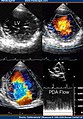
<span class="mw-page-title-main">Left ventricular hypertrophy</span> Medical condition

Left ventricular hypertrophy (LVH) is thickening of the heart muscle of the left ventricle of the heart, that is, left-sided ventricular hypertrophy and resulting increased left ventricular mass.

In cardiology, ventricular remodeling refers to changes in the size, shape, structure, and function of the heart. This can happen as a result of exercise or after injury to the heart muscle. The injury is typically due to acute myocardial infarction, but may be from a number of causes that result in increased pressure or volume, causing pressure overload or volume overload on the heart. Chronic hypertension, congenital heart disease with intracardiac shunting, and valvular heart disease may also lead to remodeling. After the insult occurs, a series of histopathological and structural changes occur in the left ventricular myocardium that lead to progressive decline in left ventricular performance. Ultimately, ventricular remodeling may result in diminished contractile (systolic) function and reduced stroke volume.

<span class="mw-page-title-main">Ventricular hypertrophy</span> Medical condition

Ventricular hypertrophy (VH) is thickening of the walls of a ventricle of the heart. Although left ventricular hypertrophy (LVH) is more common, right ventricular hypertrophy (RVH), as well as concurrent hypertrophy of both ventricles can also occur.

<span class="mw-page-title-main">Uhl anomaly</span> Medical condition

Uhl anomaly is a rare cardiac malformation that was first identified by Dr. Henry Uhl in 1952. It is characterized by the absence of the right ventricle (RV) myocardium, either entirely or partially, and the replacement of the RV myocardium by nonfunctional fibroelastic tissue that resembles parchment. As of 2010 less than 100 cases have been reported in liturature.

<span class="mw-page-title-main">Amrinone</span> Chemical compound

Amrinone, also known as inamrinone, and sold as Inocor, is a pyridine phosphodiesterase 3 inhibitor. It is a drug that may improve the prognosis in patients with congestive heart failure. Amrinone has been shown to increase the contractions initiated in the heart by high-gain calcium induced calcium release (CICR). The positive inotropic effect of amrinone is mediated by the selective enhancement of high-gain CICR, which contributes to the contraction of myocytes by phosphorylation through cAMP dependent protein kinase A (PKA) and Ca2+ calmodulin kinase pathways.

<span class="mw-page-title-main">Takotsubo cardiomyopathy</span> Sudden temporary weakening of the heart muscle

Takotsubo cardiomyopathy or takotsubo syndrome (TTS), also known as stress cardiomyopathy, is a type of non-ischemic cardiomyopathy in which there is a sudden temporary weakening of the muscular portion of the heart. It usually appears after a significant stressor, either physical or emotional; when caused by the latter, the condition is sometimes called broken heart syndrome. Examples of physical stressors that can cause TTS are sepsis, shock, subarachnoid hemorrhage, and pheochromocytoma. Emotional stressors include bereavement, divorce, or the loss of a job. Reviews suggest that of patients diagnosed with the condition, about 70–80% recently experienced a major stressor, including 41–50% with a physical stressor and 26–30% with an emotional stressor. TTS can also appear in patients who have not experienced major stressors.

<span class="mw-page-title-main">Tricuspid regurgitation</span> Type of valvular heart disease

Tricuspid regurgitation (TR), also called tricuspid insufficiency, is a type of valvular heart disease in which the tricuspid valve of the heart, located between the right atrium and right ventricle, does not close completely when the right ventricle contracts (systole). TR allows the blood to flow backwards from the right ventricle to the right atrium, which increases the volume and pressure of the blood both in the right atrium and the right ventricle, which may increase central venous volume and pressure if the backward flow is sufficiently severe.

The following outline is provided as an overview of and topical guide to cardiology, the branch of medicine dealing with disorders of the human heart. The field includes medical diagnosis and treatment of congenital heart defects, coronary artery disease, heart failure, valvular heart disease and electrophysiology. Physicians who specialize in cardiology are called cardiologists.

<span class="mw-page-title-main">Heart failure with preserved ejection fraction</span> Medical condition

Heart failure with preserved ejection fraction (HFpEF) is a form of heart failure in which the ejection fraction – the percentage of the volume of blood ejected from the left ventricle with each heartbeat divided by the volume of blood when the left ventricle is maximally filled – is normal, defined as greater than 50%; this may be measured by echocardiography or cardiac catheterization. Approximately half of people with heart failure have preserved ejection fraction, while the other half have a reduction in ejection fraction, called heart failure with reduced ejection fraction (HFrEF).

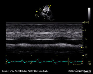
Tissue Doppler echocardiography (TDE) is a medical ultrasound technology, specifically a form of echocardiography that measures the velocity of the heart muscle (myocardium) through the phases of one or more heartbeats by the Doppler effect of the reflected ultrasound. The technique is the same as for flow Doppler echocardiography measuring flow velocities. Tissue signals, however, have higher amplitude and lower velocities, and the signals are extracted by using different filter and gain settings. The terms tissue Doppler imaging (TDI) and tissue velocity imaging (TVI) are usually synonymous with TDE because echocardiography is the main use of tissue Doppler.

Heart problems are more common in people with HIV/AIDS. Those with left ventricular dysfunction have a median survival of 101 days as compared to 472 days in people with AIDS with healthy hearts. HIV is a major cause of cardiomyopathy. The most common type of HIV induced cardiomyopathy is dilated cardiomyopathy also known as eccentric ventricular hypertrophy which leads to impaired contraction of the ventricles due to volume overload. The annual incidence of HIV associated dilated cardiomyopathy was 15.9/1000 before the introduction of highly active antiretroviral therapy (HAART). However, in 2014, a study found that 17.6% of HIV patients have dilated cardiomyopathy (176/1000) meaning the incidence has greatly increased.

<span class="mw-page-title-main">Ischemic cardiomyopathy</span> Medical condition

Ischemic cardiomyopathy is a type of cardiomyopathy caused by a narrowing of the coronary arteries which supply blood to the heart. Typically, patients with ischemic cardiomyopathy have a history of acute myocardial infarction, however, it may occur in patients with coronary artery disease, but without a past history of acute myocardial infarction. This cardiomyopathy is one of the leading causes of sudden cardiac death. The adjective ischemic means characteristic of, or accompanied by, ischemia — local anemia due to mechanical obstruction of the blood supply.

Ayodele Olajide Falase is a Nigerian cardiologist and academic. He is a former vice chancellor of the University of Ibadan. He served as a WHO Expert committee member on cardiopathies and on a WHO expert panel on cardiovascular disease. Professor Ayodele Falase got the Honorary fellowship award at the University of Ibadan 71st founder’s day held in 2019.

<span class="mw-page-title-main">Bernheim syndrome</span>

Bernheim syndrome is a presumed disorder wherein the right ventricle is severely compressed due to a shift in the ventricular septal wall of the heart, leading to heart failure. It was first described by Hippolyte Bernheim in 1910. Today, it is argued whether or not Bernheim syndrome is indeed a syndrome or a side effect of other cardiac conditions, such as left ventrical heart failure where the left ventricle is substantially enlarged, encroaching on the space of the right ventricle.

References

  1. Pignatelli RH, McMahon CJ, Dreyer WJ, Denfield SW, Price J, Belmont JW, et al. (November 2003). "Clinical characterization of left ventricular noncompaction in children: a relatively common form of cardiomyopathy". Circulation. 108 (21): 2672–8. doi: 10.1161/01.CIR.0000100664.10777.B8 . PMID   14623814.
  2. 1 2 3 4 5 Espinola-Zavaleta N, Soto ME, Castellanos LM, Játiva-Chávez S, Keirns C (September 2006). "Non-compacted cardiomyopathy: clinical-echocardiographic study". Cardiovascular Ultrasound. 4: 35. doi: 10.1186/1476-7120-4-35 . PMC   1592122 . PMID   17002802.
  3. 1 2 3 Jenni R, Oechslin E (2005). "Non-compaction of the Left Ventricular Myocardium – From Clinical Observation to the Discovery of a New Disease". European Cardiology Review. 1 (1): 23. doi: 10.15420/ECR.2005.23 . ISSN   1758-3756. S2CID   221234437.
  4. The Cardiomyopathy Association (2007-07-23). "LV Non-compaction" (website). Retrieved 2007-07-23.
  5. Maron BJ, Towbin JA, Thiene G, Antzelevitch C, Corrado D, Arnett D, et al. (April 2006). "Contemporary definitions and classification of the cardiomyopathies: an American Heart Association Scientific Statement from the Council on Clinical Cardiology, Heart Failure and Transplantation Committee; Quality of Care and Outcomes Research and Functional Genomics and Translational Biology Interdisciplinary Working Groups; and Council on Epidemiology and Prevention". Circulation. 113 (14): 1807–16. doi:10.1161/CIRCULATIONAHA.106.174287. PMID   16567565. S2CID   6660623.
  6. Vatta M, Mohapatra B, Jimenez S, Sanchez X, Faulkner G, Perles Z, et al. (December 2003). "Mutations in Cypher/ZASP in patients with dilated cardiomyopathy and left ventricular non-compaction". J. Am. Coll. Cardiol. 42 (11): 2014–27. doi:10.1016/j.jacc.2003.10.021. PMID   14662268. S2CID   25932261.
  7. A publication is expected by Leiden University Medical Centre
  8. Marakhonov AV, Brodehl A, Myasnikov RP, Sparber PA, Kiseleva AV, Kulikova OV, Meshkov AN, Zharikova AA, Koretsky SN, Kharlap MS, Stanasiuk C (June 2019). "Noncompaction cardiomyopathy is caused by a novel in-frame desmin ( DES ) deletion mutation within the 1A coiled-coil rod segment leading to a severe filament assembly defect". Human Mutation. 40 (6): 734–741. doi: 10.1002/humu.23747 . ISSN   1059-7794. PMID   30908796. S2CID   85515283.
  9. 1 2 3 Sedaghat-Hamedani F, Haas J, Zhu F, Geier C, Kayvanpour E, Liss M, et al. (2017). "Clinical genetics and outcome of left ventricular non-compaction cardiomyopathy". European Heart Journal. 38 (46): 3449–3460. doi: 10.1093/eurheartj/ehx545 . PMID   29029073.
  10. Kulikova O, Brodehl A, Kiseleva A, Myasnikov R, Meshkov A, Stanasiuk C, Gärtner A, Divashuk M, Sotnikova E, Koretskiy S, Kharlap M (2021-01-19). "The Desmin (DES) Mutation p.A337P Is Associated with Left-Ventricular Non-Compaction Cardiomyopathy". Genes. 12 (1): 121. doi: 10.3390/genes12010121 . ISSN   2073-4425. PMC   7835827 . PMID   33478057.
  11. Scott DA, Hernandez-Garcia A, Azamian MS, Jordan VK, Kim BJ, Starkovich M, Zhang J, Wong L, Darilek SA, Breman AM, Yang Y (November 2016). "Congenital heart defects and left ventricular non-compaction in males with loss-of-function variants in NONO". Journal of Medical Genetics. 54 (1): 47–53. doi:10.1136/jmedgenet-2016-104039. ISSN   0022-2593. PMID   27550220. S2CID   206998226.
  12. Chang B, Nishizawa T, Furutani M, Fujiki A, Tani M, Kawaguchi M, Ibuki K, Hirono K, Taneichi H, Uese K, Onuma Y (February 2011). "Identification of a novel TPM1 mutation in a family with left ventricular noncompaction and sudden death". Molecular Genetics and Metabolism. 102 (2): 200–206. doi:10.1016/j.ymgme.2010.09.009. ISSN   1096-7206. PMID   20965760.
  13. Kalavakunta JK, Tokala H, Gosavi A, Gupta V (2010-01-01). "Left ventricular noncompaction and myocardial fibrosis: a case report". International Archives of Medicine. 3: 20. doi: 10.1186/1755-7682-3-20 . ISSN   1755-7682. PMC   2945326 . PMID   20843341.
  14. Chin TK, Perloff JK, Williams RG, et al. (Aug 1990). "Isolated noncompaction of left ventricular myocardium. A study of eight cases". Circulation. 82 (2): 507–13. doi: 10.1161/01.cir.82.2.507 . PMID   2372897.
  15. Martínez-Baca López F, Alonso Bravo RM, Rodríguez Huerta DA (2009). "Echocardiographic features of non-compaction cardiomyopathy: missed and misdiagnosed disease". Arquivos Brasileiros de Cardiologia. 93 (2): e33–e35. doi: 10.1590/S0066-782X2009000800024 . PMID   19838476.
  16. Lorenzo Botto, MD (September 2004). "Left Ventricular Non-compacted" (PDF). Archived from the original (PDF) on 2007-02-07. Retrieved 2007-06-13.
  17. Semsarian C, Ingles J, Maron MS (2015). "New perspectives on the prevalence of hypertrophic cardiomyopathy". J Am Coll Cardiol. 65 (12): 1249–54. doi: 10.1016/j.jacc.2015.01.019 . PMID   25814232.
  18. Weiford BC, Subbarao VD, Mulhern KM (2004). "Noncompaction of the ventricular myocardium". Circulation. 109 (24): 2965–71. doi: 10.1161/01.CIR.0000132478.60674.D0 . PMID   15210614.
  19. Engberding R, Bender F (June 1984). "Identification of a rare congenital anomaly of the myocardium by two-dimensional echocardiography: persistence of isolated myocardial sinusoids". Am. J. Cardiol. 53 (11): 1733–4. doi:10.1016/0002-9149(84)90618-0. PMID   6731322.

Further reading